The UCL Optical Brain Imaging System UNIVERSITY COLLEGE LONDON
advertisement

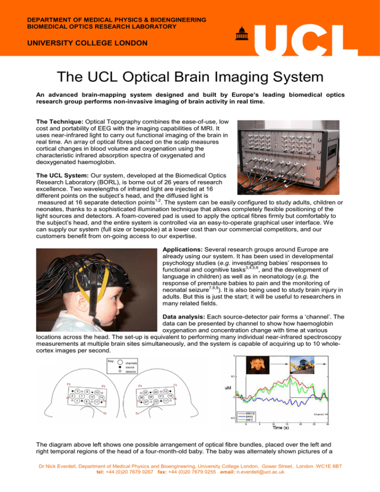
DEPARTMENT OF MEDICAL PHYSICS & BIOENGINEERING BIOMEDICAL OPTICS RESEARCH LABORATORY UNIVERSITY COLLEGE LONDON The UCL Optical Brain Imaging System An advanced brain-mapping system designed and built by Europe’s leading biomedical optics research group performs non-invasive imaging of brain activity in real time. The Technique: Optical Topography combines the ease-of-use, low cost and portability of EEG with the imaging capabilities of MRI. It uses near-infrared light to carry out functional imaging of the brain in real time. An array of optical fibres placed on the scalp measures cortical changes in blood volume and oxygenation using the characteristic infrared absorption spectra of oxygenated and deoxygenated haemoglobin. The UCL System: Our system, developed at the Biomedical Optics Research Laboratory (BORL), is borne out of 26 years of research excellence. Two wavelengths of infrared light are injected at 16 different points on the subject’s head, and the diffused light is 1,2 measured at 16 separate detection points . The system can be easily configured to study adults, children or neonates, thanks to a sophisticated illumination technique that allows completely flexible positioning of the light sources and detectors. A foam-covered pad is used to apply the optical fibres firmly but comfortably to the subject’s head, and the entire system is controlled via an easy-to-operate graphical user interface. We can supply our system (full size or bespoke) at a lower cost than our commercial competitors, and our customers benefit from on-going access to our expertise. Applications: Several research groups around Europe are already using our system. It has been used in developmental psychology studies (e.g. investigating babies’ responses to 3,4,5,6 functional and cognitive tasks , and the development of language in children) as well as in neonatology (e.g. the response of premature babies to pain and the monitoring of 7,8,9 neonatal seizure ). It is also being used to study brain injury in adults. But this is just the start; it will be useful to researchers in many related fields. Data analysis: Each source-detector pair forms a ‘channel’. The data can be presented by channel to show how haemoglobin oxygenation and concentration change with time at various locations across the head. The set-up is equivalent to performing many individual near-infrared spectroscopy measurements at multiple brain sites simultaneously, and the system is capable of acquiring up to 10 wholecortex images per second. The diagram above left shows one possible arrangement of optical fibre bundles, placed over the left and right temporal regions of the head of a four-month-old baby. The baby was alternately shown pictures of a Dr Nick Everdell, Department of Medical Physics and Bioengineering, University College London, Gower Street, London WC1E 6BT tel: +44 (0)20 7679 0267 fax: +44 (0)20 7679 0255 email: n.everdell@ucl.ac.uk woman’s face and a mechanical digger, each for several seconds, for a few minutes in total. The graph on the right plots the concentrations of oxygenated, deoxygenated, and total haemoglobin with time for one particular channel. This reveals a rise in oxyhaemoglobin concentration in response to the face stimulus, along with a slight drop in deoxyhaemoglobin concentration. Imaging: The same data can also be processed into various types of images to highlight changes in haemoglobin oxygenation or concentration. The images to the left depict the left and right temporal regions of the baby’s brain during the previous experiment. The left temporal region responds much more strongly to the face stimulus than the right side. Tried and trusted technology: We have built many systems for 10 +10 various groups around Europe, µmolar concentration including: the Ecole des Hautes change in Hb02 Etudes in Paris, the Central European University in Budapest, Manchester University, King’s College Hospital in South London, Babylab at Birkbeck College (part of the University of London) and of course our own group at UCL. One of our systems is being used to study the effect of malnutrition on the cognitive development of children in the Gambia – this project is funded by the Gates Foundation. - More Information: this can be found at our website at http://www.ucl.ac.uk/medphys/topography and we are very happy to answer any queries you might have – contact details are at the foot of the page. References: Below is a selection of peer-reviewed articles describing experimental work undertaken with the UCL system. PDFs are available at the link above. 1. Everdell N L, A P Gibson , I D C Tullis, T Vaithianathan, J C Hebden, D T Delpy (2005) "A frequency multiplexed near infrared topography system for imaging functional activation in the brain" Review of Scientific Instruments 76, 093705 2. Correia T, A Banga, N L Everdell, A P Gibson, J C Hebden (2009) "A quantitative assessment of the depth sensitivity of an optical topography system using a solid dynamic tissue-phantom" Physics in Medicine and Biology 54 6277-6286 3. Correia T, Lloyd-Fox S, N L Everdell, A Blasi, C Elwell, J C Hebden, A Gibson (2012) "Three-dimensional optical topography of brain activity in infants watching videos of human movement" Physics in Medicine and Biology 57 1135-1146 4. Lloyd-Fox S, A Blasi, N L Everdell, C E Elwell, M H Johnson (2011) "Selective cortical mapping of biological motion processing in young infants" Journal of Cognitive Neuroscience 23(9), 2521-2532 5. Lloyd-Fox S, A Blasi, A Volein, N Everdell, C E Elwell, M H Johnson (2009) "Social Perception in Infancy: a near infrared spectroscopy study" Child Development 80(4), 986-999 6. Blasi A, S Fox, N Everdell, A Volein, L Tucker, G Csibra, A P Gibson, J C Hebden, M H Johnson, C E Elwell (2007) "Investigation of depth dependent changes in cerebral haemodynamics during face perception in infants" Physics in Medicine and Biology 52, 68496864 7. Cooper R J, N L Everdell, L C Enfield, ,A P Gibson, A Worley, J C Hebden (2009) "Design and evaluation of a probe for simultaneous EEG and near-infrared imaging of cortical activation" Physics in Medicine and Biology 54, 2093-2102 8. Cooper R J, D Bhatt, N L Everdell, J C Hebden (2009) "A tissue-like, optically turbid and electrically conducting phantom for simultaneous EEG and near-infrared imaging" Physics in Medicine and Biology 54 N403-N408 9. Cooper RJ, Hebden, JC, O'Reilly H, Mitra S, Mitchell A, Everdell NL, Gibson AP, Austin T (2011) "Transient haemodynamic events in neurologically compromised infants: A simultaneous EEG and diffuse optical imaging study" Neuroimage 55(4), 1610-1616 Dr Nick Everdell, Department of Medical Physics and Bioengineering, University College London, Gower Street, London WC1E 6BT tel: +44 (0)20 7679 0267 fax: +44 (0)20 7679 0255 email: n.everdell@ucl.ac.uk
