Anti-Ikaros antibody ab77411 Product datasheet 1 Abreviews 1 Image
advertisement
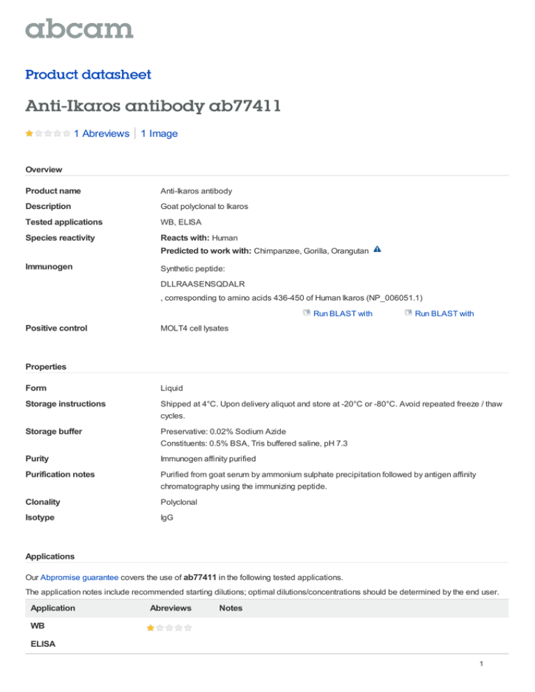
Product datasheet Anti-Ikaros antibody ab77411 1 Abreviews 1 Image Overview Product name Anti-Ikaros antibody Description Goat polyclonal to Ikaros Tested applications WB, ELISA Species reactivity Reacts with: Human Predicted to work with: Chimpanzee, Gorilla, Orangutan Immunogen Synthetic peptide: DLLRAASENSQDALR , corresponding to amino acids 436-450 of Human Ikaros (NP_006051.1) Run BLAST with Positive control Run BLAST with MOLT4 cell lysates Properties Form Liquid Storage instructions Shipped at 4°C. Upon delivery aliquot and store at -20°C or -80°C. Avoid repeated freeze / thaw cycles. Storage buffer Preservative: 0.02% Sodium Azide Constituents: 0.5% BSA, Tris buffered saline, pH 7.3 Purity Immunogen affinity purified Purification notes Purified from goat serum by ammonium sulphate precipitation followed by antigen affinity chromatography using the immunizing peptide. Clonality Polyclonal Isotype IgG Applications Our Abpromise guarantee covers the use of ab77411 in the following tested applications. The application notes include recommended starting dilutions; optimal dilutions/concentrations should be determined by the end user. Application Abreviews Notes WB ELISA 1 Application notes Peptide ELISA: Use at an assay dependent dilution. Antibody detection limit dilution: 1/32000. WB: Use at a concentration of 0.01 - 0.03 µg/ml. Detects a band of approximately 65 kDa (predicted molecular weight: 58 kDa). Not yet tested in other applications. Optimal dilutions/concentrations should be determined by the end user. Target Function Transcription regulator of hematopoietic cell differentiation. Binds gamma-satellite DNA. Binds with higher affinity to gamma satellite A. Plays a role in the development of lymphocytes, B- and T-cells. Binds and activates the enhancer (delta-A element) of the CD3-delta gene. Repressor of the TDT (terminal deoxynucleotidyltransferase) gene during thymocyte differentiation. Regulates transcription through association with both HDAC-dependent and HDAC-independent complexes. Targets the 2 chromatin-remodeling complexes, NuRD and BAF (SWI/SNF), in a single complex (PYR complex), to the beta-globin locus in adult erythrocytes. Increases normal apoptosis in adult erythroid cells. Confers early temporal competence to retinal progenitor cells (RPCs). Tissue specificity Abundantly expressed in thymus, spleen and peripheral blood Leukocytes and lymph nodes. Lower expression in bone marrow and small intestine. Involvement in disease Note=Defects in IKZF1 are frequent occurrences (28.6%) in acute lymphoblasic leukemia (ALL). Such alterations or deletions lead to poor prognosis for ALL. Sequence similarities Belongs to the Ikaros C2H2-type zinc-finger protein family. Contains 6 C2H2-type zinc fingers. Domain The N-terminal zinc-fingers 2 and 3 are required for DNA binding as well as for targeting IKFZ1 to pericentromeric heterochromatin. The C-terminal zinc-finger domain is required for dimerization. Post-translational modifications Phosphorylation controls cell-cycle progression from late G(1) stage to S stage. Hyperphosphorylated during G2/M phase. Dephosphorylated state during late G(1) phase. Phosphorylation on Thr-140 is required for DNA and pericentromeric location during mitosis. CK2 is the main kinase, in vitro. GSK3 and CDK may also contribute to phosphorylation of the C-terminal serine and threonine residues. Phosphorylation on these C-terminal residues reduces the DNA-binding ability. Phosphorylation/dephosphorylation events on Ser-13 and Ser295 regulate TDT expression during thymocyte differentiation. Dephosphorylation by protein phosphatase 1 regulates stability and pericentromeric heterochromatin location. Phosphorylated in both lymphoid and non-lymphoid tissues. Sumoylated. Simulataneous sumoylation on the 2 sites results in a loss of both HDACdependent and HDAC-independent repression. Has no effect on pericentromeric heterochromatin location. Desumoylated by SENP1. Polyubiquitinated. Cellular localization Cytoplasm and Nucleus. In resting lymphocytes, distributed diffusely throughout the nucleus. Localizes to pericentromeric heterochromatin in proliferating cells. This localization requires DNA binding which is regulated by phosphorylation / dephosphorylation events. Form There are 7 isoforms produced by alternative splicing. Anti-Ikaros antibody images 2 Anti-Ikaros antibody (ab77411) at 0.03 µg/ml + MOLT4 cell lysate in RIPA buffer at 35 µg Predicted band size : 58 kDa Observed band size : 65 kDa Western blot - Ikaros antibody (ab77411) Please note: All products are "FOR RESEARCH USE ONLY AND ARE NOT INTENDED FOR DIAGNOSTIC OR THERAPEUTIC USE" Our Abpromise to you: Quality guaranteed and expert technical support Replacement or refund for products not performing as stated on the datasheet Valid for 12 months from date of delivery Response to your inquiry within 24 hours We provide support in Chinese, English, French, German, Japanese and Spanish Extensive multi-media technical resources to help you We investigate all quality concerns to ensure our products perform to the highest standards If the product does not perform as described on this datasheet, we will offer a refund or replacement. For full details of the Abpromise, please visit http://www.abcam.com/abpromise or contact our technical team. Terms and conditions Guarantee only valid for products bought direct from Abcam or one of our authorized distributors 3
![Anti-Caspase-7 antibody [11E4] ab49733 Product datasheet Overview Product name](http://s2.studylib.net/store/data/012098602_1-ce7fa9622a832730158de0f78e12c560-300x300.png)
