Anti-PML Protein antibody ab53773 Product datasheet 13 Abreviews 4 Images
advertisement
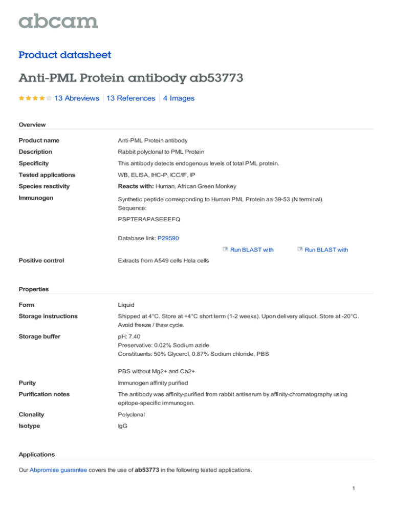
Product datasheet Anti-PML Protein antibody ab53773 13 Abreviews 13 References 4 Images Overview Product name Anti-PML Protein antibody Description Rabbit polyclonal to PML Protein Specificity This antibody detects endogenous levels of total PML protein. Tested applications WB, ELISA, IHC-P, ICC/IF, IP Species reactivity Reacts with: Human, African Green Monkey Immunogen Synthetic peptide corresponding to Human PML Protein aa 39-53 (N terminal). Sequence: PSPTERAPASEEEFQ Database link: P29590 Run BLAST with Positive control Run BLAST with Extracts from A549 cells Hela cells Properties Form Liquid Storage instructions Shipped at 4°C. Store at +4°C short term (1-2 weeks). Upon delivery aliquot. Store at -20°C. Avoid freeze / thaw cycle. Storage buffer pH: 7.40 Preservative: 0.02% Sodium azide Constituents: 50% Glycerol, 0.87% Sodium chloride, PBS PBS without Mg2+ and Ca2+ Purity Immunogen affinity purified Purification notes The antibody was affinity-purified from rabbit antiserum by affinity-chromatography using epitope-specific immunogen. Clonality Polyclonal Isotype IgG Applications Our Abpromise guarantee covers the use of ab53773 in the following tested applications. 1 The application notes include recommended starting dilutions; optimal dilutions/concentrations should be determined by the end user. Application WB Abreviews Notes 1/500 - 1/1000. Detects a band of approximately 100 kDa (predicted molecular weight: 69 kDa). ELISA 1/10000. IHC-P Use a concentration of 4 µg/ml. Perform heat mediated antigen retrieval before commencing with IHC staining protocol. ICC/IF 1/100 - 1/250. IP Use at an assay dependent concentration. PubMed: 23222847 Target Function Key component of PML nuclear bodies that regulate a large number of cellular processes by facilitating post-translational modification of target proteins, promoting protein-protein contacts, or by sequestering proteins. Functions as tumor suppressor. Required for normal, caspasedependent apoptosis in response to DNA damage, FAS, TNF, or interferons. Plays a role in transcription regulation, DNA damage response, DNA repair and chromatin organization. Plays a role in processes regulated by retinoic acid, regulation of cell division, terminal differentiation of myeloid precursor cells and differentiation of neural progenitor cells. Required for normal immunity to microbial infections. Plays a role in antiviral response. In the cytoplasm, plays a role in TGFB1-dependent processes. Regulates p53/TP53 levels by inhibiting its ubiquitination and proteasomal degradation. Regulates activation of p53/TP53 via phosphorylation at 'Ser-20'. Sequesters MDM2 in the nucleolus after DNA damage, and thereby inhibits ubiquitination and degradation of p53/TP53. Regulates translation of HIF1A by sequestering MTOR, and thereby plays a role in neoangiogenesis and tumor vascularization. Regulates RB1 phosphorylation and activity. Required for normal development of the brain cortex during embryogenesis. Can sequester herpes virus and varicella virus proteins inside PML bodies, and thereby inhibit the formation of infectious viral particles. Regulates phosphorylation of ITPR3 and plays a role in the regulation of calcium homeostasis at the endoplasmic reticulum (By similarity). Regulates transcription activity of ELF4. Inhibits specifically the activity of the tetrameric form of PKM2. Together with SATB1, involved in local chromatin-loop remodeling and gene expression regulation at the MHC-I locus. Regulates PTEN compartmentalization through the inhibition of USP7-mediated deubiquitinylation. Involvement in disease Note=A chromosomal aberration involving PML may be a cause of acute promyelocytic leukemia (APL). Translocation t(15;17)(q21;q21) with RARA. The PML breakpoints (type A and type B) lie on either side of an alternatively spliced exon. Sequence similarities Contains 2 B box-type zinc fingers. Contains 1 RING-type zinc finger. Domain Interacts with PKM2 via its coiled-coil domain. Binds arsenic via the RING-type zinc finger. Post-translational modifications Ubiquitinated; mediated by RNF4, SIAH1 or SIAH2 and leading to subsequent proteasomal degradation. 'Lys-6'-, 'Lys-11'-, 'Lys-48'- and 'Lys-63'-linked polyubiquitination by RNF4 is polysumoylation-dependent. Undergoes 'Lys-11'-linked sumoylation. Sumoylation on all three sites is required for nuclear body formation. Sumoylation on Lys-160 is a prerequisite for sumoylation on Lys-65. The PMLRARA fusion protein requires the coiled-coil domain for sumoylation. Desumoylated by SENP2 and SENP6. Arsenic induces PML and PML-RARA oncogenic fusion proteins polysumoylation and their subsequent RNF4-dependent ubiquitination and proteasomal degradation, and is used 2 as treatment in acute promyelocytic leukemia (APL). Phosphorylated in response to DNA damage, probably by ATR. Acetylation may promote sumoylation and enhance induction of apoptosis. Cellular localization Nucleus > nucleoplasm. Cytoplasm. Nucleus > PML body. Nucleus > nucleolus. Endoplasmic reticulum membrane. Early endosome membrane. Sumoylated forms localize to the PML nuclear bodies. The B1 box and the RING finger are also required for this nuclear localization. Isoforms lacking a nuclear localization signal are cytoplasmic. Detected in the nucleolus after DNA damage. Sequestered in the cytoplasm by interaction with rabies virus phosphoprotein. Anti-PML Protein antibody images ab53773 (1/200) staining PML protein in Hela cells (green). Cells were fixed with methanol, permeabilised with triton X-100/PBS and counterstained with DAPI in order to highlight the nucleus (red). Please refer to abreview for Immunocytochemistry/ Immunofluorescence - further experimental details. PML Protein antibody (ab53773) This image is courtesy of an Abreview submitted by Dr Kirk McManus Chromatin was prepared from modified murine D3 ES cells (an in vitro model of RB1 imprinted expression was generated by integrating human PPP1R26P1 into intron 2 of mouse Rb1 in murine embryonic stem cells (mESCs)). Cells were fixed with formaldehyde for 10 mins. Chromatin was sheared into fragment sizes ranging from 200 to 600 bp. For immunoprecipitation 50 μl of sheared chromatin was set aside as input and 20-40 μg of sheared chromatin in a ChIP - Anti-PML Protein antibody (ab53773) volume of 100 μl was used for antibody Image from Steenpass et al. PLoS One. 2013 8(9):e74159. Figure 3. doi: 10.1371/journal.pone.0074159. eCollection 2013 incubation. 4 µg of ab53773 to PML was used as a non-related control (black bars) and 4 µl of a rabbit polyclonal to RNAPII CTD repeat YSPTSPS (phospho S5) (shaded bars) was added as the test. The immunoprecipitated DNA was quantified at 3 positions on the the retinoblastoma (Rb1) promoter. 1,2 = Rb1_48; 3,4 = Rb1_69 and 5,6 = Rb1_74 3 All lanes : Anti-PML Protein antibody (ab53773) at 1/500 dilution Lane 1 : Extracts from A549 cells Lane 2 : Extracts from A549 cells, plus immunising peptide Predicted band size : 69 kDa Observed band size : 100 kDa Western blot - PML Protein antibody (ab53773) Ab53773 staining Human normal colon. Staining is localized to the nucleus. Left panel: with primary antibody at 4 ug/ml. Right panel: isotype control. Sections were stained using an automated Immunohistochemistry (Formalin/PFA-fixed system DAKO Autostainer Plus , at room paraffin-embedded sections) - PML Protein temperature. Sections were rehydrated and antibody (ab53773) antigen retrieved with the Dako 3-in-1 AR buffer EDTA pH 9.0 in a DAKO PT Link. Slides were peroxidase blocked in 3% H2O2 in methanol for 10 minutes. They were then blocked with Dako Protein block for 10 minutes (containing casein 0.25% in PBS), then incubated with primary antibody for 20 minutes, and detected with Dako Envision Flex amplification kit for 30 minutes. Colorimetric detection was completed with diaminobenzidine for 5 minutes. Slides were counterstained with Haematoxylin and coverslipped under DePeX. Please note that for manual staining we recommend to optimize the primary antibody concentration and incubation time (overnight incubation), and amplification may be required. Please note: All products are "FOR RESEARCH USE ONLY AND ARE NOT INTENDED FOR DIAGNOSTIC OR THERAPEUTIC USE" Our Abpromise to you: Quality guaranteed and expert technical support Replacement or refund for products not performing as stated on the datasheet Valid for 12 months from date of delivery Response to your inquiry within 24 hours We provide support in Chinese, English, French, German, Japanese and Spanish 4 Extensive multi-media technical resources to help you We investigate all quality concerns to ensure our products perform to the highest standards If the product does not perform as described on this datasheet, we will offer a refund or replacement. For full details of the Abpromise, please visit http://www.abcam.com/abpromise or contact our technical team. Terms and conditions Guarantee only valid for products bought direct from Abcam or one of our authorized distributors 5
![Anti-PML Protein antibody [C7] ab96055 Product datasheet 2 Images Overview](http://s2.studylib.net/store/data/012095338_1-4868c878433c7243c74f795acef6727f-300x300.png)
