Anti-BMP7 antibody ab56023 Product datasheet 3 Abreviews 5 Images
advertisement
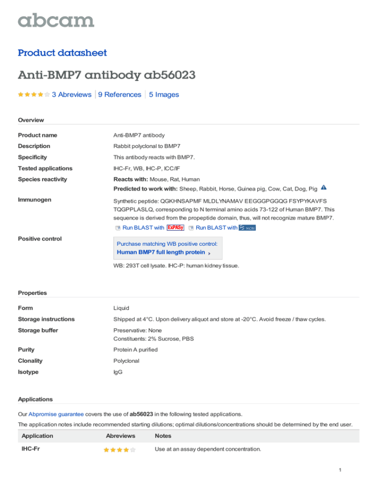
Product datasheet Anti-BMP7 antibody ab56023 3 Abreviews 9 References 5 Images Overview Product name Anti-BMP7 antibody Description Rabbit polyclonal to BMP7 Specificity This antibody reacts with BMP7. Tested applications IHC-Fr, WB, IHC-P, ICC/IF Species reactivity Reacts with: Mouse, Rat, Human Predicted to work with: Sheep, Rabbit, Horse, Guinea pig, Cow, Cat, Dog, Pig Immunogen Synthetic peptide: QGKHNSAPMF MLDLYNAMAV EEGGGPGGQG FSYPYKAVFS TQGPPLASLQ, corresponding to N terminal amino acids 73-122 of Human BMP7. This sequence is derived from the propeptide domain, thus, will not recognize mature BMP7. Run BLAST with Positive control Run BLAST with Purchase matching WB positive control: Human BMP7 full length protein WB: 293T cell lysate. IHC-P: human kidney tissue. Properties Form Liquid Storage instructions Shipped at 4°C. Upon delivery aliquot and store at -20°C. Avoid freeze / thaw cycles. Storage buffer Preservative: None Constituents: 2% Sucrose, PBS Purity Protein A purified Clonality Polyclonal Isotype IgG Applications Our Abpromise guarantee covers the use of ab56023 in the following tested applications. The application notes include recommended starting dilutions; optimal dilutions/concentrations should be determined by the end user. Application IHC-Fr Abreviews Notes Use at an assay dependent concentration. 1 Application Abreviews WB Notes Use a concentration of 1 - 5 µg/ml. Detects a band of approximately 49 kDa (predicted molecular weight: 49 kDa).Can be blocked with Human BMP7 peptide (ab111668). Good results were obtained when blocked with 5% non-fat dry milk in 0.05% PBS-T. IHC-P Use a concentration of 4 - 8 µg/ml. ICC/IF Use a concentration of 1 - 5 µg/ml. Target Function Induces cartilage and bone formation. May be the osteoinductive factor responsible for the phenomenon of epithelial osteogenesis. Plays a role in calcium regulation and bone homeostasis. Tissue specificity Expressed in the kidney and bladder. Lower levels seen in the brain. Sequence similarities Belongs to the TGF-beta family. Developmental stage Expressed in the developing eye, brain and ear during embryogenesis. Post-translational modifications Several N-termini starting at positions 293, 300, 315 and 316 have been identified by direct sequencing resulting in secretion of different mature forms (PubMed:17977014). Cellular localization Secreted. Anti-BMP7 antibody images Immunohistochemistry (Formalin/PFA-fixed paraffin-embedded sections) analysis of human kidney tissue labelling BMP7 with ab56023 at 4-8µg/ml. Arrows indicate positively labelled epithelial cells of the renal tubule. Magnification: 400X. Immunohistochemistry (Formalin/PFA-fixed paraffin-embedded sections) - Anti-BMP7 antibody (ab56023) 2 Lane 1 : Anti-BMP7 antibody (ab56023) at 1.25 µg/ml Lane 2 : Anti-BMP7 antibody (ab56023) at 2.5 µg/ml Lane 3 : Anti-BMP7 antibody (ab56023) at 5 µg/ml Lane 1 : Cell lysate prepared from BMP7 transfected 293T cells Lane 2 : Cell lysate prepared from BMP7 transfected 293T cells Western blot - BMP7 antibody (ab56023) Lane 3 : Cell lysate prepared from BMP7 transfected 293T cells Lysates/proteins at 25 µg per lane. Predicted band size : 49 kDa ICC/IF image of ab56023 stained HeLa cells. The cells were 4% PFA fixed (10 min) and then incubated in 1%BSA / 10% normal goat serum / 0.3M glycine in 0.1% PBS-Tween for 1h to permeabilise the cells and block nonspecific protein-protein interactions. The cells were then incubated with the antibody (ab56023, 1µg/ml) overnight at +4°C. The Immunocytochemistry/ Immunofluorescence BMP7 antibody (ab56023) secondary antibody (green) was Alexa Fluor® 488 goat anti-rabbit IgG (H+L) used at a 1/1000 dilution for 1h. Alexa Fluor® 594 WGA was used to label plasma membranes (red) at a 1/200 dilution for 1h. DAPI was used to stain the cell nuclei (blue) at a concentration of 1.43µM. 3 All lanes : Anti-BMP7 antibody (ab56023) at 1/500 dilution Lane 1 : Rat Brain Whole Tissue (age P8) Lysate Lane 2 : Rat Brain Whole Tissue (age P15) Lysate Lane 3 : Rat Brain Whole Tissue (age P21) Lysate Western blot - BMP7 antibody (ab56023) Ruma Raha-Chowdhury, University Of Cambridge, United Kingdom Lane 4 : Rat Brain Whole Tissue (age P21) Lysate Lysates/proteins at 20 µg per lane. Secondary HRP conjugated at 1/3000 dilution Performed under reducing conditions. Predicted band size : 49 kDa Observed band size : 49 kDa Exposure time : 30 seconds Ruma Raha-Chowdhury, University Of Cambridge, United Kingdom A number of additional non-specific bands were also observed at 17, 33, 35 & 40kDa ab56023 staining mouse e18 skeleton tissues by immunohistochemistry (frozen sections). Sections were formaldehyde fixed prior to being blocked in 20% serum for 1 hour at RT. Sample was incubated with ab56023, diluted 1/1000, for 16 hours at 4°C. Alexa fluor® 680 donkey polyclonal, diluted 1/200, was used as the secondary antibody. Immunohistochemistry (Frozen sections) - BMP7 antibody (ab56023) This image is courtesy of an anonymous Abreview Please note: All products are "FOR RESEARCH USE ONLY AND ARE NOT INTENDED FOR DIAGNOSTIC OR THERAPEUTIC USE" Our Abpromise to you: Quality guaranteed and expert technical support Replacement or refund for products not performing as stated on the datasheet 4 Valid for 12 months from date of delivery Response to your inquiry within 24 hours We provide support in Chinese, English, French, German, Japanese and Spanish Extensive multi-media technical resources to help you We investigate all quality concerns to ensure our products perform to the highest standards If the product does not perform as described on this datasheet, we will offer a refund or replacement. For full details of the Abpromise, please visit http://www.abcam.com/abpromise or contact our technical team. Terms and conditions Guarantee only valid for products bought direct from Abcam or one of our authorized distributors 5
