Comparison of Kinematics and Kinetics During Drop and Drop Jump Performance
advertisement
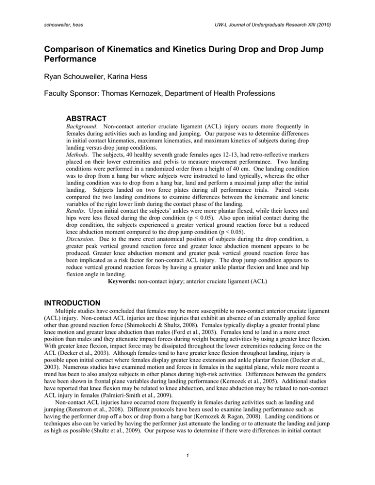
schouweiler, hess UW-L Journal of Undergraduate Research XIII (2010) Comparison of Kinematics and Kinetics During Drop and Drop Jump Performance Ryan Schouweiler, Karina Hess Faculty Sponsor: Thomas Kernozek, Department of Health Professions ABSTRACT Background. Non-contact anterior cruciate ligament (ACL) injury occurs more frequently in females during activities such as landing and jumping. Our purpose was to determine differences in initial contact kinematics, maximum kinematics, and maximum kinetics of subjects during drop landing versus drop jump conditions. Methods. The subjects, 40 healthy seventh grade females ages 12-13, had retro-reflective markers placed on their lower extremities and pelvis to measure movement performance. Two landing conditions were performed in a randomized order from a height of 40 cm. One landing condition was to drop from a hang bar where subjects were instructed to land typically, whereas the other landing condition was to drop from a hang bar, land and perform a maximal jump after the initial landing. Subjects landed on two force plates during all performance trials. Paired t-tests compared the two landing conditions to examine differences between the kinematic and kinetic variables of the right lower limb during the contact phase of the landing. Results. Upon initial contact the subjects’ ankles were more plantar flexed, while their knees and hips were less flexed during the drop condition (p < 0.05). Also upon initial contact during the drop condition, the subjects experienced a greater vertical ground reaction force but a reduced knee abduction moment compared to the drop jump condition (p < 0.05). Discussion. Due to the more erect anatomical position of subjects during the drop condition, a greater peak vertical ground reaction force and greater knee abduction moment appears to be produced. Greater knee abduction moment and greater peak vertical ground reaction force has been implicated as a risk factor for non-contact ACL injury. The drop jump condition appears to reduce vertical ground reaction forces by having a greater ankle plantar flexion and knee and hip flexion angle in landing. Keywords: non-contact injury; anterior cruciate ligament (ACL) INTRODUCTION Multiple studies have concluded that females may be more susceptible to non-contact anterior cruciate ligament (ACL) injury. Non-contact ACL injuries are those injuries that exhibit an absence of an externally applied force other than ground reaction force (Shimokochi & Shultz, 2008). Females typically display a greater frontal plane knee motion and greater knee abduction than males (Ford et al., 2003). Females tend to land in a more erect position than males and they attenuate impact forces during weight bearing activities by using a greater knee flexion. With greater knee flexion, impact force may be dissipated throughout the lower extremities reducing force on the ACL (Decker et al., 2003). Although females tend to have greater knee flexion throughout landing, injury is possible upon initial contact where females display greater knee extension and ankle plantar flexion (Decker et al., 2003). Numerous studies have examined motion and forces in females in the sagittal plane, while more recent a trend has been to also analyze subjects in other planes during high-risk activities. Differences between the genders have been shown in frontal plane variables during landing performance (Kernozek et al., 2005). Additional studies have reported that knee flexion may be related to knee abduction, and knee abduction may be related to non-contact ACL injury in females (Palmieri-Smith et al., 2009). Non-contact ACL injuries have occurred more frequently in females during activities such as landing and jumping (Renstrom et al., 2008). Different protocols have been used to examine landing performance such as having the performer drop off a box or drop from a hang bar (Kernozek & Ragan, 2008). Landing conditions or techniques also can be varied by having the performer just attenuate the landing or to attenuate the landing and jump as high as possible (Shultz et al., 2009). Our purpose was to determine if there were differences in initial contact 1 schouweiler, hess UW-L Journal of Undergraduate Research XIII (2010) kinematics, maximum kinematics, and maximum kinetics of landing performance during drop versus drop jump landing conditions. The landing techniques from the two protocols were expected to have no difference in kinetic and kinematic factors. METHODS Forty-seventh grade females ((age range 12-13 years), mean height = 159.84 (SD 6.74) cm, mean weight = 51.78 (SD 11.93) kg) completed five landings of each condition from a hang bar. The hang bar was adjusted to a height of 40 cm from each subject’s foot to the floor when the ankle was in an anatomical neutral position. Each subject provided three practice trials prior to each set of recorded trials. Subjects provided written informed consent, prior to participation, according to university campus guidelines. An eight-camera motion analysis system (Motion Analysis Corporation, Santa Rosa, CA, USA) sampling at 240 Hz and two synchronized force platforms (Bertec Corporation, Columbus, OH) sampling at 2400 Hz were used to collect kinetic and kinematic data. Three retro-reflective surface markers were placed on each segment (thighs, shanks, foot segments, and pelvis) to capture the three-dimensional coordinates of the subjects using a modified Helen Hayes marker set (see Figure 1). A 4th order recursive Butterworth low-pass filter was applied to kinematic and kinetic data at 20 Hz. Cadaveric data were used to estimate hip joint centers based on inter-anterior superior iliac spine distance pelvis coordinates. Joint centers of the knee were calculated by markers placed on the medial and lateral condyles, based on a static neutral (erect) trial. Thigh markers, the hip joint center, and the distance halfway between the femoral condyles defined a plane in line with the knee joint center. Ankle joint centers were calculated by markers placed on the medial and lateral and malleoli, based on a static neutral (erect) trial. Medial and lateral malleoli, shank markers, and the calculated knee center, defined a plane within which the ankle joint center was located. To transition between laboratory and anatomical coordinate systems, calculations were based on the static neutral trial, where zero degrees corresponded to an erect posture. In calculating ankle angle, the foot segment was at a right angle to the shank. The Euler sequence selected for the segment rotations was flexionextension, abduction-adduction, and internal-external rotation. Motion Monitor Software (Version 7.0, Innovative Sports Training, Chicago, IL, USA) was used to calculate joint moments and angles. The Motion Monitor software used the inverse dynamic approach to calculate joint moments (Kadaba et al., 1990, 1989). Figure 1. Surface retro-reflective markers placed to the subjects’ thighs, shanks, foot segments, and pelvis. Gray rectangles depict the force platforms in the measurement space. Two landing conditions were performed in a randomized order. One landing condition required a drop from a hang bar where subjects were instructed to land typically (see Figure 2), whereas the other landing condition required a drop from a hang bar, landing and a maximal jump after the initial landing (see Figure 3). Subjects landed on two force plates during all performance trials. Paired sample t-tests compared the two landing conditions to examine differences between the kinematic (ankle plantar flexion angle, knee abduction angle, knee flexion angle, 2 schouweiler, hess UW-L Journal of Undergraduate Research XIII (2010) hip abduction angle, hip flexion angle) and kinetic (ankle moment, knee abduction moment, knee flexion moment, hip abduction moment, hip flexion moment, ground reaction force) variables of the right lower limb during the contact phase of the landing. A p-value less than 0.05 reflected a difference between the two landing conditions. Hanging Position Initial Ground Contact Maximum Flexion End Figure 2. The drop landing condition where subjects were instructed to drop from a hang bar and to land typically. Hanging Position Initial Ground Contact Maximum Flexion Leave Ground In Air Second Landing End Figure 3. The drop jump landing condition where subjects were instructed to drop from a hang bar, land and perform a maximal jump after the initial landing. RESULTS Each subjects’ data were averaged for each drop landing condition for analysis. A paired t-test determined performance differences between the two landing conditions. Upon initial contact, the knee flexion angle for the drop jump landing condition was greater than the drop landing condition (p < 0.05). This indicated that the subjects’ knees showed greater flexion during the drop jump condition, whereas subjects in the drop condition showed a more extended knee position (see Figure 4). The maximum knee flexion angle, shown by subjects after initial contact, for the drop jump landing condition was also greater than during the drop landing condition (p < 0.05) (see Figure 4). This means that overall; subjects’ showed more knee flexion in the drop jump landing conditions. However, the maximum knee flexion moment for the drop jump landing condition was not different from the maximum knee flexion moment for the drop landing condition (p > 0.05) (see Figure 4). 3 schouweiler, hess UW-L Journal of Undergraduate Research XIII (2010) Maximum Knee Flexion Initial Contact Knee Flexion 100 Knee Flexion Angle (Degrees) Knee Flexion Angle (Degrees) 30 25 20 15 10 5 0 90 80 70 60 50 40 30 20 10 0 Maximum Knee Flexion Moment Knee Flexion Moment (N-m) 0 -20 -40 -60 -80 -100 -120 -140 Figure 4. The peak mean values for knee flexion angle at initial contact, maximum flexion, and knee flexion moment. The white bars represent the mean knee flexion angles during the drop landing condition while the grey bars represent the mean knee flexion values during the drop jump landing condition. The standard deviation is shown by the vertical lines in the middle of the graphs. The stars in the upper right corner of the graphs indicate a significant difference between the two landing conditions (p<0.05). During initial contact, the knee adduction angle was not different during the drop landing condition when compared to the drop jump landing condition (p > 0.05) (see Figure 5). The maximum knee abduction angle was greater in the drop jump landing condition than the drop landing condition (p < 0.05) (see Figure 5). The maximum knee abduction moment was also greater in the drop jump landing condition than the drop landing condition (p < 0.05) (see Figure 5). Maximum Knee Abduction 7 Knee Abduction Angle (Degrees) Knee Abduction Angle (Degrees) Initial Contact Knee Adduction 6 5 4 3 2 1 0 -1 4 8 6 4 2 0 -2 -4 -6 -8 -10 -12 -14 schouweiler, hess UW-L Journal of Undergraduate Research XIII (2010) Knee Abduction Moment (N-m) Maximum Knee Abduction Moment 35 30 25 20 15 10 5 0 Figure 5. The peak mean values of knee abduction between the initial contact, maximum abduction, and the maximum knee abduction moment were displayed. The white bars represent the mean knee abduction values during the drop landing condition, while the grey bars represent the mean knee abduction values during the drop jump landing condition. The standard deviation of the data set is also shown by the vertical lines in the middle of the graphs. The stars in the upper right corner of the graphs indicate a difference between the two landing conditions (p < 0.05). During initial contact, the mean ankle plantar flexion angle was greater in the drop landing condition than the mean drop jump landing condition (see Figure 6). The mean maximum ankle dorsiflexion angle was greater during the drop jump landing condition than the drop landing condition (see Figure 6). In addition, the average maximum ankle dorsiflexion moment was greater during the drop landing condition than the drop jump landing condition (see Figure 6). Maximum Ankle Dorsiflexion Ankle Dorsiflexion Angle (Degrees) Ankle Plantar Flexion Angle (Degrees) Initial Contact Ankle Plantar Flexion 0 -5 -10 -15 -20 -25 -30 -35 -40 45 40 35 30 25 20 15 10 5 0 Ankle Dorsiflexion Moment (N-m) Maximum Ankle Dorsiflexion Moment 0 -0.2 -0.4 -0.6 -0.8 -1 -1.2 Figure 6. The graphs display the mean values of abduction between the initial contact, maximum dorsiflexion, and the maximum ankle dorsiflexion moment. Where the white bars represent are the mean ankle dorsiflexion/plantar flexion values during the drop landing condition, while the grey bars represent the mean ankle dorsiflexion/plantar flexion values during the drop jump landing condition. The standard deviation of the data set is also shown by the vertical lines in the middle of the graphs. The stars in the upper right corner of the graphs indicate a significant difference between the two landing conditions (p < 0.05). 5 schouweiler, hess UW-L Journal of Undergraduate Research XIII (2010) The maximum ground reaction force for the drop landing condition was greater than the drop jump landing condition (see Figure 7). Maximum Ground Reaction Force Ground Reaction Force (N) 1200 1000 800 600 400 200 0 Figure 7. The graph displays the mean values of maximum ground reaction force. Where the white bars represent the mean maximum ground reaction force for the drop landing condition, while the grey bars represent the mean maximum ground reaction force for the drop jump landing condition. The standard deviation of the data set is also shown by the vertical lines in the middle of the graphs. The star in the upper right corner of the graph indicates a significant difference between the two landing conditions (p < 0.05). DISCUSSION The landing techniques from the two protocols were not expected to have different kinetic and kinematic factors. Our results showed that there were differences in some of the kinetic and kinematic factors such as initial knee flexion angle, initial hip flexion angle, initial ankle plantar flexion angle, maximum knee flexion angle, maximum knee adduction angle, maximum hip flexion angle, maximum hip abduction angle, maximum ankle dorsiflexion angle, maximum knee abduction moment, maximum ankle dorsiflexion moment, and the vertical ground reaction force. This indicates that drop landing protocol used influences many of kinetic and kinematic factors. The drop landing condition showed reduced hip and knee flexion angles, and a greater ankle plantar flexion angle upon initial contact with the ground. These three factors have been reported to contribute to greater stiffness of the lower extremities (Shultz et al., 2009). A more erect (stiff) landing may cause the quadriceps muscles to be more activated placing a greater anterior force on the tibia. During the drop jump landing condition, there was greater knee flexion, hip flexion, and a less ankle plantar flexion. Due to the greater flexion of the lower extremities during the drop jump landing condition, the vertical ground reaction forces were reduced. The ground reaction force was lower during the drop jump landing condition. Greater peak vertical ground reaction force has been linked to a greater incidence of non-contact ACL injury (Hewett, 2009). Therefore, it may be more likely that the drop landing condition poses mechanics that are more risky for the ACL. During the drop jump condition, maximum knee flexion angle, maximum knee adduction angle, maximum hip flexion angle, maximum hip abduction angle, maximum ankle dorsiflexion angle, maximum knee abduction moment, and maximum ankle dorsiflexion moment were all greater. These factors indicated that the overall flexion of the lower extremities was greater during the drop jump condition. This study may indicate that greater lower extremity flexion may be the reason for the greater ankle dorsiflexion moment, as well as the greater knee abduction moment found. Knee abduction has been related to increased risk of ACL injury (Hewett, 2009). Another relevant finding was a greater external ankle dorsiflexion moment during the drop jump landing condition. This indicated that the plantar flexors of the ankle were producing a larger moment during the drop jump. Activation of the ankle plantar flexors has been implicated in applying a shear force to the tibia loading the ACL (Decker et al., 2003). It appeared that the drop jump condition may pose a more risky type of landing in terms of ACL injury risk. LIMITATIONS For a clearer understanding of non-contact ACL injury, we need to better understand when the mechanism of an ACL tear occurs. It may be also beneficial to measure muscle activation during the landing conditions. With a 6 schouweiler, hess UW-L Journal of Undergraduate Research XIII (2010) more clear understanding of non-contact ACL injury it may be possible to teach proper landing technique to reduce the risk of ACL injury. ACKNOWLEDGEMENTS We would like to thank all of the seventh grade females from Lincoln, Longfellow, and West Salem Middle Schools for participating in our study. We would also like to thank Dr. Thomas Kernozek for his guidance and support throughout the study. REFERENCES Decker, M.J., Torry, M.R., Wyland, D.J., Sterett, W.I., & Steadman, R.J. (2003). Gender differences in lower extremity kinematics, kinetics and energy absorption during landing. Clin Biomech, 18(7), 662-669. Ford, K.R., Myer, G.D., & Hewett, T.E. (2003). Valgus knee motion during landing in high school female and male basketball players. Med Sci Sports Exerc, 35(10), 1745-1750. Hewett, T. (2009). Neuromuscular intervention targeted to mechanisms of acl load in female. ClinicalTrialsFeeds.org, Retrieved from http://clinicaltrialsfeeds.org/clinical-trials/show/NCT01034527 Kadaba, M.P., Ramakrishnan, H.K., Wooten, M.E., Gainey, J., Gorton, G., Cochran, V. (1989). Repeatability of kinematic, kinetic, and electromyography data in normal adult gait. J. Orthop. RES. 7, 849-860. Kernozek, T.W., & Ragan, R.J. (2008). Estimation of anterior cruciate ligament tension from inverse dynamics data and electromyography in females during drop landing. Clin Biomech, 23(10), 1279-1286. Kernozek, T.W., Torry, M.R., Van Hoof, H., Cowley, H., & Tanner, S. (2005). Gender differences in frontal and sagittal plane biomechanics during drop landings. Med Sci Sports Exerc, 37(6), 1003-1012. Palmieri-Smith, R.M., McLean, S.G., Ashton-Miller, J.A., & Wojtys, E.M. (2009). Association of quadriceps and hamstrings cocontraction patterns with knee joint loading. J Athl Train, 44(3), Retrieved from http://www.ncbi.nlm.nih.gov/pmc/articles/PMC2681209/ doi: PMC2681209. Renstrom, P., Ljungqvist, A., Arendt, E., Beynnon, B., & Fukubayashi, T. (2008). Non-contact acl injuries in female athletes: an international olympic committee current concepts statement. Br J Sports Med, 42(6), 394-412. Shimokochi, Y., & Shultz, S.J. (2008). Mechanisms of non-contact anterior cruciate ligament injury. Journal of Athletic Training, 43(4), 396-408. Shultz, S.J., Schmitz, R.J., Nguyen, A.D., & Levine, B.J. (2009). Joint laxity is related to lower extremity energetics during a drop jump landing. Med Sci Sports Exerc, Epub ahead of print. 7
