A NOTE ON A MATHEMATICAL MODEL FOR THE INITIATION OF... ANGIOGENESIS WITH AN APPLICATION TO METASTASIS AND A REMARK ON
advertisement
A NOTE ON A MATHEMATICAL MODEL FOR THE INITIATION OF TUMOR
ANGIOGENESIS WITH AN APPLICATION TO METASTASIS AND A REMARK ON
THE ROLE OF AMPLIFICATION IN SIGNAL TRANSDUCTION
HOWARD A. LEVINE & BRIAN D. SLEEMAN & MARIT NILSEN-HAMILTON
Abstract. Concerning the model we presented in [6], we correct an oversight, simplify a derivation and
make two observations, one concerning metastasis and the other concerning cell signal amplification and
transduction.
In [6] a mathematical model developed in [5] to describe the initiation of capillary formation in tumor
angiogenesis was extended to include the role of pericytes and macrophages in regulating angiogenesis.
The model also included a mechanism whereby anti-angiogenesis factors either directly inhibit angiogenesis
or cause endothelial cells to express inhibitors which arrest angiogenesis by inhibiting the protease expressed
by VEGF in response to tumor emitted growth factor. The model is based on the observation that that
at least one anti-angiogenesis factor, thrombospondin, can prevent the degradation of fibronectin F by
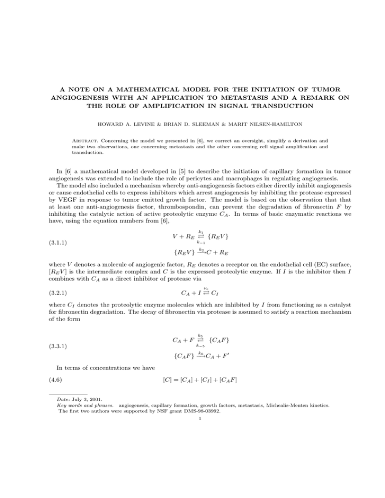
inhibiting the catalytic action of active proteolytic enzyme CA . In terms of basic enzymatic reactions we
have, using the equation numbers from [6],
k1
V + RE {RE V }
(3.1.1)
k−1
k
2
{RE V } −→C
+ RE
where V denotes a molecule of angiogenic factor, RE denotes a receptor on the endothelial cell (EC) surface,
[RE V ] is the intermediate complex and C is the expressed proteolytic enzyme. If I is the inhibitor then I
combines with CA as a direct inhibitor of protease via
(3.2.1)
νe
CA + I CI
where CI denotes the proteolytic enzyme molecules which are inhibited by I from functioning as a catalyst
for fibronectin degradation. The decay of fibronectin via protease is assumed to satisfy a reaction mechanism
of the form
k5
CA + F {CA F }
(3.3.1)
k−5
k
6
0
{CA F } −→C
A+F
In terms of concentrations we have
(4.6)
[C] = [CA ] + [CI ] + [CA F ]
Date: July 3, 2001.
Key words and phrases. angiogenesis, capillary formation, growth factors, metastasis, Michealis-Menten kinetics.
The first two authors were supported by NSF grant DMS-98-03992.
1
2
HOWARD A. LEVINE & BRIAN D. SLEEMAN & MARIT NILSEN-HAMILTON
where
[CI ] = νe [I][CA ],
[CA F ] = ν3 [CA ][F ].
The term [CA F ] in (4.6) was inadvertently omitted from [6]. From (4.6) it follows that
(0.1)
[CA ] =
[C]
.
1 + νe [I] + ν3 [F ]
Based on the parameter values νe = 1.0 × 10³ , ν3 = 0.002 given in Table I of [6], we have
[CA ] ≈
[C]
.
1 + νe [I]
In addition, we need to know that the partial derivative, [Ca ]x , is likewise small when the inhibitor concentration is large. A routine calculation gives:
(0.2)
∂[CA ]
[C]x
[CA ](νe [I]x + ν3 [F ]x )
=
−
∂x
1 + νe [I] + ν3 [F ]
(1 + νe [I] + ν3 [F ])2
[C]x
[CA ](νe [I]x )
≈
−
1 + νe [I]
1 + νe [I]
where we have neglected ν3 [F ] in comparison with 1 + νe [I]. This is permissible since as [I] increases, [F ]
returns to its equilibrium value, and regularity considerations dictate that the solution should return to its
equilibrium values. (See [4] for details.)
Thus the corrected form for [CA ], (0.1), would not have changed the results of our numerical experiments
significantly.
In [5, 6], the biochemical kinetics are based on the enzymatic reaction
k1
S + E {ES}
(0.3)
k−1
k
2
{ES} −→P
+ E.
That is, a substrate S reacts with an enzyme E to form a complex [SE] which in turn is converted into a
product P and the enzyme E. The law of mass action together with the Michaelis-Menten pseudo steady
state hypothesis was then invoked to obtain systems of ordinary differential equations governing enzymatic
dynamics.
The standard derivation using the Michaelis-Menten hypothesis leads to the rate equations
(0.4)
d[S]
Kcat [E](0)[S](t)
=−
dt
Km + [S](t)
d[P ]
Kcat [E](0)[S](t)
=
dt
Km + [S](t)
where Kcat = k2 and Km = (k−1 + k2 )/k1 .
The assumption here is that the concentration of enzyme, [E](0) is fixed. This need not always be the
case. For example, E itself could be a product from another kinetic reaction.
Another example for which [E] varies with time occurs during contact inhibition of growth in which
the rate of production of E (receptor) could change due to changes in cellular function such as decreased
synthesis, down-regulation, or desensitization of receptors. We view the cells at the edges of the capillary
as being in a different cellular state than the cells at the tip of the capillary, the former as contact inhibited
and the latter as released from contact inhibition. Consequently the density of available receptors in each
state will be different and will vary with time (as well as with position).
TUMOR ANGIOGENESIS AND METASTASIS
3
To include these possibilities, we need to modify (0.4). (In [6] we did this for the second possibility, the
argument was, while rigorous, not completely transparent.)
If, for example, enzyme is being supplied at a rate dEr (t)/dt and Er (0) = 0, then we have, from mass
action:
d[ES]
= −(k−1 + k2 )[ES] + k1 [E][S]
dt
d[E]
d
= Er (t) + (k−1 + k2 )[ES] − k1 [E][S]
dt
dt
(0.5)
d[S]
= k−1 [ES] − k1 [E][S]
dt
d[P ]
= k2 [ES].
dt
The Michaelis-Menten hypothesis states that the first of these equations is in steady state so that
k 1 [S](t)[E](t)
[S](t)[E](t)
(0.6)
=
[ES](t) =
(k−1 + k2 )
Km
while the second equation then integrates to give
(0.7)
[E](t) + [SE](t) = Er (t) + [E](0)
. Eliminating [SE](t) between (0.6) and (0.7), we find that
(0.8)
[E](t) =
Km (Er (t) + [E](0))
Km + [S](t)
and consequently
(0.9)
d[S]
Kcat (Er (t) + [E](0))[S](t)
=−
dt
Km + [S](t)
d[P ]
Kcat (Er (t) + [E](0))[S](t)
=
dt
Km + [S](t)
As an example of the case for which the enzyme is coming from another reaction pair of the type (0.3). It is
known that tP A, tissue plasminogen activator, degrades plasminogen to produce plasmin (P A). Plasmin, in
its turn, degrades tissue fibronectin via another reaction pair of the type (0.3). We are currently developing
this idea along the lines considered in [7] so we shall not further discuss these kinetics here. They are
complicated by the further consideration that among the products of the second reaction pair is tissue
plasminogen activator inhibitor (tP AI).
As a second example, consider the situation with fibronectin. The protease degrades fibronectin according
to the kinetics:
k3
C + F {CF }
(0.10)
k−3
k
4
{CF } −→C
+ F0
and consequently, in so far as the degradation of the basement lamina is concerned, by the above argument
the fibronectin rate equation is:
(0.11)
K 2 f (x, t)c(x, t)
∂f
= − cat2
+ S(f, η)
∂t
Km + f (x, t)
4
HOWARD A. LEVINE & BRIAN D. SLEEMAN & MARIT NILSEN-HAMILTON
2
the protease concentration c(x, t) coming from (3.1.1). The kinetic constants here are given by Kcat
= k4
2
and Km = (k−3 + k4 )/k3 . (Here S(f, η) is a source term whose form need not concern us here. We took it
to be of logistic form in [5, 6, 7].) We used (0.11) in these papers also.
As a final example, consider (3.1.1). Here the VEGF receptor protein tyrosine kinase plays the role of the
enzyme. We write
[RE ](t) = ([RE ](t) − RE (0)) + RE (0)
where we now consider the term [RE ](t) − RE (0) = Er (t) to be the excess or deficit of receptors over the
number RE (0) of receptors available per liter of EC in a normal capillary due to crowding or dispersion of
endothelial cells. Then, in terms of endothelial cell density and available growth factor distributed along the
capillary wall as in ([6]) we have
∂v
∂t
∂c
∂t
Vm (x, t)v η(x, t)
+ vr (x, t)
1 +v
Km
η0
Vm (x, t)v η(x, t)
1 +v
Km
η0
= −
=
1
where we have set Vm (x, t) = Kcat
η0 δ(x, t) where δ(x, t) is the number of receptors per endothelial cell at
1
1
(x, t) and where we have used the notation Kcat
= k2 and Km
= (k−1 + k1 )/k1 . This is precisely (2.1.12) in
[6] when we take δ constant. (We also used this in [5].) In actuality, these must be modified to be of the
form
Vm (x, t)v η(x, t)
∂v
= − 1
+ σ1 η − µv + vr (x, t)
∂t
Km + v η0
∂c
Vm (x, t)v η(x, t)
(0.12)
=
− µ1 c
1 +v
∂t
Km
η0
since growth factor and protease decay with half lives of ln 2/µ and ln 2/µ1 respectively. However, the
constants σ1 , µ, µ1 were not available to us when we prepared [6].
This in turn leads us to an observation or thought experiment about metastasis. This observation is
undoubtedly a simplification as to what is happening in vivo. The model described in [6] can be viewed as
follows. The source term vr (x, t) is to be thought of as the rate of supply at the capillary wall of VEGF
supplied by the secondary tumor just inside the ECM (extra cellular matrix.) The anti-angiogenesis factor
can be thought of as coming from a primary tumor at some remote distance from the secondary tumor rather
than, as in [6], where it was assumed to have been supplied intravenously. Then, once the primary tumor
is removed from the host and the inhibitor it had supplied is cleared from the blood stream, the process of
angiogenesis at the capillary wall can begin again. (It is useful in all of this to remember also that inhibitor
has a very long half life (measured in hours) while growth factor has a very short half life (measured in
minutes) [9, 11].) A model for gradient driven anti-angiogenesis has been proposed in [1]. However, as the
authors state, no experimental evidence exists for the hypothesis that endothelial cells move chemotactically in response to inhibitors such as angiostatin and endostatin. The model above does not suffer from this
problem since EC can move as a result of the action of protease. If one decreases the availability of active
protease, then one reduces the the ability of EC to move.
Finally, It is known that an endothelial cell will produce, in response to a single molecule of growth factor,
several, nv say, molecules of protease. This means that the mechanism (3.1.1) should read
k1
V + RE (0.13)
k−1
k
{RE V }
2
{RE V } −→n
v C + RE .
TUMOR ANGIOGENESIS AND METASTASIS
5
(This is not the same as mechanisms which describe phenomena for which a single enzyme E will bind
cooperatively to several (ns ) molecules of substrate S such as occurs in the binding of several molecules of
oxygen to hemoglobin and which leads to an expression for the reaction velocity given by Hill’s equation.
The number ns is known as the Hill constant. See [8, 10].) The mechanism (0.13) is not to be viewed as
stoichiometric. Rather, we view it as an over all mechanism for a complex sequence of reactions which leads
to the statement that
d[C]
(0.14)
= nv k2 [RE V ].
dt
In other words, a single molecule of growth factor will cause protease to be produced at a rate nv times faster
than one would obtain from a simplified Michealis-Menten reaction for which only one protease molecule is
produced in the time it takes the cell to regenerate a receptor of the initial type in much the same way that
a single light activated rhodopsin molecule will induce the opening of many ion channels in the rods of the
eye. See [2] for example.1 The number nv will be quite large thus allowing a very small input of growth
factor to induce a large output of enzyme. Thus, we must write, in place of (0.12):
(0.15)
∂v
∂t
∂c
∂t
Vm (x, t)v η(x, t)
+ σ1 η − µv + vr (x, t)
1 +v
Km
η0
nv Vm (x, t)v η(x, t)
− µ1 c
1 +v
Km
η0
= −
=
In [5, 6, 7] we took nv = 1 for illustrative purposes since it was not available to us. It is actually much larger
than unity, thus facilitating a much lower value of the source term vr (x, t) of tumor supplied growth factor
than we took in order to achieve the same effect on the breakdown of the capillary wall. This fact also allows
1
1
one to justify the approximation Km
+ v ≈ Km
which is often used in some models of tumor angiogenesis
without justification.
References
[1] Anderson, A. R. A., Chaplain M. A. J., Garcia-Reimbert, C., Vargas, C. A. A gradient driven mathematical model
of anti-angiogenesis, Math. Comp. Modelling 32, 1141-1152, (2000)
[2] Chabre, M., Vuong, T. M., Kinetics and energetics of the rhodopsin-transducin-cGMP phosphodiesterase cascade of
visual transduction, Biochim Biophys Acta, 1101(2), 260-3, (1992)
[3] Eckhard, G. Angiogenesis inhibitors as cancer therapyHosp. Prac., 25(5), (2001).
[4] Fontelos, M. A., Friedman, A., Mathematical analysis of a model for the initiation of angiogenesis,
SIAM. J. Math. Anal., (submitted)
[5] Levine, H. A., Sleeman, B. D., Nilsen-Hamilton, M., Mathematical modeling of the onset of capillary formation
initiating angiogenesis. J. Math. Biol., 42(3), 195-238, (2001)
[6] Levine, H. A., Sleeman, B. D., Nilsen-Hamilton, M., A Mathematical model for the roles of pericytes and
macrophages in the onset of angiogenesis: I. The Role of protease inhibitors in preventing angiogenesis, Math Biosci.,
168, 77-115, (2000)
[7] Levine, H. A., Pamuk, S., Sleeman, B. D and Nilsen-Hamilton, M., Mathematical modeling of capillary formation
and development in tumor angiogenesis: penetration into the Stroma Bull. Math. Biol, (in press).
[8] Murray, J. D., Mathematical Biology, Biomathematics Texts, Springer-Verlag, (1989).
[9] Ramanujan, S., Koenig, G. C., Padera, T. P., Stoll, B. R., Jain, R.K.,Local imbalance of proangiogenic and
antiangiogenic factors: a potential mechanism of focal necrosis and dormancy in tumors, Cancer Res. 60(5) 1442-8,
(2000)
[10] Voet, D., Voet, J. G., Biochemistry, Second Ed. Wiley, (1995).
1 This
statement too, is a simplification of the events involved in signal transduction. A nice cartoon which gives an overview
of the mechanism can be found in [3]. However, for our purposes, the above statement suffices. A precise mathematical
description of the actual biochemical events leading from a molecule of growth factor to nv molecules of protease within an
endothelial cell is quite involved. Suffice it to say here that a single molecule of growth factor initiates a cascade of signaling
and amplification events involving G-proteins, transcription factors and DNA which lead the synthesis of several molecules of
protease and a cell receptor of the initial type . Once this cascade has been initiated, the growth factor is rendered inert along
a second pathway. The sequence of kinetic events is very long and the rate constants for each step are not known.
6
HOWARD A. LEVINE & BRIAN D. SLEEMAN & MARIT NILSEN-HAMILTON
[11] Zetter, B. R., Angiogenesis and tumor metastasis ,Review, Annu Rev Me, 49, 407-22, (1998).
Author addresses
H.A. Levine
Department of Mathematics
Iowa State University,
Ames, Iowa, 50011
United States of America
halevine@iastate.edu
and
B.D. Sleeman
School of Mathematics
University of Leeds
Leeds LS2 9JT
England, U.K.
bds@amsta.leeds.ac.uk
and
Marit Nilsen-Hamilton
Department of Biochemistry, Biophysics and Molecular Biology
Iowa State University
Ames, Iowa, 50011
United States of America
marit@iastate.edu
