Chapter 3: Biological Psychology Lecture Preview
advertisement

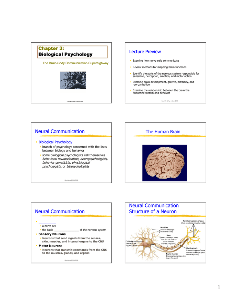
Chapter 3: Biological Psychology The Brain Brain--Body Communication Superhighway Lecture Preview Examine how nerve cells communicate Review methods for mapping brain functions Id Identify tif th the parts t off th the nervous system t responsible ibl ffor sensation, perception, emotion, and motor action Examine brain development, growth, plasticity, and reorganization Examine the relationship between the brain the endocrine system and behavior Copyright © Allyn & Bacon 2008 Neural Communication Copyright © Allyn & Bacon 2008 The Human Brain Biological Psychology branch of psychology concerned with the links between biology and behavior some biological psychologists call themselves behavioral neuroscientists, neuropsychologists, behavior geneticists, physiological psychologists, or biopsychologists Revision 2006 PSB Neural Communication Revision 2006 PSB Neural Communication Structure of a Neuron __________ a nerve cell the basic _________________ of the nervous system Sensory Neurons Neurons that send signals from the senses, skin, muscles, and internal organs to the CNS Motor Neurons Neurons that transmit commands from the CNS to the muscles, glands, and organs Revision 2006 PSB Revision 2006 PSB 1 Neurons: The Brain’s Communicators Cell body - makes proteins, replenishes molecules l l vital it l to t cellll function f ti Separated from outside by neuronal membrane Neurons cells specialized for communication ____________ – the process of the neuron specialized to receive “information” from adjacent cells (receptor sites, increased surface area/volume yielding enough area for perhaps 10,000 synapses). __________ – the process of the neuron specialized for the conduction of the nerve impulse (all or none law), clusters of axons form nerve bundles. Revision 2006 PSB Copyright © Allyn & Bacon 2008 Neuron with a Myelin Sheath Neural Communication Dendrite the bushy, branching extensions of a neuron that ______________ messages and conduct impulses toward the cell body Axon the extension of a neuron, ending in branching terminal fibers, through which messages are ______ to other neurons or to muscles or glands Myelin [MY-uh-lin] Sheath Copyright © Allyn & Bacon 2008 Resting Potential All neurons are _________ or charged cells. More _________________ reside inside the cell relative to the extracellular fluids. The resting potential is -70 mv. Neurons defend this resting potential. Revision 2006 PSB a layer of fatty cells segmentally encasing the fibers of many neurons enables vastly greater transmission speed of neutral impulses Revision 2006 PSB Ions Like charged molecules ______ each other. pp charged g ions attract each other. Opposite Nature's impulse is to distribute the ions so that they would become balanced on both sides of the cell membrane. If the ion concentrations were balanced on both sides of the cell membrane the neuron would noRevision longer 2006 PSBbe polarized. 2 Nerve cells are metabolically expensive The brain is about 2% of a person's body weight, but it consumes about 20% of the calories we consume every day. Neurons are continuously in the process of restoring themselves to their _______________ (-70 mv), and this requires the expenditure of calories. Revision 2006 PSB Neural Communication Neural Communication __________ Potential generated by the movement of positively charged atoms in and out of channels in the axon’s membrane An electrical impulse that surges along an axon, caused by an influx of positive ions in the neuron Revision 2006 PSB Voltage Across the Membrane During an Action Potential Cell body end of axon Direction of neural impulse: toward Revision 2006 PSBaxon terminals Copyright © Allyn & Bacon 2008 Neural Communication Nerve Impulse The action potential travels in _____ direction Starts at the beginning of the axon (axon hillock) Ends at the termination of the axon Revision 2006 PSB Synapse [SIN-aps] junction between the axon tip of the sending neuron and the dendrite or cell body of the receiving neuron tiny gap at this junction is called the synaptic gap or cleft Revision 2006 PSB 3 Neurons: The Brain’s Communicators How neurons communicate Axon - sending portion of neuron Axon terminal - end of axon which contains synaptic _________ with _______________ Revision 2006 PSB Copyright © Allyn & Bacon 2008 Synaptic Transmission PRS The flow of activation from one cell to the next (in most portions of the brain) is in one direction only, and cannot be reversed. T/F Revision 2006 PSB How neurons communicate Impulse releases neurotransmitter from axon terminals. gap. Neurotransmitter enters synaptic gap Neurotransmitter _______________ on the receiving neuron. Revision 2006 PSB Revision 2006 PSB Excitation & Inhibition Neurotransmitters are released at the axon terminals and they disturb the membrane of the postsynaptic cell so that ions flow across the cell membrane membrane. _____________ synapse – the net flow of ions make the cell less negative or depolarized. _____________ synapse – the net flow of ions make the cell more negative or hyperpolarized. Revision 2006 PSB 4 The All or None Law Number of Synapses? For the post-synaptic neuron to produce a nerve impulse it must become sufficiently depolarized to reach its ____________ (about - 40 mv). A typical neuron may have 10,000 synapses contacting the cell body (soma) and dendrites. Approximately half the synapses are excitatory, and the other half produce inhibition. The relative activation of these two contrasting influences determine if the neuron will fire or not. Revision 2006 PSB Excitatory synapses cause the neuron to become depolarized and shift it towards its threshold. Inhibitory synapses cause the neuron to become hyperpolarized and shift it away from its threshold. Revision 2006 PSB Neural networks are dynamic Drugs & Behavior The balance between inhibition and excitation may shift. Neurons may ________ and become slow to restore the resting potential. Neurons may exhaust their supply of ____________________. New receptor sites may be developed. Some drugs block the release of neurotransmitters (botox). Some drugs bind to receptor sites and block their activation by neurotransmitters (Cobra venom). Some drugs disturb the re-uptake or synthesis of neurotransmitters. Revision 2006 PSB Revision 2006 PSB Neural Communication Neurotransmitter molecule Receptor site on receiving neuron PRS High rates of activation of a pre-synaptic neuron always result in high rates of activation in the post-synaptic neuron. Receiving cell membrane T/F Agonist mimics neurotransmitter Antagonist blocks neurotransmitter Revision 2006 PSB Revision 2006 PSB 5 Speed of Neural Conduction The speed of conduction ranges from 0.5 m/sec to about 100 m/sec. Thick axons exhibit faster conduction. Myelinated axons exhibit faster conduction. White matter – myelinated axons, grey matter is composed of cell bodies and unmyelinated neurons. Revision 2006 PSB Glial Cells: Supporting Roles Glia - support cells of the nervous system Form myelin sheath covering of axons Form blood-brain barrier to protect the brain Respond to injury Remove debris Copyright © Allyn & Bacon 2008 Serotonin Pathways Serotonin pathways are involved with mood regulation. Serotonin Pathways Revision 2006 PSB Cooperative Learning Some portions of the brain do not become myelinated until a child becomes 5-6 years of age. Meet M t with ith your study t d group members b and d discuss how a child’s behavior may likely differ due to the presence or absence of myelin. You have 60 seconds. Revision 2006 PSB Neurotransmitters Different pathways in the brain may use different _______________. Sending neurons “classically” always release the same neurotransmitter. Receiving neurons may have synapses from different pathways employing different neurotransmitters. Over 100 neurotransmitters have been discovered in the brain, and it is likely that many new ones will be discovered. Revision 2006 PSB Dopamine Pathways Dopamine pathways are involved with are involved with diseases like schizophrenia and Parkinson’s disease. Revision 2006 PSB Dopamine Pathways 6 PRS PRS All the synaptic pathways in the human brain use the same neurotransmitter. The speed of transmission of a nerve impulse is dependent upon the presence (or absence of myelin), and the diameter of the axon. T/F T/F Revision 2006 PSB Cooperative Group Challenge The Rules Left Group vs. Right Group All the members of each group receive points for each question q the group g p bonus p answers correctly. The group (left or right) that answers the most questions correctly wins additional bonus points. Revised 2006 PSB Revision 2006 PSB Cooperative Group Challenge 1. 2. 3. 4. 5. 6. 7. 8. blood-brain barrier dendrite axon synaptic vesicles synaptic cleft cell body myelin sheath resting potential Revision 2006 PSB Cooperative Group Challenge Cooperative Group Challenge Q1. The central region of the neuron which manufactures new cell components is called the _____. Q2. The receiving ends of a neuron extending from the cell body like a tree branch are called the _____. Revision 2006 PSB Revision 2006 PSB 7 Cooperative Group Challenge Cooperative Group Challenge Q3. The space between two connecting neurons where neurotransmitters are released is called the _____. Q4. _____ are long extensions from the cell body of the neuron that transmit messages from one neuron to another. Revision 2006 PSB Revision 2006 PSB Cooperative Group Challenge Cooperative Group Challenge Q5. _____ small spheres within the axon that contain chemical messages specialized for communication. Q6. The brain’s ability to protect itself from infection and high hormone levels is through the _____. Revision 2006 PSB Revision 2006 PSB Cooperative Group Challenge Cooperative Group Challenge Q7. The autoimmune disease multiple sclerosis is linked to the destruction of the glial cells wrapped around the axon – called the _____. Q8. The electrical charge difference measured across the membrane of a neuron when it is not being stimulated is called the _____. Revision 2006 PSB Revision 2006 PSB 8 Schizophrenia and Neurotransmitters Stein and Wise Norepinephrine Theory Deficit in ________ directed thinking. Deficit in the capacity to experience _______________. A pathological gene leads to the reduction in the synthesis of a brain enzyme dopamine-B-hydroxylase (DBH) which converts dopamine into norepinephrine. Stein & Wise These affected fibers release __________ at the synapse instead of norepinephrine. There is too much dopamine in the brain. Some of the excess dopamine is converted extracellulary into 6-hydroxydopamine (6-OHDA). 6-OH-DA is a neurotoxin that selectively destroys adjacent norepinephrine synapses that are still healthy. Revision 2006 PSB Revision 2006 PSB Schizophrenia Evidence Schizophrenia exhibits a heritable factor. Brains of schizophrenics show a reduction in the level of the DBH enzyme. 6 6-OH-DA OH DA abolishes brain ________________. 6-OH-DA induces _________________. 6-OH-DA is linked to the production of unusual body odor. 6-OH-DA may be converted into a hallucinogen (2-hydroxy 4,5 dimethoxyphenethanolamine). Revision 2006 PSB Other researchers believe that schizophrenia is associated with excess p , but it mayy not involve the dopamine, norepinephrine pathway. Schizophrenia is a complicated disorder, different patients exhibit different symptoms, and different mechanisms may account for the variability in symptoms between patients. Revision 2006 PSB The Nervous System PRS Schizophrenia is believed to be due to an imbalance in one or more neurotransmitters. T/F Revision 2006 PSB Nervous System the body’s speedy, electrochemical communication system consists of all the nerve cells of the peripheral and central nervous systems Revision 2006 PSB 9 The Nervous System The Nervous System __________ Nervous System(CNS) The network of nerves contained within the brain and spinal p cord __________ Nervous System(PNS) The PNS comprises the somatic and autonomic nervous systems Revision 2006 PSB Revision 2006 PSB The Nervous System The Nervous System Nerves Interneurons neural “cables” containing many axons part of the peripheral nervous system connect the central nervous system with muscles, glands, and sense organs Sensory Neurons neurons that carry incoming information from the sense receptors to the central nervous system Revision 2006 PSB The Autonomic Nervous System CNS neurons that internally communicate and intervene between the sensory inputs and motor outputs Motor Neurons carry outgoing information from the CNS to muscles and glands Somatic Nervous System the division of the peripheral nervous system that controls the body’s skeletal muscles Revision 2006 PSB The Autonomic Nervous System: Sympathetic and Parasympathetic Branches __________ division - active during emotional arousal; activates fight-or-flight responses _______________ division - active during rest and digestion Work in opposition to each other: when one is active, the other is passive Copyright © Allyn & Bacon 2008 Copyright © Allyn & Bacon 2008 10 The Brain PRS The CNS is that portion of the nervous system housed within the skull and spinal cord. Lesion tissue destruction a brain lesion is a naturally or experimentally caused destruction of brain tissue T/F Revision 2006 PSB Revision 2006 PSB Brain--Mapping Methods Brain Electroencephalogram (EEG) 3. Electrical stimulation and recording of the nervous system an amplified recording of the waves of electrical activity that sweep across the brain’s surface these waves are measured by electrodes placed on the scalp EEG 4. Brain scans a. CT and MRI - structural imaging b. PET and fMRI - functional imaging 5. Magnetic stimulation and recording a. Transcranial Magnetic Stimulation (TMS) b. Magnetoencephalography (MEG) Revision 2006 PSB Copyright © Allyn & Bacon 2008 Tools of Behavioral Neuroscience Electroencephalogram (EEG) An instrument used to measure electrical activity in the brain through electrodes placed on the scalp Revision 2006 PSB The Brain CT (computed tomography) Scan a series of x-ray photographs taken from different angles and combined by computer into a composite representation of a slice through the body; also called CAT scan PET (positron ( it emission i i tomography) t h ) Scan S a visual display of brain activity that detects where a radioactive form of glucose goes while the brain performs a given task MRI (magnetic resonance imaging) a technique that uses magnetic fields and radio waves to produce computer-generated images that distinguish among different types of soft tissue; allows us to see structures within the brain Revision 2006 PSB 11 PET Scan MRI Scan MRI (magnetic resonance imaging) uses magnetic fields and radio waves to produce computer‐ generated images that generated images that distinguish among different types of brain tissue. Top images show ventricular enlargement in a schizophrenic patient. Bottom image shows brain regions when a participants lies. Radioactive isotopes (small amounts) are placed in the blood. Sensors detect radioactivity. Different tasks show distinct activity patterns. Revision 2006 PSB Central Nervous System ______ brains, one on the left and one on the right. Brain structures were named for familiar objects Revision 2006 PSB Regions of the Brain Cerebral Cortex Limbic System Brain Stem Cortex – tree bark Cerebellum – little brain Pons – bridge Thalamus – inner chamber Revision 2006 PSB Revision 2006 PSB The Four Lobes of the Cerebral Cortex Regions R i of the Brain Revision 2006 PSB Revision 2006 PSB 12 The Forebrain (_________ Cortex) 1. Cerebral Cortex outermost covering, contains: Neocortex - most recently developed cortex Cerebral hemispheres left and right Corpus callosum connects the two hemispheres Copyright © Allyn & Bacon 2008 The Forebrain (Cerebral Cortex) The Forebrain (Cerebral Cortex) 2. ________ Lobe Motor cortex - sends signals to muscles Prefrontal cortex executive functions Injury: Broca’s area & aphasia Phineas Gage & personality change Copyright © Allyn & Bacon 2008 Representation of the Body Mapped onto the Motor and Sensory Areas of the Cerebral Cortex 3. ____________ Lobe perception of space, object shape and orientation, actions of others, numbers – Integrates vision, vision touch, touch motor information Somatosensory cortex pressure, temperature, pain Injury: acalculia, contralateral neglect Copyright © Allyn & Bacon 2008 The Forebrain (Cerebral Cortex) 4. ___________ Lobe hearing, language comprehension, autobiographical memories Auditory cortex Injury: Wernicke’s area and aphasia Copyright © Allyn & Bacon 2008 The Forebrain (Cerebral Cortex) 5. _____________ Lobe vision Visual cortex Sensory Cortical Hierarchies Association Cortex (e.g., conscious perception of visual scene) Sensory cortex (e.g., visual ctx) Sensory info (e.g., light) Copyright © Allyn & Bacon 2008 Copyright © Allyn & Bacon 2008 13 Selected Areas of the Cerebral Cortex PRS The frontal lobe is responsible for visual perception. T/F Revision 2006 PSB Copyright © Allyn & Bacon 2008 The Limbic System The Brain and Emotion ________ system - emotional center of the brain networked with the autonomic nervous system to influence blood pressure, heart and the endocrine system, etc. Information about our internal state Copyright © Allyn & Bacon 2008 Copyright © Allyn & Bacon 2008 The Brain and Emotion: Limbic Circuits 1. Hypothalamus - maintains internal bodily states by overseeing the endocrine and autonomic nervous systems (e.g., releases hormones to influence hunger, sexual motivation) 2. Amygdala - excitement, arousal, fear, social signals related to emotion Copyright © Allyn & Bacon 2008 Hypothalamus neural structure lying below (hypo) the thalamus; directs several maintenance activities eating drinking body temperature helps govern the endocrine system via the __________ gland is linked to emotion 14 The Brain and Emotion: Limbic Circuits The Limbic System 3. Cingulate Cortex - active during emotional expression – knowledge of socially appropriate behavior – regulates autonomic nervous system Electrode implanted in reward center 4. Hippocampus - spatial memory (e.g., place cells), fear conditioning Injury: problem forming new memories Revision 2006 PSB Copyright © Allyn & Bacon 2008 Brainstem PRS The limbic system is involved in regulation of affect and emotion. T/F Revision 2006 PSB Copyright © Allyn & Bacon 2008 Cerebellum The Brainstem Medulla Vital involuntary functions Pons Sleep and arousal Reticular formation Sleep, arousal, attention Cerebellum Motor coordination Cerebellum [sehr-uh-BELL-um] the “little brain” attached to the rear of the brainstem it helps coordinate voluntary movement and balance Revision 2006 PSB 15 The Brain The Cerebral Cortex Brainstem the oldest part and central core of the brain, beginning g g where the spinal p cord swells as it enters the skull responsible for ________________ functions Medulla [muh-DUL-uh] base of the brainstem controls heartbeat and breathing Neocortex regulates reading, talking, problem solving. Subcortical regions regulate sleep, thermoregulation g and other vegetative g processes. Paradox: the functions of the neocortex which define us as unique individuals are ____________, during a trauma, such as lack of oxygen, blood flow is shunted away from the neocortex to preserve subcortical structures. Revision 2006 PSB Revision 2006 PSB PRS Specialization and Integration The brainstem is responsible for high level cognitive processes. Brain activity when hearing, seeing, and speaking words T/F Revision 2006 PSB Language Processing _______ Area Located in the left hemisphere, directs the muscle movements t in i speech production Wernicke’s Area Located in the left hemisphere, involved in the comprehension of language Revision 2006 PSB Revision 2006 PSB The Cerebral Cortex Broca’s Area an area of the left frontal lobe that directs the muscle movements involved in speech ___________ Area an area of the left temporal lobe involved in language comprehension and expression Revision 2006 PSB 16 The Cerebral Cortex Aphasia impairment of language, usually caused by left hemisphere damage either to Broca’s area (impairing speaking) or to Wernicke Wernicke’ss area (impairing understanding) PRS For most individuals the left hemisphere is dominant for language processing. T/F Revision 2006 PSB Revision 2006 PSB Textbook Assignment 3-3. Brain Reorganization Complete the assess your knowledge assignment on page 146. __________________________________ __________________________________ __________________________________ __________________________________ __________________________________ __________________________________ __________________________________ Revision 2006 PSB _________ Brain Changes During Development and Experience the brain’s capacity for modification, difi ti as evident id t in i brain b i reorganization following damage (especially in children) and in experiments on the effects of experience on brain development Revision 2006 PSB Neural Plasticity Neural plasticity - the nervous system’s ability to ___________: 1. Before birth and until maturation is complete (early adulthood) adulthood). Cell division, division migration, migration connections: a. b. c. d. Growth of dendrites and axons Synaptogenesis Pruning Myelination Early Brain Development 2. During learning: Long-Term Potentiation (LTP), enriched environments Copyright © Allyn & Bacon 2008 Increased branching of dendrites with enrichment Copyright © Allyn & Bacon 2008 17 Neural Plasticity (cont’d) 3. Following Injury and Degeneration 4. Stem Cells: cells that have potential to b become a variety i t off specialized i li d cells ll 5. Neurogenesis: production of new neurons in the adult brain Plasticity Richer environments lead to heavier, thicker brains, more synapses, and better learning. The cost of plasticity is the case of the phantom limb. Revision 2006 PSB Copyright © Allyn & Bacon 2008 Living life to the full, the girl with only half a brain takes her first steps. She was suffering from an incurable genetic condition for which the only possible hope was to have half her brain removed. d Doctors warned her parents that their daughter would never walk, talk, cry or smile like a normal child - even if she survived. Revision 2006 PSB Revision 2006 PSB Cooperative Learning Answer In some cases enormous amounts of brain tissue may be removed with “acceptable” or minimal negative consequences, in other instances a modest amount of brain damage produces enormous effects. Meet with your group and discuss why the outcomes are so different in these cases. You have 60 seconds. Revision 2006 PSB __________________________________ __________________________________ __________________________________ __________________________________ __________________________________ __________________________________ __________________________________ __________________________________ Revision 2006 PSB 18 Splitting the Brain Corpus Callosum A procedure in which the two hemispheres of the brain are isolated by cutting the _________ __________ (mainly those of the corpus callosum) between them. A bundle of nerve fibers that connects the left and right h hemispheres i h Martin M. Rother C Courtesy of Terence Williams, University of Iowa Corpus Callosum If surgically severed for treatment of epilepsy, hemispheres cannot communicate directly. Revision 2006 PSB Split Brain Patients Textbook Assignment 3-3. Which object would you perceive when these two words are flashed to different hemispheres? Which object would a split-brain subject perceive? Submit your answers to your TA. The information highway from the eye to the brain With the corpus callosum severed, objects (apple) presented in the right visual field can be named. Objects (pencil) in the left visual field cannot. Revision 2006 PSB Sperry’s Split Brain Experiment Brain Functioning Split-brain subjects could not name objects shown only to the right hemisphere. If asked to select these objects with their left hand, they succeeded. The left hemisphere controls speech, the right does not. Revision 2006 PSB Revision 2006 PSB 19 The Endocrine System: Hormonal Regulation PRS Surgically isolation of the left and right hemispheres is sometimes performed to reduce the severity of epileptic seizures. T/F Revision 2006 PSB Hierarchy of Control over the Endocrine System 1. Pituitary gland “master gland” controls other bodily glands and is under control of the ___________ Copyright © Allyn & Bacon 2008 The Endocrine System 2. Adrenal glands - release adrenaline and cortisol during physical and psychological stress – activated by the sympathetic nervous system Copyright © Allyn & Bacon 2008 The Endocrine System 3. Sexual reproductive glands Testes in males produce testosterone Ovaries in females produce estrogen However, both sexes release some sex hormone associated with the opposite sex Copyright © Allyn & Bacon 2008 Copyright © Allyn & Bacon 2008 Endocrine System Hormones are like a neurotransmitter that are distributed to receptor sites via the __________ supply. Hormones orchestrate and coordinate responses in i a diverse di number b off structures. _______ delay between release of the hormone and the response, and a ____ return to baseline. Revision 2006 PSB 20 PRS Stress Responses The neurotransmitter and endocrine system work together to coordinate behavioral responses. T/F The sympathetic branch of the autonomic nervous system innervates the adrenal gland and causes the release of adrenalin. Neurons from the hypothalamus innervate the pituitary gland and cause the release of ACTH, and this hormone causes the adrenal gland to release three additional hormones involved in the body’s stress response. Revision 2006 PSB Stress Responses Pupils dilate, blood pressure increases, heart rate increases, muscles tense, digestion is terminated, catabolic processes required for the _____ __________ of energy are activated. ti ted Because the endocrine system is sluggish (slower to start and stop) emotions and feeling’s of the body’s response continue after the emergence is resolved. Revision 2006 PSB Revision 2006 PSB PRS Behavioral responses strongly influenced by the endocrine system are relatively slow to start and to return to baseline. T/F Revision 2006 PSB 21


