20.0 Physiology 20.1 Introduction
advertisement
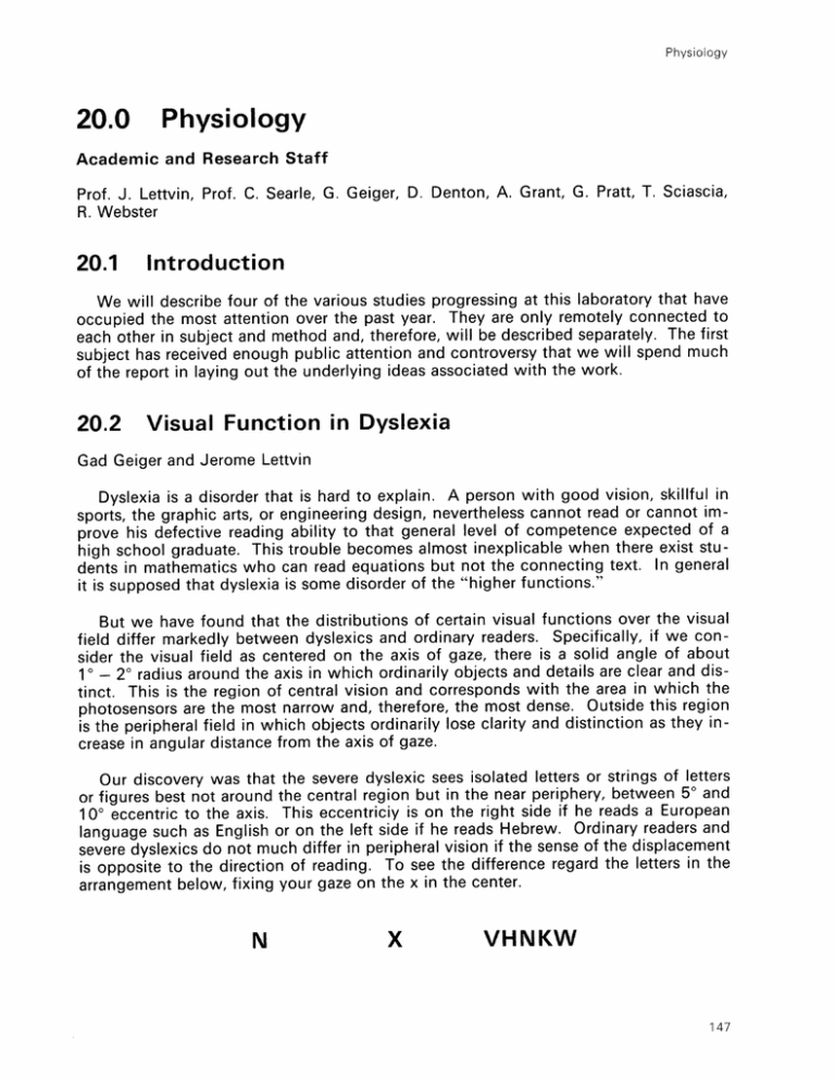
Physiology 20.0 Physiology Academic and Research Staff Prof. J. Lettvin, Prof. C. Searle, G. Geiger, D. Denton, A. Grant, G. Pratt, T. Sciascia, R. Webster 20.1 Introduction We will describe four of the various studies progressing at this laboratory that have occupied the most attention over the past year. They are only remotely connected to each other in subject and method and, therefore, will be described separately. The first subject has received enough public attention and controversy that we will spend much of the report in laying out the underlying ideas associated with the work. 20.2 Visual Function in Dyslexia Gad Geiger and Jerome Lettvin Dyslexia is a disorder that is hard to explain. A person with good vision, skillful in sports, the graphic arts, or engineering design, nevertheless cannot read or cannot improve his defective reading ability to that general level of competence expected of a high school graduate. This trouble becomes almost inexplicable when there exist students in mathematics who can read equations but not the connecting text. In general it is supposed that dyslexia is some disorder of the "higher functions." But we have found that the distributions of certain visual functions over the visual field differ markedly between dyslexics and ordinary readers. Specifically, if we consider the visual field as centered on the axis of gaze, there is a solid angle of about 1 0 - 20 radius around the axis in which ordinarily objects and details are clear and distinct. This is the region of central vision and corresponds with the area in which the photosensors are the most narrow and, therefore, the most dense. Outside this region is the peripheral field in which objects ordinarily lose clarity and distinction as they increase in angular distance from the axis of gaze. Our discovery was that the severe dyslexic sees isolated letters or strings of letters best not around the central region but in the near periphery, between 50 and figures or 100 eccentric to the axis. This eccentriciy is on the right side if he reads a European language such as English or on the left side if he reads Hebrew. Ordinary readers and severe dyslexics do not much differ in peripheral vision if the sense of the displacement is opposite to the direction of reading. To see the difference regard the letters in the arrangement below, fixing your gaze on the x in the center. N X VHNKW 147 Physiology The letter N is the same distance from the X on right and the X on the left. But while the letter is reasonably clear on the left, it is not clear on the right. When looking directly at the letter string you see the array clearly even if your gaze is held fixed on any letter in the string. For the severe dyslexic the array is confused on direct gaze in the same way it is confused in your vision when you are fixing on the X. On the other hand, it is clearer for him when he fixes on the X than when he regards it directly. Reviewing the literature we found that visual function tests have almost uniformly been done on readers. College students are cheaply had subjects. But the results have been supposed not only as description of the norm but evidence of a specific built-in organization of the visual field. Prior to our study of dyslexics, we had questioned this concept. We felt that the information necessary to identify a letter in the interior of an eccentric string was not lost in early visual processing and could be retrieved. The confusion that you see in the interior of the eccentrically viewed string is defined as "lateral masking" by Boumarn. Existence of nearby flanking letters addles the perception of a letter. We felt that lateral masking was a learned operation, a weighting function that, applied to the peripheral field, reduced an aggregate of forms to a texture. We took a texture to be that property of an aggregate that is had from discerning textural elements, e.g., the "textons" of B. Julesz, but not perceptually providing them with that connectivity that determines form. It is as if the aggregate has more a statistical rather than a formal description. To a significant degree we made the point that lateral masking is a perceptual strategy by showing that one could "demask" the laterally masked letters in the periphery. In the case of the dyslexic, the visual strategy is to laterally mask in the center of the visual field and attend what is the case away from that center. This, which we call the "hunter" strategy, is what all of us use in driving through fast two-way traffic or playing in competitive sports, e.g., tennis or football. We know more what to do from the ambience of the ball than from watching the ball. When we read, we use the "scribe" stragegy in which we laterally mask in the visual periphery so as to attend the center of vision. We easily switch between these strategies. The severe dyslexic seems frozen in the "hunter" mode and so cannot learn conventional reading. Indeed, practicing to read with central vision reinforces the perceptual block. Geiger's experiments tested the learning of a new strategy by showing how a 24 year-old severe dyslexic, scarcely at the third grade level in reading despite repeated attempts at remediation in the past, could be brought to tenth grade level in four months by training him to read in the peripheral field by blocking the text in the central field. This procedure, now repeated on four more cases, has pro tem proved effective. But what is more important is that the tests we designed for examining central versus peripheral vision have emerged not only as diagnostic, but sensitive enough to follow the improvement and degradation in reading ability of those prone to dyslexia. This claim is borne out by finding subjects who are readers in the morning and dyslexic at night so that we can track repeatedly the clinical variation by test and correlate it with the gain and loss of ability to read. We have records now on such a case. 148 RLE Progress Report Number 130 Physiology The report as given here is cursory and somewhat popularized. The data supporting this approach are in two papers already published. Rather more is said in two other papers now being prepared for publication in which a more extended set of results are given, but this would take too much space in this note. 20.3 Physiology of Vision in the Frog Arthur Grant and Jerome Lettvin The major visual center of the frog, certainly the largest, is the tectum, a paired structure on top of the midbrain. Each tectal lobe is directly connected only to the opposite eye: there is no splitting of the output of each eye as in our visual projection to cortex. Associated with each tectal lobe is a relatively small accessory nucleus, n. isthmi, that receives output only from that tectal lobe, but projects back to both tectal lobes. Ipsilaterally the axons of n. isthmi end on the dendrites of those cells that send axons to nucleus isthmi. Its topology closely resembles that of a layered "nerve net." In the past this laboratory has shown that the crossed projection of n. isthmi is responsible for "binocular" vision in the frog. We could not account for the ipsilateral projection. Then E. Gruberg found that if n. isthmi is ablated on one side, the frog becomes visually indifferent to both prey and threat in the image seen by the opposite eye. Yet, when the frog is made to jump it avoids obstacles, and goes over barriers and does not hit walls. We had begun recording in the tectum many years ago. Several new features have emerged this last year from Mr. Grant's investigations. First and foremost is the demonstration that the records taken from single units in the superficial neuropil do not represent the firing of the terminal bunches of primary optic nerve fibers. Instead they appear to be active responses of special dendritic appendages of certain tectal cells. An extraordinary anatomical feature of these appendages is that they form a mutually synapsing net between the tectal cells all across the tectum. This identification of the signal sources at single nodes in the net changes considerably the current view of the visual processes in amphibia. But the second feature that has come to light seems to be that the crossed n. isthmi fibers terminate on these nodes as well. For the first time we can compare at the same recording site some of the processed output of one tectum with the optic nerve input to the other. It is premature to assert the results, but what is emerging is that, however the tectum handles its input data, it preserves almost slavishly at the output the several categories attributable to different types of retinal feature detectors. That is, the output can pe parsed easily in terms of combinations of input. 20.4 Image Processing in the Photo-receptors Gill Pratt, Robert Webster, Jerome Lettvin A great deal is now known about the mechanism of transduction of light by rods and cones and the first steps of amplifying the transduction into signal. But if the receptors 149 Physiology are taken as discrete sensors feeding the nervous part of the retina, a distinct problem emerges. First of all, the point spread function on the retina through the optical system of the eye under optimal focus and aperture is quite large, several receptors in width - much worse than occurs with the cheapest cameras. Second, receptors of the same type, e.g., rods or cones are laterally connected among themselves ohmically in a resistive net. Each receptor is a node in such a net. And the space constant of the resistive connection is larger than the point-spread function. However, at the retinal output the ganglion cells, in their response, reflect sensitivity to details in the image as if the image had somehow been sharpened in a way familiar to modern image processing. The existence of the resistive net between the receptors would be corruptive if each receptor signaled the effects of light on it as a current. However, if the measured signal from the outer segment drive a voltage follower in the inner segment of each rod or cone, then the nodes in the net become voltage sources. The feedback current used to clamp the voltage to that which is set by the light signal, now measures the signal level difference between any receptor and its immediate neighbors because of the resistive coupling. It this feedback current is that which drives the output signal of the receptor, the image at the output of the receptor layer has been sharpened in the same way as is used in conventional image processing. We have modelled this system succesfully in a computer program and are now doing experiments on actual retinas to test the hypothesis. 20.5 Voltage Control of Cell Membrane Campbell Searle and Jerome Lettvin The mechanism of voltage control is still unknown. A large body of data from voltage clamp experiments gives the empirics of the rate processes of this control on the ionic channels of nerve. However the chemical operation has stayed obscure. We have modeled a system in which the heads of phosphatidyl serine molecules in the membrane trimerize reversibly in the presence of Ca2+ , with three Ca2+ binding three heads in a triangle. This is an excellent electro-mechanical transducer with adsorption of Ca2+ sensitive to boundary density of Ca2 + and to the local electric field; desorption sensitive to the ionic strength of the solution and the local receptive field. This model fits the known data, especially the rate processes, remarkably well and accounts for the voltage-sensitive component of membrane capacitance. We are preparing the study for publication. 150 RLE Progress Report Number 130
