Color Coding the Cell
advertisement

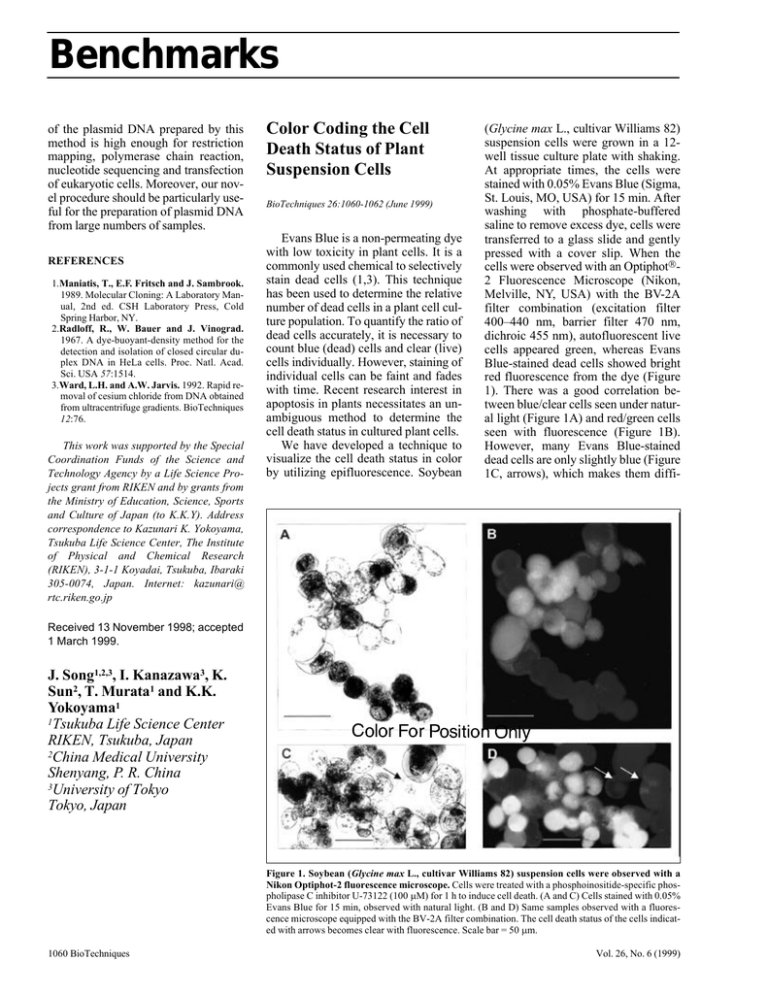
Benchmarks of the plasmid DNA prepared by this method is high enough for restriction mapping, polymerase chain reaction, nucleotide sequencing and transfection of eukaryotic cells. Moreover, our novel procedure should be particularly useful for the preparation of plasmid DNA from large numbers of samples. REFERENCES 1.Maniatis, T., E.F. Fritsch and J. Sambrook. 1989. Molecular Cloning: A Laboratory Manual, 2nd ed. CSH Laboratory Press, Cold Spring Harbor, NY. 2.Radloff, R., W. Bauer and J. Vinograd. 1967. A dye-buoyant-density method for the detection and isolation of closed circular duplex DNA in HeLa cells. Proc. Natl. Acad. Sci. USA 57:1514. 3.Ward, L.H. and A.W. Jarvis. 1992. Rapid removal of cesium chloride from DNA obtained from ultracentrifuge gradients. BioTechniques 12:76. This work was supported by the Special Coordination Funds of the Science and Technology Agency by a Life Science Projects grant from RIKEN and by grants from the Ministry of Education, Science, Sports and Culture of Japan (to K.K.Y). Address correspondence to Kazunari K. Yokoyama, Tsukuba Life Science Center, The Institute of Physical and Chemical Research (RIKEN), 3-1-1 Koyadai, Tsukuba, Ibaraki 305-0074, Japan. Internet: kazunari@ rtc.riken.go.jp Color Coding the Cell Death Status of Plant Suspension Cells BioTechniques 26:1060-1062 (June 1999) Evans Blue is a non-permeating dye with low toxicity in plant cells. It is a commonly used chemical to selectively stain dead cells (1,3). This technique has been used to determine the relative number of dead cells in a plant cell culture population. To quantify the ratio of dead cells accurately, it is necessary to count blue (dead) cells and clear (live) cells individually. However, staining of individual cells can be faint and fades with time. Recent research interest in apoptosis in plants necessitates an unambiguous method to determine the cell death status in cultured plant cells. We have developed a technique to visualize the cell death status in color by utilizing epifluorescence. Soybean (Glycine max L., cultivar Williams 82) suspension cells were grown in a 12well tissue culture plate with shaking. At appropriate times, the cells were stained with 0.05% Evans Blue (Sigma, St. Louis, MO, USA) for 15 min. After washing with phosphate-buffered saline to remove excess dye, cells were transferred to a glass slide and gently pressed with a cover slip. When the cells were observed with an Optiphot2 Fluorescence Microscope (Nikon, Melville, NY, USA) with the BV-2A filter combination (excitation filter 400–440 nm, barrier filter 470 nm, dichroic 455 nm), autofluorescent live cells appeared green, whereas Evans Blue-stained dead cells showed bright red fluorescence from the dye (Figure 1). There was a good correlation between blue/clear cells seen under natural light (Figure 1A) and red/green cells seen with fluorescence (Figure 1B). However, many Evans Blue-stained dead cells are only slightly blue (Figure 1C, arrows), which makes them diffi- Received 13 November 1998; accepted 1 March 1999. J. Song1,2,3, I. Kanazawa3, K. Sun2, T. Murata1 and K.K. Yokoyama1 1Tsukuba Life Science Center RIKEN, Tsukuba, Japan 2China Medical University Shenyang, P. R. China 3University of Tokyo Tokyo, Japan Figure 1. Soybean (Glycine max L., cultivar Williams 82) suspension cells were observed with a Nikon Optiphot-2 fluorescence microscope. Cells were treated with a phosphoinositide-specific phospholipase C inhibitor U-73122 (100 µM) for 1 h to induce cell death. (A and C) Cells stained with 0.05% Evans Blue for 15 min, observed with natural light. (B and D) Same samples observed with a fluorescence microscope equipped with the BV-2A filter combination. The cell death status of the cells indicated with arrows becomes clear with fluorescence. Scale bar = 50 µm. 1060 BioTechniques Vol. 26, No. 6 (1999) Benchmarks cult to detect. In contrast, with fluorescence, these cells appear bright red (Figure 1D, arrows). We have successfully used this technique on cultured suspension cells of two additional plant species, Medicago truncatula and Nicotiana tabacum. Unlike in soybean and M. truncatula cells, the green autofluorescence in N. tabacum was mainly from nuclei rather than the whole cells. However, the dead cells appeared clearly red as observed in other species tested. Fluorescein diacetate (FDA), a compound commonly used in determining cell viability, was tested in soybean cell suspensions. In this method, intracellular esterases cleave FDA, producing fluorescein in live cells (2). We observed that the strong fluorescence from live cells could spill over to non-fluorescent dead cells, making it difficult to clearly distinguish dead cells from live cells, especially when cells are aggregated. Evans Blue does not have this problem. In addition, unlike in the FDA staining technique our method allows us to count dead and live cells at the same time with fluorescence. Therefore, this technique is especially useful when accurate and quick counts of both dead and live cells are necessary. REFERENCES 1.Gaff, D.F. and O. Okong’o-Ogola. 1971. The use of nonpermeating pigments for testing the survival of cells. J. Exp. Bot. 22:756-758. 2.Rotman, B. and B.W. Papermaster. 1966. Membrane properties of living cells as studied by enzymatic hydrolysis of fluorogenic esters. Proc. Natl. Acad. Sci. USA 55:134-141. 3.Turner, J.G. and A. Novacky. 1974. The quantitative relation between plant and bacterial cells involved in the hypersensitive reaction. Phytopathology 64:885-890. This work was supported by the Samuel Roberts Noble Foundation. We thank Xin S. Ding, Yiming Bao, Ping Xu and Yongqing Liu for technical assistance, and Darla F. Boydston and Cuc K. Ly for providing photographs. We also thank Hiroaki Katagi and Chun-lin Su for the suspension cells of Medicago truncatula and Nicotiana tabacum, respectively. We extend our thanks to Richard A. Dixon and Christian Dammann for critically reading the manuscript. Address correspondence to Dr. Madan K. Bhattacharyya, The Samuel Roberts Noble Foundation, P.O. Box 2180, 2510 Sam Noble Parkway, Ardmore, OK 73402, USA. Internet: mkbhattach@noble.org Received 3 December 1998; accepted 17 March 1999. Toshiro Shigaki and Madan K. Bhattacharyya The Samuel Roberts Noble Foundation Ardmore, OK, USA


