Recent Glimpses of the Elusive Spindle Matrix Kristen M. Johansen* ABSTRACT Jørgen Johansen
advertisement
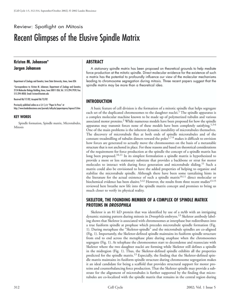
[Cell Cycle 1:5, 312-314, September/October 2002]; © 2002 Landes Bioscience Review: Spotlight on Mitosis Recent Glimpses of the Elusive Spindle Matrix Kristen M. Johansen* Jørgen Johansen Department of Zoology and Genetics; Iowa State University; Ames, Iowa USA *Correspondence to: Kristen M. Johansen; Department of Zoology and Genetics; 3154 Molecular Biology Building; Ames, Iowa 50011 USA; Tel.: 515.294.7959; Fax: 515.294.4858; Email: kristen@iastate.edu Received 06/17/02; Accepted 06/12/02 Previously published online as a Cell Cycle “Paper In Press” at http://www.landesbioscience.com/journals/cellcycle/papersinpress/inpress15.htm KEY WORDS Spindle formation, Spindle matrix, Microtubules, Mitosis ABSTRACT A stationary spindle matrix has been proposed on theoretical grounds to help mediate force production at the mitotic spindle. Direct molecular evidence for the existence of such a matrix has the potential to profoundly influence our view of the molecular mechanisms leading to chromosome segregation during mitosis. Three recent papers suggest that the spindle matrix may be more than a theoretical idea. INTRODUCTION A basic feature of cell division is the formation of a mitotic spindle that helps segregate each set of the duplicated chromosomes to the daughter nuclei.1 The spindle apparatus is a complex molecular machine known to be made up of polymerized tubulin and various associated motor proteins.2 While numerous models have been proposed for how the spindle apparatus may transmit forces none of these models have been completely satisfying.1,3-6 One of the main problems is the inherent dynamic instability of microtubules themselves. The discovery of microtubule flux at both ends of spindle microtubules and of the constant treadmilling of tubulin dimers toward the poles1,7-9 makes it difficult to envision how forces are generated to actually move the chromosomes on the basis of a metastable structure that is not anchored in place. For these reasons and based on theoretical considerations of the requirement for force production at the spindle the concept of a spindle matrix has long been proposed.10,11 In its simplest formulation a spindle matrix is hypothesized to provide a more or less stationary substrate that provides a backbone or strut for motor molecules to interact with during force generation and microtubule sliding.11 Such a matrix could also be envisioned to have the added properties of helping to organize and stabilize the microtubule spindle. Although there have been some tantalizing hints in the literature for the actual existence of such a spindle matrix10,11 direct molecular or biochemical evidence has been elusive.3,12 However, the results from three recent studies13-15 reviewed here breathe new life into the spindle matrix concept and promises to bring us much closer to verify its physical reality. SKELETOR, THE FOUNDING MEMBER OF A COMPLEX OF SPINDLE MATRIX PROTEINS IN DROSOPHILA Skeletor is an 81 kD protein that was identified by use of a mAb with an intriguing dynamic staining pattern during mitosis in Drosophila embryos.13 Skeletor antibody labeling shows that Skeletor is associated with chromosomes at interphase but redistributes into a true fusiform spindle at prophase which precedes microtubule spindle formation (Fig. 1). During metaphase the “Skeletor-spindle” and the microtubule spindles are co-aligned (Fig. 1). Importantly, the Skeletor-defined spindle maintains its fusiform spindle structure from end to end across the metaphase plate during anaphase when the chromosomes segregate (Fig. 1). At telophase the chromosomes start to decondense and reassociate with Skeletor where the two daughter nuclei are forming while Skeletor still defines a spindle in the midregion (Fig. 1). Thus, the Skeletor-defined spindle exhibits all the properties predicted for the spindle matrix.13 Especially, the finding that the Skeletor-defined spindle matrix maintains its fusiform spindle structure during chromosome segregation makes it an ideal candidate for being a scaffold that provides structural support for motor proteins and counterbalancing force production. That the Skeletor spindle may provide a substrate for the alignment of microtubules is further supported by the finding that microtubules are co-localized with the spindle matrix that remains in the central region during 312 Cell Cycle 2002; Vol. 1 Issue 5 RECENT GLIMPSES OF THE ELUSIVE SPINDLE MATRIX midbody formation at telophase. This alignment of microtubules at the midbody region is largely unaccounted for by most other models but could be explained if the Skeletor spindle as hypothesized by Walker et al.13 helps in organizing microtubule fibers. However, Skeletor encodes a low-complexity protein with no obvious motifs making it unlikely that Skeletor itself is a structural component of the spindle matrix but rather that it is a member of a multi-protein complex.13 Consequently, in order to identify other members of such a complex Wang et al.16 performed yeast two-hybrid interaction assays as well as coimmunoprecipitation analysis. In preliminary experiments using these approaches a novel chromodomain containing protein, which was named Chromator, was found to interact with Skeletor.16 A mAb generated against Chromator showed that it has a dynamic cell cycle specific pattern similar but not identical to that of Skeletor.16 A third protein that in immunolabelings completely co-localizes with Skeletor during mitosis was also identified.16 This protein displays coiled-coil motifs suggesting it may comprise a structural component of the spindle matrix. Analysis of a P-element insertion into this gene suggests that a reduction in protein levels encoded by this locus results in spindle abnormalities and chromosome segregation defects and that the protein is essential for viability. Thus, these findings indicate that Skeletor is not alone but may be the founding member of an interacting complex of nuclear components that act in concert to form a spindle matrix. The identification of several of these other players and the availability of P-element insertions in some of their genes promises to pave the way for a direct structural as well as functional analysis of the spindle matrix in Drosophila. THE KINESIN EG5 AND MICROTUBULE FLUX IN XENOPUS EXTRACT SPINDLES Using a very different approach Kapoor and Mitchison14 studied the distribution and dynamics of the motor protein Eg5 in Xenopus spindles relative to that of the microtubules. Eg5 previously had been shown to localize throughout the spindle and play a key role in establishing its bipolar organization.17,18 Kapoor and Mitchison14 applied the recently developed “fluorescence speckle microscopy” (FSM) technique19 that relies on random variation of incorporation of normal and fluorescently-tagged subunits within a macromolecular assembly (such as a microtubule) to create a “speckled” pattern. These speckles can then provide fiduciary marks serving as reference points to follow movement or flux of the structure over time using simple and non-perturbing CCD camera imaging approaches. By applying FSM to compare the mobility of fluorescently speckled microtubules with that of the microtubule-binding motor protein Eg5 Kapoor and Mitchison14 made the surprising discovery that the vast majority of Eg5 in the spindle is static despite the continuous poleward flux observed for microtubules. A number of important controls showed that the recombinant, fluorescently-tagged Eg5 behaved similarly to the endogenous Eg5: 1. in biophysical assays the recombinant protein displayed the proper Stokes radius, 2. it promoted microtubule gliding with the same velocities, 3. its localization to the spindle was indistinguishable from that of wild-type, and 4. it was capable of rescuing bipolar spindle formation from Eg5-depleted extracts. Furthermore, the fact that excess Eg5 added to control experiments did not alter spindle assembly or microtubule dynamics in any measurable way suggests that the assays were not being influenced by www.landesbioscience.com Figure 1. The dynamics of Skeletor spindle matrix formation and distribution during mitosis as compared to the microtubule spindle. Confocal images were obtained from syncytial Drosophila blastoderm embryos labeled with mAb 1A1 for Skeletor (red), with anti-tubulin antibody (green), and with Hoechst for DNA (blue). The composite images of the labelings (comp) are shown to the left. The labelings show that in late prophase, when the microtubules have not yet entered the nuclear space, the Skeletor spindle is already aligned within the nucleus. During metaphase the two spindles are co-extensive, although the Skeletor spindle appears broader than the microtubule spindle. At anaphase the Skeletor spindle persists as an intact spindle extending across the metaphase plate as the chromosomes segregate to the poles. During telophase the chromosomes have segregated and as they decondense, they reassociate with Skeletor at the poles. However, the Skeletor spindle persists in the central region where midbody formation of the microtubules is found to take place in alignment with the remaining Skeletor spindle structure. variations in Eg5 protein level. Thus, this raises the conundrum of how a microtubule-binding motor protein could remain fixed in place on a constantly treadmilling spindle. One possible explanation would be if the motor protein was “walking” on the microtubule in the opposite direction to its poleward flux at an equal but opposite rate of speed. However, this explanation is unlikely since when monastrol, a specific inhibitor of Eg5 motility, was included in the assays similar results were observed. Had the motor been counteracting the poleward microtubule flux, the Eg5 should have been carried poleward with the microtubules once its motor was inactivated. Also unlikely is that Eg5 itself associates with a class of stationary microtubules as there was no evidence from the FSM measurements of a proportionate number of static microtubules. Thus, the authors suggest the immobility of Eg5 on mitotic spindles may be due to an interaction with a static spindle matrix.14 Interestingly, the static behavior observed for Eg5 in the above studies appears inconsistent with results reported by Wilde et al.20 who used fluorescently-tagged antibodies against Eg5 to speckle-label asters emanating from centrosomes added to cytostatic factor-arrested Xenopus egg extracts in the presence of Ran-GTP. In these studies the Eg5-speckles moved towards the plus end of micro- Cell Cycle 313 RECENT GLIMPSES OF THE ELUSIVE SPINDLE MATRIX tubules at ~2.8 µm/min. Possibilities for this apparent discrepancy considered by Kapoor and Mitchison14 are that these studies were performed on asters, not bipolar spindles, and that the antibody labeling could potentially block interaction sites of Eg5 with the static spindle matrix. In addition, it should be noted given the nuclear origin of the spindle matrix as reported in Walker et al.13 that the spindle matrix may not be present in Ran-GTP induced asters. Thus, there may be no fixed scaffold in this preparation, at least at its early stages. This supports the idea that the immobilization of Eg5 during normal spindle morphogenesis may require its association with the nuclear-derived spindle matrix which would only be present in the bipolar spindle preparation. Whereas many models for Eg5 function have previously posited a role in microtubule cross-linking, the static behavior observed by Kapoor and Mitchison14 encourage a reevaluation of these models and open up the exciting possibility that Eg5 may instead help to organize the spindle by promoting interactions between the microtubules and a spindle matrix. These experiments do not address the biochemical nature of the inferred spindle matrix but the authors14 point out that something homologous to the Skeletor defined spindle matrix in Drosophila would have the required properties. FIN1P FORMS NON-MICROTUBULE FILAMENTS IN YEAST NUCLEI A third glimpse of a potential spindle matrix protein came from studies in yeast. In a two-hybrid interaction screen to identify binding partners of the S. cerevisiae 14-3-3 homolog van Hemert et al.15 identified a coiled-coil protein, Fin1p, that forms a filamentous structure extending between the two nuclei of dividing cells. Intriguingly, this structure was observed to colocalize with the spindle microtubules at this stage. Furthermore, the distribution and organization of Fin1p changes dynamically during the cell cycle as Fin1p in non-dividing cells is nuclear but not filamentous and distinct from the microtubules. The ability to form filaments appears inherent to the Fin1 protein since 6xHis-tagged Fin1p isolated from yeast extract could self-assemble into 10 nm filamentous structures independently of microtubules or other proteins in vitro.15 The finding of a coiled-coil protein that can form filaments independently of microtubules yet co-localizes with the microtubules during mitosis hints towards a potential presence of a stationary scaffold in the yeast mitotic spindle as well. The significance or role of this scaffold in yeast is not yet clear as current studies suggest that yeast spindle microtubules do not “treadmill” but rather assemble and disassemble from their plus ends.21 Nonetheless, the majority of microtubules in yeast metaphase spindles are still highly dynamic and curiously, during anaphase, the entire spindle reorients itself relative to the mother-bud neck while simultaneously elongating.21 Thus, in yeast questions pertain not only to how force production is generated but also to how mitotic apparatus repositioning can be effected on rapidly remodeling structures such as microtubules. Whether Fin1p may play a role in these processes will be of great interest to probe in future studies. The fact that the fin1 null mutant is viable15 may indicate redundancy and that there are other components of a potential yeast spindle matrix yet to be discovered. 314 CONCLUSION Studies using preparations spanning the evolutionary spectrum from lower eukaryotes to vertebrates have provided new and intriguing evidence that a spindle matrix may be a general feature of mitosis. Especially, the identification of several potential spindle matrix molecules in Drosophila together with P-element mutations in their genes should provide an avenue for genetic and biochemical experiments directly testing the functional concept of the spindle matrix's role in chromosome segregation and force production. These experiments have the potential to finally resolve whether the spindle matrix is a physical reality or a mirage. Acknowledgments We thank all members of our laboratory for critical reading of the manuscript and for helpful comments. The authors’ work on the spindle matrix is supported by NSF grant MCB0090877. References 1. Mitchison TJ, Salmon ED. Mitosis: a history of division. Nature Cell Biol 2001; 3:E17-21. 2. Karsenti E, Vernos I. The mitotic spindle: a self-made machine. Science 2001; 294:543-7. 3. Scholey JM, Rogers GC, Sharp DJ. Mitosis, microtubules, and the matrix. J Cell Biol 2001; 154:261-6. 4. Wittmann T, Hyman A, Desai A. The spindle: a dynamic assembly of microtubules and motors. Nature Cell Biol 2001; 3:E28-34. 5. Kapoor TM, Compton DA. Searching for the middle ground: mechanisms of chromosome alignment during mitosis. J Cell Biol 2002; 157:551-6. 6. Bloom K. Yeast weighs in on the elusive spindle matrix: new filaments in the nucleus. Proc Natl Acad Sci USA 2002; 99:4757-9. 7. Mitchison TJ. Polewards microtubule flux in the mitotic spindle: evidence from photoactivation of fluorescence. J Cell Biol 1989; 109:637-52. 8. Sawin KE, Mitchison TJ. Poleward microtubule flux in mitotic spindles assembled in vitro. J Cell Biol 1991; 112:941-54. 9. Sawin KE, Mitchison TJ. Microtubule flux in mitosis is independent of chromosomes, centrosomes, and antiparralel microtubules. Mol Biol Cell 1994; 5:217-226. 10. Pickett-Heaps JD, Tippit DH, Porter KR. Rethinking mitosis. Cell 1982; 29:729-44. 11. Pickett-Heaps JD, Forer A, Spurck T. Traction fibre: toward a “tensegral” model of the spindle. Cell Motil Cytoskel 1997; 37:1-6. 12. Wells WA. Searching for a spindle matrix. J Cell Biol 2001; 154:1102-4. 13. Walker DL, Wang D, Jin Y, Rath U, Wang Y, Johansen J, Johansen KM. Skeletor, a novel chromosomal protein that redistributes during mitosis provides evidence for the formation of a spindle matrix. J Cell Biol 2000; 151:1401-11. 14. Kapoor TM, Mitchison TJ. Eg5 is static in bipolar spindles relative to tubulin: evidence for a static spindle matrix. J Cell Biol 2001; 154:1125-33. 15. van Hemert MJ, Lamers GEM, Klein DCG, Oosterkamp TH, Steensma HY, van Heusden GPH. The Saccharomyces cerevisiae Fin1 protein forms cell cycle-specific filaments between spindle pole bodies. Proc Natl Acad Sci USA 2002, 99:5390-93. 16. Wang D, Blacketer MJ, Rath U, Xu Y, Johansen J, Johansen KM. Mitotic requirement for a functional complex of spindle matrix proteins in Drosophila. Mol Biol Cell 2001;12:7a. 17. Sawin KE, Mitchison TJ. Mutations in the kinesin-like protein Eg5 disrupting localization to the mitotic spindle. Proc Natl Acad Sci USA 1995; 92:4289-93. 18. Kapoor TM, Mayer TU, Coughlin ML, Mitchison TJ. Probing spindle assembly mechanisms with monastrol, a small molecule inhibitor of the mitotic kinesin, Eg5. J Cell Biol 2000; 150:975-88. 19. Waterman-Storer CM, Salmon ED. How microtubules get fluorescent speckles. Biophys J 1998; 75:2059-69. 20. Wilde A, Lizarraga SB, Zhang L, Wiese C, Gliksman NR, Walczak CE, Zheng Y. Ran stimulates spindle assembly by altering microtubule dynamics and the balance of motor activities. Nature Cell Biol 2001; 3:221-7. 21. Maddox PS, Bloom KS, Salmon ED. The polarity and dynamics of microtubule assembly in the budding yeast Saccharomyces cerevisiae. Nature Cell Biol 1999; 2:36-41. Cell Cycle 2002; Vol. 1 Issue 5


