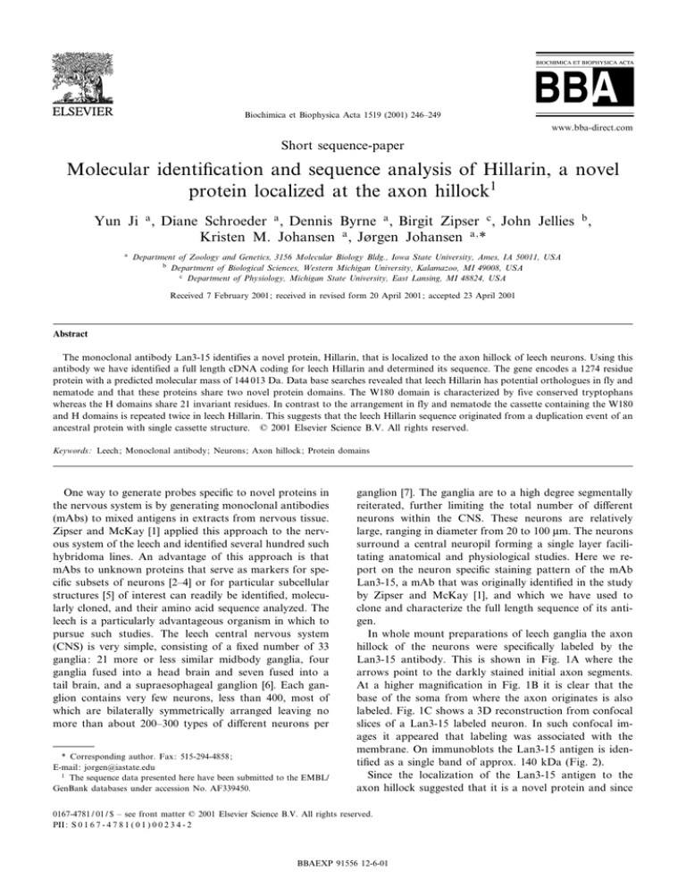
Biochimica et Biophysica Acta 1519 (2001) 246^249
www.bba-direct.com
Short sequence-paper
Molecular identi¢cation and sequence analysis of Hillarin, a novel
protein localized at the axon hillock1
Yun Ji a , Diane Schroeder a , Dennis Byrne a , Birgit Zipser c , John Jellies b ,
Kristen M. Johansen a , JÖrgen Johansen a; *
a
Department of Zoology and Genetics, 3156 Molecular Biology Bldg., Iowa State University, Ames, IA 50011, USA
b
Department of Biological Sciences, Western Michigan University, Kalamazoo, MI 49008, USA
c
Department of Physiology, Michigan State University, East Lansing, MI 48824, USA
Received 7 February 2001; received in revised form 20 April 2001 ; accepted 23 April 2001
Abstract
The monoclonal antibody Lan3-15 identifies a novel protein, Hillarin, that is localized to the axon hillock of leech neurons. Using this
antibody we have identified a full length cDNA coding for leech Hillarin and determined its sequence. The gene encodes a 1274 residue
protein with a predicted molecular mass of 144 013 Da. Data base searches revealed that leech Hillarin has potential orthologues in fly and
nematode and that these proteins share two novel protein domains. The W180 domain is characterized by five conserved tryptophans
whereas the H domains share 21 invariant residues. In contrast to the arrangement in fly and nematode the cassette containing the W180
and H domains is repeated twice in leech Hillarin. This suggests that the leech Hillarin sequence originated from a duplication event of an
ancestral protein with single cassette structure. ß 2001 Elsevier Science B.V. All rights reserved.
Keywords : Leech; Monoclonal antibody; Neurons; Axon hillock; Protein domains
One way to generate probes speci¢c to novel proteins in
the nervous system is by generating monoclonal antibodies
(mAbs) to mixed antigens in extracts from nervous tissue.
Zipser and McKay [1] applied this approach to the nervous system of the leech and identi¢ed several hundred such
hybridoma lines. An advantage of this approach is that
mAbs to unknown proteins that serve as markers for speci¢c subsets of neurons [2^4] or for particular subcellular
structures [5] of interest can readily be identi¢ed, molecularly cloned, and their amino acid sequence analyzed. The
leech is a particularly advantageous organism in which to
pursue such studies. The leech central nervous system
(CNS) is very simple, consisting of a ¢xed number of 33
ganglia: 21 more or less similar midbody ganglia, four
ganglia fused into a head brain and seven fused into a
tail brain, and a supraesophageal ganglion [6]. Each ganglion contains very few neurons, less than 400, most of
which are bilaterally symmetrically arranged leaving no
more than about 200^300 types of di¡erent neurons per
* Corresponding author. Fax: 515-294-4858 ;
E-mail : jorgen@iastate.edu
1
The sequence data presented here have been submitted to the EMBL/
GenBank databases under accession No. AF339450.
ganglion [7]. The ganglia are to a high degree segmentally
reiterated, further limiting the total number of di¡erent
neurons within the CNS. These neurons are relatively
large, ranging in diameter from 20 to 100 Wm. The neurons
surround a central neuropil forming a single layer facilitating anatomical and physiological studies. Here we report on the neuron speci¢c staining pattern of the mAb
Lan3-15, a mAb that was originally identi¢ed in the study
by Zipser and McKay [1], and which we have used to
clone and characterize the full length sequence of its antigen.
In whole mount preparations of leech ganglia the axon
hillock of the neurons were speci¢cally labeled by the
Lan3-15 antibody. This is shown in Fig. 1A where the
arrows point to the darkly stained initial axon segments.
At a higher magni¢cation in Fig. 1B it is clear that the
base of the soma from where the axon originates is also
labeled. Fig. 1C shows a 3D reconstruction from confocal
slices of a Lan3-15 labeled neuron. In such confocal images it appeared that labeling was associated with the
membrane. On immunoblots the Lan3-15 antigen is identi¢ed as a single band of approx. 140 kDa (Fig. 2).
Since the localization of the Lan3-15 antigen to the
axon hillock suggested that it is a novel protein and since
0167-4781 / 01 / $ ^ see front matter ß 2001 Elsevier Science B.V. All rights reserved.
PII: S 0 1 6 7 - 4 7 8 1 ( 0 1 ) 0 0 2 3 4 - 2
BBAEXP 91556 12-6-01
Y. Ji et al. / Biochimica et Biophysica Acta 1519 (2001) 246^249
247
Fig. 1. Immunohistochemical labeling of leech central neurons with Lan3-15 Hillarin antibody. (A) The axon hillock of neurons in the anterior packages labeled with Lan3-15 antibody in a whole mount preparation of a leech midbody ganglion. The arrows point to examples of the darkly labeled initial axon segments whereas a few representative somata are indicated by an s. The gray hue of the somata represents the level of weak non-speci¢c
background labeling. Scale bar equals 125 Wm. (B) Higher magni¢cation micrograph of a single labeled neuron. The arrow points to the initial axon
segment. The soma (s) is unlabeled except for the base where the axon originates. Scale bar equals 20 Wm. (C) Stereo pair of a 3D reconstruction from
confocal z-sections of a Lan3-15 labeled neuron on a scale comparable to that in B. The labeling is visualized using HRP-conjugated secondary antibody in A and B and by using TRITC-conjugated secondary antibody in C.
the organization and physiology of this region of the neuron are of particular interest we decided to determine the
molecular identity of the Lan3-15 antigen. To this end we
screened 1U106 plaques of a leech random primed expression vector library [3] with the antibody and several positive partial cDNA clones were identi¢ed. Subsequently,
the cDNA library was screened with radiolabeled nucleotide probe generated from the 5P end of the original cDNA
clones. From this screen several overlapping cDNA clones
were isolated which are likely to encompass the entire
coding sequence since the predicted sequence has a 5P
ATG start codon just downstream from an in-frame
TGA stop codon. The predicted sequence is for a protein
containing 1274 residues (Fig. 3) with a calculated molecular mass of 144 013 Da. Due to its localization to the
axon hillock we have named this protein Hillarin. Several
lines of evidence suggest that the Hillarin cDNA corresponds to the antigen recognized by the antibody: (1) all
clones identi¢ed by the Lan3-15 antibody in the original
screen which were analyzed proved to be derived from the
same gene, making non-speci¢c cross-reactivity with an
unrelated gene product unlikely; (2) the predicted molecular mass of Hillarin of 144 013 Da is close to the relative
molecular mass of the Lan3-15 antigen of 140 kDa as
estimated by SDS^PAGE (Fig. 2).
The complete sequence of Hillarin is shown in Fig. 3.
Using the National Center for Biotechnology and Infor-
Fig. 2. Immunoblot of SDS^PAGE fractionated leech CNS proteins labeled with Lan3-15. The antibody recognizes a single band with an apparent molecular mass of 140 kDa. The migration of molecular weight
markers is indicated to the right.
BBAEXP 91556 12-6-01
248
Y. Ji et al. / Biochimica et Biophysica Acta 1519 (2001) 246^249
Fig. 3. The complete predicted amino acid sequence of leech Hillarin. Leech Hillarin is a 1274 residue protein with a calculated molecular mass of
144 013 Da.
mation BLAST server the Hillarin sequence was compared
with known and predicted sequences from other organisms
in the data bases [8]. From these searches two hitherto
uncharacterized potential orthologous sequences were
identi¢ed in £y (CG13435) and nematode (Z47811), respectively (Fig. 4). Furthermore, the Hillarin sequence
was analyzed for similar sequences within itself by the
computer program COMPARE (part of the GCG suite
of programs). This dot plot analysis (data not shown)
revealed that Hillarin has two di¡erent conserved domains
that are repeated twice (Fig. 4C). Interestingly, the £y and
the nematode sequences only have one cassette of these
domains (Fig. 4C). This suggests that the Hillarin sequence may have been derived from a duplication event
of an ancestral protein with the single cassette structure.
The sequences of the conserved domains of Hillarin are
aligned with their £y and nematode counterparts in Fig.
4A,B, respectively. The ¢rst domain is comprised of 180
amino acids and is characterized by ¢ve tryptophans in
conserved positions (Fig. 4A, asterisks) wherefore we
have named this domain W180. The two W180 domains
within leech Hillarin are 44% identical on the amino acid
level whereas the £y and nematode W180 domains are
71% identical. This domain can also be found in proteins
of unicellular organisms such as the cyanobacterium Synechocystis (D90900). The second type of domain found in
Hillarin is characterized by 21 invariant amino acids
among the leech, £y, and nematode sequences (Fig. 4B)
and we have named it the H domain since it was ¢rst
described in Hillarin. Outside the invariant residues there
is less conservation of amino acids compared to the W180
domain as re£ected by the only 28% identity between the
two Hillarin H domains. The H domains of the £y and
nematode are 58% identical. In addition, sequences potentially homologous to the two types of Hillarin domains are
present in the draft of the human genome (NCBI accession No. NT_022711).
The molecular cloning of Hillarin and the identi¢cation
of two potential orthologues in £y and nematode, two
animals amenable to genetic manipulation, promise to
provide new insight into the function of W180 and H
domain containing proteins.
The authors wish to thank Anna Yeung for excellent
technical assistance. This work was supported by NIH
Grant NS28857 to J.Jo.
References
[1] B. Zipser, R. McKay, Nature 289 (1991) 549^554.
[2] K.K. Briggs, A.J. Silvers, K.M. Johansen, J. Johansen, J. Cell Biol.
129 (1995) 1355^1362.
[3] Y. Huang, J. Jellies, K.M. Johansen, J. Johansen, J. Cell Biol. 138
(1997) 394^402.
[4] Y. Xu, B. Bolton, B. Zipser, J. Jellies, K.M. Johansen, J. Johansen,
J. Neurobiol. 40 (1999) 244^253.
[5] K.M. Johansen, J. Johansen, J. Neurobiol. 27 (1995) 227^239.
[6] K.J. Muller, J.G. Nicholls, G.S. Stent, Neurobiology of the Leech,
Cold Spring Harbor Laboratory Press, Cold Spring Harbor, NY,
1981.
[7] E.R. Macagno, J. Comp. Neurol. 190 (1980) 283^302.
[8] S.F. Altschul, T.L. Madden, A.A. Scha«¡er, J. Zhang, Z. Zhang, W.
Miller, D.J. Lipman, Nucleic Acids Res. 25 (1997) 3389^3402.
BBAEXP 91556 12-6-01
Y. Ji et al. / Biochimica et Biophysica Acta 1519 (2001) 246^249
249
Fig. 4. Sequence comparison of leech Hillarin with related proteins in £y and nematode. (A) Alignment of the two W180 domains in leech Hillarin
with the corresponding domains in orthologues from £y and nematode. The ¢ve conserved tryptophans characterizing this domain are indicated by asterisks. Shared amino acids as well as conservative changes (Y/F, D/E, L/V/I, and R/K) are in white type face outlined in black. (B) Alignment of the
two H domains in leech Hillarin with the corresponding domains in orthologues from £y and nematode. The 21 invariant residues characterizing this
domain are indicated by asterisks. Shared amino acids as well as conservative changes (Y/F, D/E, L/V/I, and R/K) are in white type face outlined in
black. (C) Diagram and alignment of the domain composition of leech Hillarin as compared to the £y and nematode orthologues. Sequence identity on
the amino acid level between the W180 and H domains in leech, £y, and nematode is indicated in percent.
BBAEXP 91556 12-6-01

