Functional Connectivity Networks Associated With Dorsal and Ventral Striatum
advertisement
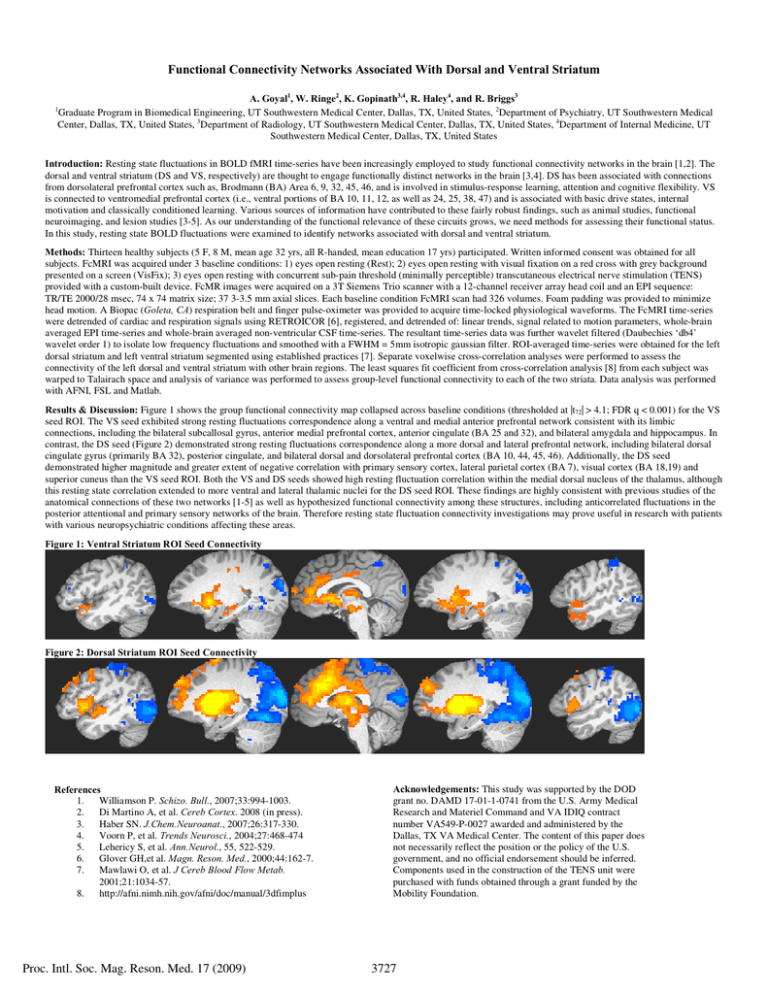
Functional Connectivity Networks Associated With Dorsal and Ventral Striatum A. Goyal1, W. Ringe2, K. Gopinath3,4, R. Haley4, and R. Briggs3 Graduate Program in Biomedical Engineering, UT Southwestern Medical Center, Dallas, TX, United States, 2Department of Psychiatry, UT Southwestern Medical Center, Dallas, TX, United States, 3Department of Radiology, UT Southwestern Medical Center, Dallas, TX, United States, 4Department of Internal Medicine, UT Southwestern Medical Center, Dallas, TX, United States 1 Introduction: Resting state fluctuations in BOLD fMRI time-series have been increasingly employed to study functional connectivity networks in the brain [1,2]. The dorsal and ventral striatum (DS and VS, respectively) are thought to engage functionally distinct networks in the brain [3,4]. DS has been associated with connections from dorsolateral prefrontal cortex such as, Brodmann (BA) Area 6, 9, 32, 45, 46, and is involved in stimulus-response learning, attention and cognitive flexibility. VS is connected to ventromedial prefrontal cortex (i.e., ventral portions of BA 10, 11, 12, as well as 24, 25, 38, 47) and is associated with basic drive states, internal motivation and classically conditioned learning. Various sources of information have contributed to these fairly robust findings, such as animal studies, functional neuroimaging, and lesion studies [3-5]. As our understanding of the functional relevance of these circuits grows, we need methods for assessing their functional status. In this study, resting state BOLD fluctuations were examined to identify networks associated with dorsal and ventral striatum. Methods: Thirteen healthy subjects (5 F, 8 M, mean age 32 yrs, all R-handed, mean education 17 yrs) participated. Written informed consent was obtained for all subjects. FcMRI was acquired under 3 baseline conditions: 1) eyes open resting (Rest); 2) eyes open resting with visual fixation on a red cross with grey background presented on a screen (VisFix); 3) eyes open resting with concurrent sub-pain threshold (minimally perceptible) transcutaneous electrical nerve stimulation (TENS) provided with a custom-built device. FcMR images were acquired on a 3T Siemens Trio scanner with a 12-channel receiver array head coil and an EPI sequence: TR/TE 2000/28 msec, 74 x 74 matrix size; 37 3-3.5 mm axial slices. Each baseline condition FcMRI scan had 326 volumes. Foam padding was provided to minimize head motion. A Biopac (Goleta, CA) respiration belt and finger pulse-oximeter was provided to acquire time-locked physiological waveforms. The FcMRI time-series were detrended of cardiac and respiration signals using RETROICOR [6], registered, and detrended of: linear trends, signal related to motion parameters, whole-brain averaged EPI time-series and whole-brain averaged non-ventricular CSF time-series. The resultant time-series data was further wavelet filtered (Daubechies ‘db4’ wavelet order 1) to isolate low frequency fluctuations and smoothed with a FWHM = 5mm isotropic gaussian filter. ROI-averaged time-series were obtained for the left dorsal striatum and left ventral striatum segmented using established practices [7]. Separate voxelwise cross-correlation analyses were performed to assess the connectivity of the left dorsal and ventral striatum with other brain regions. The least squares fit coefficient from cross-correlation analysis [8] from each subject was warped to Talairach space and analysis of variance was performed to assess group-level functional connectivity to each of the two striata. Data analysis was performed with AFNI, FSL and Matlab. Results & Discussion: Figure 1 shows the group functional connectivity map collapsed across baseline conditions (thresholded at |t72| > 4.1; FDR q < 0.001) for the VS seed ROI. The VS seed exhibited strong resting fluctuations correspondence along a ventral and medial anterior prefrontal network consistent with its limbic connections, including the bilateral subcallosal gyrus, anterior medial prefrontal cortex, anterior cingulate (BA 25 and 32), and bilateral amygdala and hippocampus. In contrast, the DS seed (Figure 2) demonstrated strong resting fluctuations correspondence along a more dorsal and lateral prefrontal network, including bilateral dorsal cingulate gyrus (primarily BA 32), posterior cingulate, and bilateral dorsal and dorsolateral prefrontal cortex (BA 10, 44, 45, 46). Additionally, the DS seed demonstrated higher magnitude and greater extent of negative correlation with primary sensory cortex, lateral parietal cortex (BA 7), visual cortex (BA 18,19) and superior cuneus than the VS seed ROI. Both the VS and DS seeds showed high resting fluctuation correlation within the medial dorsal nucleus of the thalamus, although this resting state correlation extended to more ventral and lateral thalamic nuclei for the DS seed ROI. These findings are highly consistent with previous studies of the anatomical connections of these two networks [1-5] as well as hypothesized functional connectivity among these structures, including anticorrelated fluctuations in the posterior attentional and primary sensory networks of the brain. Therefore resting state fluctuation connectivity investigations may prove useful in research with patients with various neuropsychiatric conditions affecting these areas. Figure 1: Ventral Striatum ROI Seed Connectivity Figure 2: Dorsal Striatum ROI Seed Connectivity References 1. Williamson P. Schizo. Bull., 2007;33:994-1003. 2. Di Martino A, et al. Cereb Cortex. 2008 (in press). 3. Haber SN. J.Chem.Neuroanat., 2007;26:317-330. 4. Voorn P, et al. Trends Neurosci., 2004;27:468-474 5. Lehericy S, et al. Ann.Neurol., 55, 522-529. 6. Glover GH,et al. Magn. Reson. Med., 2000;44:162-7. 7. Mawlawi O, et al. J Cereb Blood Flow Metab. 2001;21:1034-57. 8. http://afni.nimh.nih.gov/afni/doc/manual/3dfimplus Proc. Intl. Soc. Mag. Reson. Med. 17 (2009) Acknowledgements: This study was supported by the DOD grant no. DAMD 17-01-1-0741 from the U.S. Army Medical Research and Materiel Command and VA IDIQ contract number VA549-P-0027 awarded and administered by the Dallas, TX VA Medical Center. The content of this paper does not necessarily reflect the position or the policy of the U.S. government, and no official endorsement should be inferred. Components used in the construction of the TENS unit were purchased with funds obtained through a grant funded by the Mobility Foundation. 3727
