What the Eye Can See
advertisement

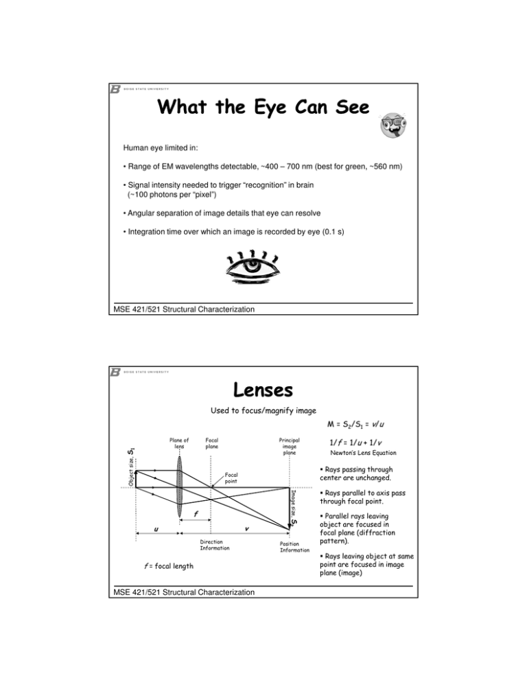
What the Eye Can See Human eye limited in: • Range of EM wavelengths detectable, ~400 – 700 nm (best for green, ~560 nm) • Signal intensity needed to trigger “recognition” in brain (~100 photons per “pixel”) • Angular separation of image details that eye can resolve • Integration time over which an image is recorded by eye (0.1 s) MSE 421/521 Structural Characterization Lenses Used to focus/magnify image M = S2/S1 = v/u Focal plane Object size, S1 Plane of lens Principal image plane Newton’s Lens Equation Rays passing through center are unchanged. Focal point Image size, f Direction Information f = focal length MSE 421/521 Structural Characterization S2 v u 1/f = 1/u + 1/v Position Information Rays parallel to axis pass through focal point. Parallel rays leaving object are focused in focal plane (diffraction pattern). Rays leaving object at same point are focused in image plane (image) Maxwell’s Conditions for a Perfect Lens All rays leaving one point on the object must meet at one point in the image - no aberrations. If points on the object lie on a plane perpendicular to the lens axis then points in the image must also lie in a plane perpendicular to the lens axis - no field/image curvature. Ratio of distances in object must be the same as those in the image - no distortion. MSE 421/521 Structural Characterization Real vs Virtual Images f v Real Image u Virtual Image 0<u<f Virtual Not inverted Magnified (M < 0, v < 0) u=f -- -- Image at infinity – no image f < u ≤ 2f Real Inverted Magnified (M ≥ 1) u > 2f Real Inverted Demagnified (0 < M < 1) MSE 421/521 Structural Characterization f Diffraction Barrier or Diffraction Limit Resolved Unresolved d1/2 d1 d1 Airy disc George Biddell Airy (1801 – 1892) MSE 421/521 Structural Characterization Resolution The Abbe Criterion θ θ θ θ Diffraction Limit: If grating is too narrow, θ is too large and only zero-order beams contribute to image. object aperture d1/2 = rd = 0.5λ µ sinβ β lens Ernst Karl Abbe (1840 - 1905) MSE 421/521 Structural Characterization µ = refractive index β = semi-angle/half-angle µ sinβ = numerical aperture Resolution The Rayleigh Criterion Resolved Rayleigh Criterion d1/2 d1 Unresolved 15% drop in intensity d1 Diffraction Limit: d1/2 = rd = δ = aperture object 0.61λ µsinβ µ = refractive index β β = semi-angle/half-angle µ sinβ = numerical aperture lens Lord Ravleigh (John William Strutt) (1877 - 1919) MSE 421/521 Structural Characterization Resolution Rayleigh: d1/2 = rd = δ = 0.61λ µsinβ Abbe: Sparrow: d1/2 = rd = δ = 0.5λ µ sinβ 0.47λ d1/2 = rd = δ = µsinβ The distance at which the first maximum in one Airy disc coincides with the first minimum of the adjacent Airy disc. The finest spacing that can be imaged with both the undiffracted and first-order diffracted waves. The distance at which there is no longer a dip in peak intensity between two adjacent Airy discs, but rather constant brightness across the region between the peaks. http://www.youtube.com/watch?v=0ULRY7RscOY MSE 421/521 Structural Characterization The Eye as Microscope? - cornea: n = 1.376 - vitreous humor ~ water: n = 1:33 - lens: n = 1:39 - 1:41 index varies: high in center, low at edges - iris: variable aperture stop diameter: 2-8 mm - retina: detector - resolution…? MSE 421/521 Structural Characterization The Eye as Microscope? Sensitivity: Fully expanded pupil can see I ≤ 10-10 W/m2 from point source Power = IA = (10-10 W/m2)[π(4mm)2] ~ 10-14 W Maximum irradiance (sunlight) ~ 250 mW/m2 Pupil area ~ π(1mm)2 So maximum power ~ 1mW (damage threshold for laser) Dynamic range: 10-14 to 10-6 W (8 orders of magnitude) (instantaneous range: ~ 5 orders of magnitude) Best artificial detectors: photographic film & high-end CCDs dynamic range 4 orders of magnitude (10 worse than eye!) MSE 421/521 Structural Characterization - cornea: n = 1.376 - vitreous humor ~ water: n = 1:33 - lens: n = 1:39 - 1:41 index varies: high in center, low at edges - iris: variable aperture stop diameter: 2-8 mm - retina: detector - resolution…? Astigmatism Differences in optical properties of lens from point to point → Rays in the horizontal plane are focused at a different point from those traveling in the vertical plane. Disc of least confusion lens rast = β∆f β must be minimised (unlike for rd) Astigmatism can also be observed with a non-astigmatic lens for object points far off the optic axis MSE 421/521 Structural Characterization Chromatic Aberration Rays of different wavelength are refracted by different amounts in the lens and so are brought to focus at different places. Shorter wavelengths refract more. blue green red This effect is caused by an energy spread in the electron beam (voltage instabilities) or inelastic scattering in the sample. rc = Ccβ (δE/E0) Cc and β must be minimised (unlike for rd) Cc (JEOL 2100) = 1.4 mm δE ~ 20 eV MSE 421/521 Structural Characterization Problem increases with thickness Spherical Aberration Rays passing near the centre of the lens are focused at a different point from rays passing through the edges. Marginal focus Axial focus β Disc of least confusion, r = ¼rs Gaussian image plane, r = rs rs = Csβ 3 (position of image when very small apertures are used - paraxial rays) Cs and β (strong dependence) must be minimised (unlike for rd) Cs (JEOL 2100) = 1 mm MSE 421/521 Structural Characterization Distortion If the magnification of the lens varies from the centre to the edge, the result is a distorted image. Rectangular object Barrel distortion Pin-cushion distortion The overall effect of aberrations on resolution In the electron microscope it is very difficult to correct for achromatic aberrations, especially Cs. Net resolution: ropt = rd2 + rs2 then: dopt (JEOL 2100) = 2.3 Å W&C says 0.77 βopt = 0.67λ1/4Cs-1/4 ropt = 1.21λ3/4Cs1/4 MSE 421/521 Structural Characterization 0.7 ≤ x ≤ 1.21 W&C says 0.91 Depth of Field/Focus Range of object/image distances over which the object can be placed/image can be viewed without the eye detecting any change in sharpness MSE 421/521 Structural Characterization Depth of Field/Focus Range of object/image distances over which the object can be placed/image can be viewed without the eye detecting any change in sharpness aperture lens In EM, the small β necessary to reduce spherical aberration results in a large depth of field/focus by default (SEM) despite the small λ. β d1 d1 α Dim Depth of field : Dob = 0.61λ µ sinβtanβ ≈ 0.61λ β2 increase (but increases rd) decrease (but increases rd) by inserting a smaller aperture (For EM, µ = 1 and sinβ ≈ tanβ ≈ β) Depth of focus : Dim = DobM2 Optical microscopy is a compromise between resolution and depth of field/focus, but in EM, reducing β can improve resolution by reducing aberrations. MSE 421/521 Structural Characterization


![Zone Axis Zone Axis Identification Zone axis [mnp] Weiss Zone Law:](http://s2.studylib.net/store/data/010544623_1-33810755141931bcda93061771d9f563-300x300.png)
