Cochlear dissection after insertion of a perimodiolar type electrode array... standard Helix). Segments of the osseous lamina and basilar membrane...
advertisement
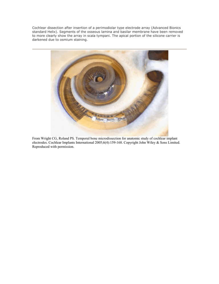
Cochlear dissection after insertion of a perimodiolar type electrode array (Advanced Bionics standard Helix). Segments of the osseous lamina and basilar membrane have been removed to more clearly show the array in scala tympani. The apical portion of the silicone carrier is darkened due to osmium staining. From Wright CG, Roland PS. Temporal bone microdissection for anatomic study of cochlear implant electrodes. Cochlear Implants International 2005;6(4):159-168. Copyright John Wiley & Sons Limited. Reproduced with permission.
