NER: Nano-Grating Force Sensor for Measurement of Neuron
advertisement
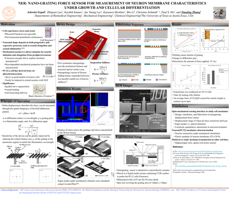
NER: NANO-GRATING FORCE SENSOR FOR MEASUREMENT OF NEURON MEMBRANE CHARACTERISTICS UNDER GROWTH AND CELLULAR DIFFERENTIATION Ashwini Gopal1, Zhiquan Luo2, Karthik Kumar1, Jae Young Lee3, Kazunori Hoshino1, Bin Li2, Christine Schmidt1, 3, Paul S. Ho2, and Xiaojing Zhang1 Departments of Biomedical Engineering1, Mechanical Engineering2, Chemical Engineering3The University of Texas at Austin,Texas, USA Motivation Device Design • Cell experiences stress and strain Physical/Chemical cues (growth) Environmental perturbation (robustness) Cell Structure [1] • Neuronal shape depend on both progressive and regressive processes such as axonal elongation and axonal elimination [2-3] • Mechanical tension is a direct stimulus for neurite initiation and elongation from peripheral neurons Classical concepts fail to explain mechanotransduction[4] Suspension Stiffness • Two symmetric nanogratings Size-dependant mechanical properties have not been 3 provide mechanical balance and 2 Ew t characterized maximal optical surface area. k s 3 • PC12-a cell line derived from rat Ls • Nanogratings consist of flexure Flexure Stiffness pheochromocytoma folding beams suspended between [5] PC12 Cells •Serve as good models of neuron cells 3 8 Ew t two parallel cantilevers of known •Easily be induced to extend neurites with NGF k f [7] 3 stiffness . Lf • Significance guided nerve regeneration Simulation Results wound healing cell based drug delivery Basic structure of neuron [6] Fabrication Sequence x: Experimental values *: Calibration Values •Probing causes motion of grating •Change in diffraction spot •Determines the amount of force applied. (F=kx) SEM Images • Experiment was conducted on 10-15 cells • Time for testing cells-30mins • An average force of 22+8μN caused the neurite length to contract up to 6μm Principle of Operation • Probe displacement, therefore the force, can be measured through the spatial frequency of far-field diffraction pattern. m psin sin Conclusions m is diffraction order,λ is wavelength, p is grating pitch, α is illumination angle, and θ is diffraction angle. p m m p 1 p 2 2 Results Absence of stress across the gratings and stress concentrated on the flexure beams • Sensitivity of the device can be greatly improved by reducing the critical feature size, p, of the grating to the nanometer regime to match the illumination wavelength. Experimental Setup Opto-mechanical sensing interface to study cell mechanics • Design, simulation, and fabrication of nanograting displacement-force sensor • Displacement range of 10µm & force sensitivity 8N/m • Eigen modes vs. optical detection • Localized, quantitative interactions in microenvironment Neuron(PC12) mechanics characterization • Neurite contraction under mechanical stimulation • Elastic modulus of neuron membrane 425±30 Pa Platform to study mechano-transduction in other cell lines • Hippocampal cells, spinal cord motor neuron Reference: Eigen modes under mechanical vibration were simulated using CoventorWare™ . • Nanograting sensor is attached to a piezoelectric actuator • Placed in a liquid media (serum containing F12K media) to probe the PC12 cells (Neurons) • Illuminated with a 635 nm He-Ne laser diode • Spot size covering the grating area of 120μm x 120μm. [1] See: http://www.uic.edu/classes/bios/bios100/lecturesf04am/cytoskeleton.jpg [2] Cowan, W.M, Fawcett, J. W, O'Leary, D. D. M, and Stanfield, B. B “Regressive events in neumgenesis.” Science. 225, p1258-1265, 1984. [3] Purves, D, and Lichtman, J. W, “Elimination of synapses in the developing nervous system.”, Science, 210, p153-157, 1980. [4] McKintosh F C, Kas J and Janmey P A 1995 Phys. Rev. Lett. 75 4425 [5] See: http://scienceblogs.com/purepedantry/2006/07/background_to_the_20_year_coma.ph p [6] See: http://www.med.osaka-u.ac.jp/pub/molonc/www/Esignal.html [7] Stephen D. Sentura “ Microsystem Design”,2001 Acknowledgement: NSF Nanoscale Exploratory Research Award (NER), ECS-0609413, the Welch Foundation and the Strategic Partnership for Research in Nanotechnology (SPRING), and NSF NNIN Facilities at UT Austin and Stanford Center for Integrated Systems (Grant # 0335765 and 9731293 respectively)

