C7- A View of the Cell
advertisement
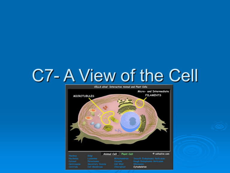
C7- A View of the Cell A View of the Cell 7-1 Discovery of Cells 7-2 Plasma Membrane 7-3 Discovery of Cells Anton van Leeuwenhoek used the first microscope early in the 1600s. It revealed the world of microorganisms and cells which are the basic unit of an organism. Discovery of Cells Robert Hooke coined the term cell after observing cork under a microscope. Hooke’s illustration of cork Discovery of Cells 1838 Matthias Schleiden observed plant cells. Theodor Schwann studied animal cells. Ideas were summarized in the Cell Theory. Cell Theory 1. All organisms are made of one or more cells. 2. The cell is the basic unit of structure and organization in organisms. 3. All cells come from preexisting cells. Electron Microscopes Light Microscopes 1500X Dev new EM in 1930s & 40s Uses beam of electrons to multiply image 500,000X Lets us see internal cell structure Scanning vs. Transmission EM Scan surface of cell for 3D image Study internal structure of cell Two Basic Cell Types All cells contain specialized structures called organelles. Eukaryotes have membrane-bound organelles Prokaryotes do not Some eukaryotes are unicellular like amoebas. Two Basic Cell Types Robert Brown observed eukaryotes had a prominent structure. Rudolf Virchow said it was responsible for cell division. Eventually the structure was called the nucleus or cell’s control center. 7.2 Plasma Membrane Responsible for controlling what goes in and out of the cell. Plasma Membrane Maintains homeostasis by selective permeability keeping some things in and keeping others out Plasma Membrane Structure Phospholipid Bilayer Glycerol backbone, 2 fatty acids & a phosphate group make a phospholipid. Water is attracted to the head but not to the lipid fatty acids. Plasma Membrane Structure Fluid Mosaic Model because phospholipids ripple like water with the currents. Protein molecules float like boats on the surface in a mosaic pattern. Flexible Other components Cholesterol stabilizes the phospholipids by keeping the the tails from sticking together Transport proteins span the membrane to move needed substances in and wastes out of the cell. 7.3 Eukaryotic Cell Structure Each component has a specific function. They work together to help the cell survive. Cell wall fairly rigid part of plant cells, bacteria & protists made of cellulose. Eukaryotic Cell Structure Nucleus Cell Control Contains directions for making proteins in chromatin. Strands of genetic material, DNA Chromatin condenses to chromosomes before cell division Eukaryotic Cell Structure Nucleolus Makes ribosomes Ribosomes are production site of proteins made by direction of DNA They’re made of RNA and must travel to the cytoplasm to build proteins. The nucleolus makes ribosomes which are the site of protein synthesis. Ribosomes must travel out of the nucleus to the Cytoplasm where they build the proteins according to DNAs direction. Eukaryotic Cell Structure Cytoplasm Clear, gelatinous fluid inside a cell Ribosomes and RNA pass through nuclear pores in the nuclear envelope to enter the cytoplasm Eukaryotic Cell Structure Assembly, transport & storage Endoplasmic reticulum site of cellular chemical reactions Highly folded membrane which ribosomes attach to (Rough ER) do protein synthesis Eukaryotic Cell Structure Each protein has a specific function. Smooth ER has no ribosomes but makes and stores lipids. After proteins are made they’re sent to the Golgi Apparatus Eukaryotic Cell Structure Golgi Apparatus Modifies proteins & sorts them into packages (vesicles) for transport to their destination Eukaryotic Cell Structure Vacuoles membrane bound compartment for temporary storage Stores food, enzymes & other needed materials. Animal cells may also have vacuoles usually smaller. Eukaryotic Cell Structure Lysosomes contain digestive enzymes. Used to digest worn out organelles, food particles or engulfed viruses or bacteria. The membrane prevents enzymes from destroying the cell. Eukaryotic Cell Structure Energy Transformers Chloroplasts capture light energy & convert it to chemical energy. Eukaryotic Cell Structure
