File
advertisement

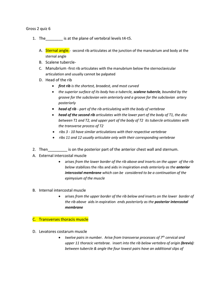
Gross 2 quiz 6 1. The________ is at the plane of vertebral levels t4-t5. A. Sternal angle.- second rib articulates at the junction of the manubrium and body at the sternal angle B. Scalene tubercleC. Manubrium -first rib articulates with the manubrium below the sternoclavicular articulation and usually cannot be palpated D. Head of the rib first rib is the shortest, broadest, and most curved the superior surface of its body has a tubercle, scalene tubercle, bounded by the groove for the subclavian vein anteriorly and a groove for the subclavian artery posteriorly head of rib - part of the rib articulating with the body of vertebrae head of the second rib articulates with the lower part of the body of T1, the disc between T1 and T2, and upper part of the body of T2 its tubercle articulates with the transverse process of T2 ribs 3 - 10 have similar articulations with their respective vertebrae ribs 11 and 12 usually articulate only with their corresponding vertebrae 2. Then_________ is on the posterior part of the anterior chest wall and sternum. A. External intercostal muscle arises from the lower border of the rib above and inserts on the upper of the rib below stabilizes the ribs and aids in inspiration ends anteriorly as the anterior intercostal membrane which can be considered to be a continuation of the epimysium of the muscle B. Internal intercostal muscle arises from the upper border of the rib below and inserts on the lower border of the rib above aids in expiration ends posteriorly as the posterior intercostal membrane C. Transverses thoracis muscle D. Levatores costarum muscle twelve pairs in number. Arise from transverse processes of 7th cervical and upper 11 thoracic vertebrae. insert into the rib below vertebra of origin (brevis): between tubercle & angle the four lowest pairs have an additional slips of muscle into the 2nd rib below their vertebrae of origin (longi): crossing the rib below its vertebrae of origin. action – raise ribs to increase thoracic cavity 3. Which if the following supplies the first and second internal spaces, posteriorly? a. Internal thoracic artery anterior intercostal arteriesthe first 5 or 6 interspaces receive blood from the internal thoracic branch of the subclavian artery b. Superior epigastric artery c. Supreme intercostal artery d. Musculo phrenic artery the remaining interspaces receive blood from the musculophrenic branch of the internal thoracic artery 4. Which of the following constitute true ribs? a. Ribs 1-7 b. Ribs 8-12 five remaining ribs are called false ribs ribs 8, 9, 10 articulate with the rib above through their costal cartilages c. Ribs 11-12 ribs 11 and 12 don't articulate with anything anteriorly so are called floating ribs articulations with the sternum d. Ribs 1-8 5. Which of the following has a cutaneous branch to the arm from the T2? a. Spinal accessory nerve b. Intercostalbrachial nerve c. Phrenic nerve phrenic nerves pass anterior to both subclavian arteries and roots of the lungs, pass between the pericardium and mediastinal pleura to reach the diaphragm parietal pleura is innervated byintercostal nerves phrenic ne d. Vagus nerve vagus nerves pass anterior to the subclavian arteries but posterior to the root of the lungs right recurrent laryngeal nerve passes under the right subclavian artery ascends to the larynx left recurrent laryngeal nerve passes under the aortic arch immediately to left of the ligamentum arteriosum to ascend to the larynx cardiac nerves descend from the cervical sympathetic trunk and vagus nerve to reach the cardiac plexuses around the aortic arch 6. The circumflex branch is off the________. a. Right coronary artery acute marginal branch runs along the acute margin sinoatrial nodal artery (60% from the proximal portion of the right coronary artery, 40% from the beginning of the left circumflex artery posterior interventricular branch or posterior descending artery (PDA) - runs in the posterior interventricular sulcus. right posterolateral branches supply the posterior inferior wall of ventricle atrioventricular nodal branch - in 80% of population b. Left coronary artery c. Pulmonary artery right ventricle - receives blood from the right atrium and pumps it to the lungs via the pulmonary artery . right pulmonary artery - at root of the lung it is anterior / inferior to superior lobar (epiarterial) bronchus,left pulmonary artery at root of the lung it is anterior / superior to superior lobar bronchus d. Bronchial artery bronchial arteries2 left branches (superior and inferior) and 1 right branch the left branches come directly off the aorta right branch comes off an intercostal artery or one of the left bronchial arteries supply blood to bronchi, areolar tissue, bronchial lymph nodes, and esophagus nutrition is its most important duty for the alveoli since they already have air 7. The trabeculated surface in the surface inside right atrum called. a. Sinus venarum right atrium receives deoxygenated blood from the body (via the superior and inferior vena cava) and the heart (via the coronary sinus, mostly); its interior has sinus venarum - smooth part between the two vena cava b. pectinate muscles c. Trabeculae carne trabecula carnae - thick muscle bundles giving the ventricle its rugged texture d. Conus arteriosus 8. The right atrioventricular valve is a ______. a. Chordinae tendinae chorda tendinae - fibrous cords attaching papillary muscles to the valve cusps b. Semilunar valve pulmonary valvelocated near the left side of the sternum at the level of the 3rd costal cartilage listen in the 2nd intercostal space on the left aortic valve located near the left side of the sternum at the level of the 3rd intercostal space listen in 2nd intercostal space on the right semilunar valves -each has three cusps with nodules pulmonary valve has anterior, right, and left cusps aortic valve - has posterior, right and left cusps: above each is a dilation called c. Miral valve mitral valve located near the left side of the sternum at the level of the 4th costal cartilage listen in the 5th intercostal space about 3½ inches to the left of the midline d. Tricuspid valve 9. The ________ is at the junction of the superior vena cava and the right atrium. a. Papillary muscles papillary muscles - conical projections of ventricular muscle related to the cusps (anterior, posterior, and septal) b. Fossa ovale fossa ovale and limbus of the fossa ovale c. Sinuaterial node d. Bundle of his atrioventricular bundle (Bundle of His) runs below the membranous part of the interventricular septum divides into the right and left bundle branches right bundle branch cord-like and runs on the moderator band connects with the Purkinje fiber network of the right ventricle left bundle branch wider than the right bundle branch divides into anterior and posterior fascicles on left surface of the interventricular septum connects with the Purkinje fiber network 10. In the right lung, the ________separates the upper lobe from the middle lobe. a. Horizontal fissure b. Oblique fissure separates the upper and middle lobes from the lower lobe starts at the fifth rib or higher posteriorly, follows the sixth rib to reach the diaphragmatic border anteriorly c.Lingual left lung - an oblique fissure separates the lung into two lobes upper lobe - served by four segments: apico-posterior, anterior, superior lingular, and inferior lingular lower lobe - served by five segments (named the same as the right lung) c. Lateral and medial bronchopulmonary segment bronchopulmonary segment is a part of a lung served by one segmental bronchus and its accompanying segmental artery can be separated from the rest of the lung by blunt dissection in normal living lung veins tend to be intersegmental


