Step 1: Observing the Cow Eye
advertisement
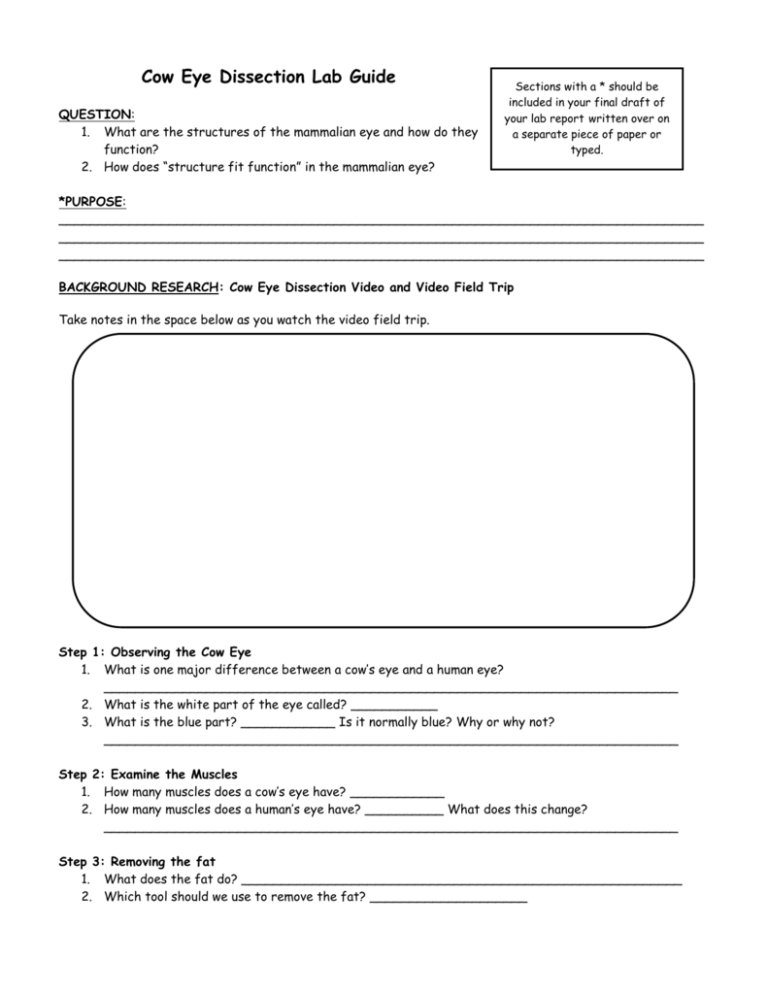
Cow Eye Dissection Lab Guide QUESTION: 1. What are the structures of the mammalian eye and how do they function? 2. How does “structure fit function” in the mammalian eye? Sections with a * should be included in your final draft of your lab report written over on a separate piece of paper or typed. *PURPOSE: __________________________________________________________________________________ __________________________________________________________________________________ __________________________________________________________________________________ BACKGROUND RESEARCH: Cow Eye Dissection Video and Video Field Trip Take notes in the space below as you watch the video field trip. Step 1: Observing the Cow Eye 1. What is one major difference between a cow’s eye and a human eye? _________________________________________________________________________ 2. What is the white part of the eye called? ___________ 3. What is the blue part? ____________ Is it normally blue? Why or why not? _________________________________________________________________________ Step 2: Examine the Muscles 1. How many muscles does a cow’s eye have? ____________ 2. How many muscles does a human’s eye have? __________ What does this change? _________________________________________________________________________ Step 3: Removing the fat 1. What does the fat do? ________________________________________________________ 2. Which tool should we use to remove the fat? ____________________ Step 4: Cutting the Cornea 1. What does the cornea do? It _____________ the eye. Light _____________ when it hits the cornea. 2. Where do we make our first incision? _____________________________ 3. What comes out? __________________ ____________________ 4. What does this liquid do? (Two functions) ____________________________________________________________________________ ______________________________________________________________________ Step 5: Cutting the Sclera 1. Where do we make our 2nd incision? ______________________________________________ 2. What tool do we use to cut after we have made our starting incision? _____________________ 3. What is the crunching sound we hear when the scientist cuts the cornea with the scalpel? _________________________________________________________________________ Step 6: Removing the Iris 1. What is the iris? ____________________________________________________________ 2. What does the iris also contain? _________________ 3. What is the pupil? ___________________________________________________________ 4. How does the iris/pupil change in response to light? ____________________________________________________________________________ ______________________________________________________________________ 5. What did the scientist use to remove the iris from the top half of the cow eye? _____________ 6. What is a major difference between a cow’s eye and a human’s eye? _________________________________________________________________________ Step 7: Removing the Lens 1. What is the jellylike substance around the lens? _________________ ___________________ 2. Why is it clear? _____________________________________________________________ 3. The ____________________ ___________________ is a mixture of protein and _________________. 4. What is the fluid’s function? ___________________________________________________ Step 8 1. 2. 3. and 9: Looking through the Lens The lens can be compared to an _______________ because it grows layers every year. The lens’ job is to help ________________________________________________________ The lens acts like a ____________________ Step 10 and 11: Observing the Retina 1. Where is the retina located? __________________________________________________ 2. The ______________ is attached to the back of the eye at just one spot. Because there are no light sensitive cells at that spot, you can’t see anything that lands in that place on the retina. This is what is called your ____________________. At the blind spot, all the nerves from the retina join to form the ______________________. Step 12: Observing the Optic Nerve 1. What does the optic nerve look like in this video? ____________________________________ 2. The optic nerve carries _______________ to the brain. Step 13: Observing the Tapetum 1. What is the shell-like layer at the back of the cow’s eye? ______________________________ 2. Do humans have this layer? _________________ 3. What does the back of the human eye look like? Why? ____________________________________________________________________________ ______________________________________________________________________ *MATERIALS Safety goggles Preserved cow’s eye Forceps Dissection scalpels Dissection scissors Dissection tray Vinyl or latex gloves Apron or tee-shirt Paper towels Plastic trash bag *PROCEDURE AND *DATA: Read the description below and answer the questions. Then, write a summarized version of the procedure for your lab report. You will include the answers to your questions in the Data Section of your final draft. 1. Locate the cornea, sclera, and optic nerve. A. The white part of the eye, the sclera, is a tough, outer covering of the eyeball. The sclera gives the eye its shape and helps to protect the delicate inner parts. B. The blue covering over the front of the eye is the cornea. When the cow was alive, the cornea was clear. Together with the lens, the cornea refracts light and helps the eye to focus. The cornea gives a larger contribution to the total refraction than the lens. The curvature of the cornea is fixed while that of the lens is changeable. In your cow’s eye, the cornea may be cloudy. C. You may be able to look through the cornea and see the iris, the colored part of the eye, and the pupil, the dark oval in the middle of the iris. At the back of the eye is the optic nerve. To see the separate fibers that make up the optic nerve, pinch the nerve with a pair of scissors or your fingers. If you squeeze the optic nerve, you may get some white goop. That is myelin, the fatty layer that surrounds each fiber of the nerve. It is the nerve that transmits visual information from the eye to the brain. 1. List two functions of the sclera. _______________________________________________________________________ _______________________________________________________________________ 2. List two functions of the cornea. _______________________________________________________________________ _______________________________________________________________________ 3. What is the function of the optic nerve? _______________________________________________________________________ _______________________________________________________________________ 2. Examine the fat and muscle surrounding the eyeball. A. Without moving your head, look up. Look down. Look all around. Six muscles attached to your eyeball move your eye so you can look in different directions. Cows have only four muscles that control their eyes. They can look up, down, left, and right, but they can’t roll their eyes like you can. Locate the externally attached muscles. These muscles control eye movement and help focus images. B. Although the muscles of each individual eye work as a team, the eyes themselves do not focus or work together until months after birth. However, one eye remains dominant. Form a circle with your thumb and index finger. Hold that position and place your hand in front of you. With both eyes, look at something through the circle. Continue to hold that position and close one eye; then open it. Close the other eye. The eye which is still able to view the object through the circle is your dominant eye. C. If you reach up and feel around your eye, you’ll feel the bone of your skull. There’s yellow fat surrounding your eyeball to keep it from bumping up against the bone and getting bruised. 1. What is the function of the externally attached muscles? ___________________________________ ___________________ 2. Which of your eyes is dominant? ____________________ 3. What is the purpose of the layer of fat? ______________________________________________ _________________________ 3. Cut away the fat and muscle. 4. Remove the cornea A. Use a scalpel to make an incision in the cornea. (Careful — don’t cut yourself!) Cut until the clear liquid under the cornea is released. That clear liquid is the aqueous humor. It’s made of mostly of water and keeps the shape of the cornea. B. Use the scalpel to make an incision (cut) through the sclera in the middle of the eye. C. Use your scissors to cut around the middle of the eye, cutting the eye in half. You’ll end up with two halves. D. On the front half will be the cornea. The cornea is made of pretty tough stuff—it helps protect your eye. It also helps you see by bending the light that comes into your eye. E. Once you have removed the cornea, place it on the board (or cutting surface) and cut it with your scalpel or razor. Listen. Hear the crunch? That’s the sound of the scalpel crunching through layers of clear tissue. The cow’s cornea has many layers to make it thick and strong. When the cow is grazing, blades of grass may poke the cow’s eye—but the cornea protects the inner eye. 5. The next step is to pull out the iris. A. The iris is between the cornea and the lens. It may be stuck to the cornea or it may have stayed with the back of the eye. Find the iris and pull it out. It should come out in one piece. B. You can see that there’s a hole in the center of the iris. That’s the pupil, the hole that lets light into the eye. The iris contracts or expands to change the size of the pupil. In dim light, the pupil opens wide to let light in. In bright light, the pupil shuts down to block light out. C. The back of the eye is filled with a clear jelly. That’s the vitreous humor, a mixture of protein and water. It’s clear so light can pass through it. It also helps the eyeball maintain its shape. The vitreous humor is attached to the lens. 6. Now you want to remove the lens. A. It’s a clear lump about the size and shape of a squashed marble. The lens is a transparent structure in the eye that, along with the cornea, helps to refract and focus light. B. A ring of tiny ciliary muscles, located along the inner side of the iris, connects the lens to the middle layer of the eye. Ciliary muscles contract to change the curvature of the lens. C. The lens of the cow’s eye feels soft on the outside and hard in the middle. Hold the lens up and look through it. In a living organism, it is completely transparent. (You cow lens may not be transparent.) To focus on closer objects, it gets fatter so it can refract more light. D. Put the lens down on a newspaper and look through it at the words on the page. If your lens is transparent, it should magnify. 1. What is the function of the lens? _______________________________________________________________________ 2. Describe the iris and explain its function. _______________________________________________________________________ _______________________________________________________________________ 3. Describe the pupil. _______________________________________________________________________ _______________________________________________________________________ 4. If you enter a very bright room after being in the dark, what would happen to your pupils – get larger or get smaller? _______________________________ 7. Now it’s time to examine the retina. A. If the vitreous humor is still in the eyeball, empty it out. B. On the inside of the back half of the eyeball, you can see some blood vessels that are part of a thin fleshy film. That film is the retina. Before you cut the eye open, the vitreous humor pushed against the retina so that it lay flat on the back of the eye. It may be all pushed together in a wad now. C. The retina is made of cells that can detect light. The eye’s lens uses the light that comes into the eye to make an image, a picture made of light. That image lands on the retina. The cells of the retina react to the light that falls on them and send messages to the brain. D. Use your finger to push the retina around. The retina is attached to the back of the eye at just one spot. Can you find that spot? That’s the place where nerves from all the cells in the retina come together. All these nerves go out the back of the eye, forming the optic nerve, the bundle of nerves that carries messages from the eye to the brain. The brain uses information from the retina to make a mental picture of the world. E. The spot where the retina is attached to the back of the eye is called the blind spot. Because there are no light-sensitive cells (photoreceptors) at that spot, you can’t see anything that lands in that place on the retina. 8. Check out the tapetum. A. Under the retina, the back of the eye is covered with shiny, blue-green stuff. This is the tapetum. It reflects light from the back of the eye. Have you ever seen a cat’s eyes shining in the headlights of a car? Cats, like cows, have a tapetum. A cat’s eye seems to glow because the cat’s tapetum is reflecting light. If you shine a light at a cow at night, the cow’s eyes will shine with a blue-green light because the light reflects from the tapetum. Humans do not have this tapetum. 1. Why does the optic nerve cause a blind spot? _______________________________________________________________________ _____________________________________________________________________ 9. Find your blind spot. A. To find your blind spot, use the two dots below. Hold one hand over your left eye and look directly at the left-hand dot. At first, you can see both dots even though you're looking directly at only one. As you slowly move the page closer to your eyes, the right-hand dot disappears! If you move your eye, the dot will reappear, but as long as you focus on the first dot, the second will be invisible. Move even closer and the missing dot reappears. You’ve found your blind spot! *CONCLUSION: Write a reflection ESSAY with the following paragraphs. Use your notes from class to help you with this assignment. DESCRIPTION No attempt (1) Emerging (2) Target (3) Paragraph 1: Introduce our dissection. What did we dissect? Why did we dissect this? Conclude with a thesis statement that introduces the theme of “structure fits function”. Paragraph 2: Briefly describe how a camera works. Be sure to address the following structures: 1. Lens (outer and inner lenses) 2. Aperture and Shutter 3. Film Paragraph 3: Compare the function of the following eye structures to the structures found in the camera and their functions. 1. Cornea 2. Iris 3. Pupil 4. Lens 5. Retina Paragraph 4: Describe the path that light takes through the eye to eventually form a signal that our brain interprets as an image. Paragraph 5: A major theme in life science is the idea that the structures in living organisms fit their function. Refer to the terms: transparent, translucent, and opaque when addressing the following terms. 1. How is the structure or shape of the lens well-suited to its job?. 2. How are the aqueous humor and the vitreous fluid suited to their jobs? Paragraph 6: What did you learn from this dissection? Did you learn anything from this dissection that you would not have been able to learn from just looking at a diagram? Explain your response. Did you enjoy the cow eye dissection? Why or why not? COW EYE DISSECTION LAB REPORT CHECKLIST Excellent (4) TITLE PAGE (2 POINTS) Brief, concise, and descriptive title Full Name, Period, Date Centered on a separate page PURPOSE (2 POINTS) Answers the question, “Why are we doing this lab?” In your own words Written in complete sentences MATERIALS (2 POINTS) A complete list of all the materials used in the lab. PROCEDURES (10 POINTS) Numbered Step-by-Step instructions (summarized from lab guide) Procedures are repeatable because they are written clearly with grammatically correct sentences. DATA (11 POINTS) Questions included on lab guide are answered in complete sentences on a separate piece of paper (11 questions total). Questions are completed accurately and thoughtfully. CONCLUSION (24 POINTS) Follows the directions included on the Conclusion Rubric for this 6-paragraph reflection. Reflection is free of spelling mistakes and grammatical errors. COMPONENTS (2 POINTS) Typed in 12-point Times New Roman font or written VERY NEATLY in blue or black ink. Title Page, Purpose, Materials, Procedure, Data, and Conclusion Section presented in that order. Pages are numbered. Student turns in this lab guide with final draft of lab report. The FINAL COW EYE DISSECTION LAB REPORT is due MONDAY, DEC. 13th
