ME 495a SENIOR DESIGN PROPOSAL
advertisement
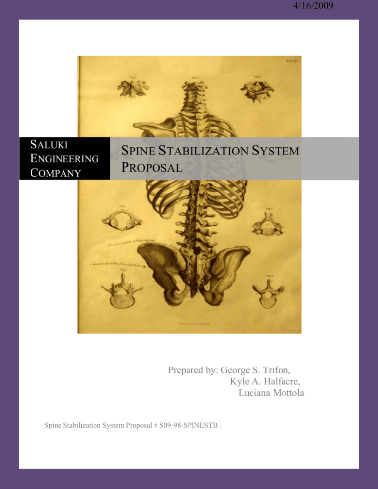
4/16/2009 SALUKI ENGINEERING COMPANY SPINE STABILIZATION SYSTEM PROPOSAL Prepared by: George S. Trifon, Kyle A. Halfacre, Luciana Mottola Spine Stabilization System Proposal # S09-98-SPINESTB | Saluki Engineering Company Spine Stabilization System Proposal # S09-98-SPINESTB Spine Stabilization System Design Group 98 Saluki Engineering Company (SEC) Southern Illinois University Carbondale Mail code 6603 Carbondale, Illinois 62901 Dr. Ajay Mahajan Southern Illinois University Carbondale Mail code 6603 Carbondale, Illinois 62901 To Whom It May Concern: The following proposal is in response to your request for an innovative spine stabilization system, without the use of pedicle screws. We appreciate the opportunity to present our design proposal, and we appreciate the time you are taking in the consideration of our proposal. As most medical professionals are well aware, there is an abundance of spine stabilization systems currently on the market. Orthopedic surgeons have a wide variety to choose from to correct just about any back problem that exists. These devices come in all varieties of shapes and sizes, with all kinds of functions and applications, but they all have one thing in common: they are all permanent and cause a considerable amount of trauma to the surrounding tissue. Also, the vertebrae that are being supported or fused are generally drilled and tapped, cut and or trimmed. Even more, the disk is never given a chance to recover and possibly regain some of its function ability. Essentially the disk is either removed or eventually encased in bone. We are confident that our new system will not only eliminate the need for any screws, but it will eliminate the need for any cutting, drilling or tapping of bone altogether. The new system will also reduce the trauma sustained by the surrounding tissue. This is done by: (1) reducing the number of ligaments that need to be cut (to access the required area), and (2) reducing the time it takes to perform the procedure. This will be accomplished by reducing the number of instruments necessary to insert the device (thus shortening and simplifying the procedure), thereby reducing exposure time and the risk of human error and infection. In addition, other benefits include the retention of some or all mobility (a.k.a. dynamic stabilization), modifiability, reparability and upgradeability. Essentially the system will allow surgeons to add to the existing hardware if more levels need stabilization, increase level of support if necessary, and repair or replace individual components of the hardware (that are not fused to bone). All this will be done while leaving the disk in place and allowing it to heal (if possible) to act as a redundant system. After reviewing existing patents we have determined that our design will not infringe upon any patents, in that it is entirely original and unique. Such a system is possible because of the recent advances in material science technology and bone growth stimulation techniques. Our system incorporates pseudo elastic alloys to support and keep the device in place while the utilization of bone morphogenetic material induces bone growth into the device to make it stable and permanent, yet minimally invasive. Page | 2 Saluki Engineering Company Spine Stabilization System Proposal # S09-98-SPINESTB Sincerely, George S. Trifon George S. Trifon SPINESTB Project Manager Page | 3 Saluki Engineering Company Spine Stabilization System Proposal # S09-98-SPINESTB EXECUTIVE SUMMARY The novel spine stabilization system (SPINESTB) consists of an estimated six specific subsystems (may include more at a future time). The function principle is; a bilateral compression of the inferior and superior rims of the vertebral body, with simultaneous vertical expansion forces of respective regions, essentially encompassing approximately 60% to 80% of the Annulus Fibrosus (the outer membrane of the vertebral disk). This action displaces the forces on the disk to the contact regions of the SPINESTB system which transfers them to a series of “Vertical Force Applicators” (pseudo elastic alloys set to simulate a response similar to that of a healthy disk). The shape of the contact regions allow for stabilization of the vertebrae while allowing natural flexion and tension of the spine. The contact regions are designed to then fuse to the vertebral body and become a permanent part of the vertebrae. After that any component is able to be replaced or upgraded as necessary through a quick minimally invasive procedure. Each SPINESTB unit is placed between two vertebrae and can act alone or in conjunction with other SPINESTB units, with semi rigid vertical stabilizers to create a fully dynamic spine stabilization system of multiple levels. The six subsystems are labeled as such; “contact site,” “contact pad,” “lateral brackets,” “rear linkage,” “vertical force applicators” (VFAs) and, “horizontal force applicators” (HFAs). Each subsystem contains one or more individual components that are defined by their location in relation to the placement of the SPINESTB system. A compilation of the research performed thus far, that was required for gaining an understanding of the biological systems involved and affected by the SPINESTB device, is listed in the following Literature Review section of the proposal along with a collection of relevant existing devices with descriptions and a list of patents with descriptions to compare how the SPINESTB device is fundamentally different from all existing devices. Detailed information on the time spent progression of events on the research and development of the SPINESTB system is also included in the following proposal. Most of the materials have already been located, so the estimated cost of the project at this time is time itself. Page | 4 Saluki Engineering Company Spine Stabilization System Proposal # S09-98-SPINESTB RESTRICTION NON-DISCLOSURE OF INFORMATION The information provided in or for this proposal is the confidential, proprietary property of the Saluki Engineering Company of Carbondale, Illinois, USA. Such information may be used solely by the party to whom this proposal has been submitted by Saluki Engineering Company and solely for the purpose of evaluating this proposal. The submittal of this proposal confers no right in, or license to use, or right to disclose to others for any purpose, the subject matter, or such information and data, nor confers the right to reproduce, or offer such information for sale. All drawings, specifications, and other writings supplied with this proposal are to be returned to Saluki Engineering Company promptly upon request. The use of this information, other than for the purpose of evaluating this proposal, is subject to the terms of an agreement under which services are to be performed pursuant to this proposal. Page | 5 Saluki Engineering Company Spine Stabilization System Proposal # S09-98-SPINESTB TABLE OF CONTENTS Contents INTRODUCTION (GT) ................................................................................................................. 7 LITERATURE REVIEW ............................................................................................................. 12 LITERATURE REVIEW SURVEY (KH) ................................................................................... 51 PROJECT DESCRIPTION (GT) .................................................................................................. 53 DESIGN BASIS (GT)................................................................................................................... 54 SUBSYSTEM DESCRIPTIONS .................................................................................................. 55 Contact Pad/Contact Site (KH) ......................................................................................... 55 Axial Linkage (KH) .......................................................................................................... 55 Lateral Bracket (LM) ........................................................................................................ 55 Vertical/Horizontal Force Actuator (GT) ......................................................................... 55 PROJECT ORGANIZATION CHART (LM) .............................................................................. 57 RESPONSIBILITY APPROVAL SUPPORT INFORMATION CHART (LM)......................... 58 ACTION ITEM LIST (KH) .......................................................................................................... 59 TIMELINE (LM) .......................................................................................................................... 60 LIST OF RESOURCES NEEDED (KH) ..................................................................................... 61 RESUMES .................................................................................................................................... 62 Page | 6 Saluki Engineering Company Spine Stabilization System Proposal # S09-98-SPINESTB INTRODUCTION Upon consideration of undertaking the Novel Spine Stabilization System project, a clear distinction needs to be made. The distinction, that although the spine is an integral part of a complex biological system (which is dynamic as well as reactive) the project is being approached from a mechanical engineering perspective. With that being said; a lot of time and effort has been devoted to learning and understanding the spinal anatomy, functions, properties, and behavior. The spinal column is the only thing keeping human beings upright and vertical. The spine provides a rigid structure to erect and provide stability to the upper torso. The spine has a wide range of motion for all the different movements that humans are capable of making. When the spine is injured or its routine functions are impaired, the results can be extremely painful and disabling. Back pain affects both male and female alike. The majority of back pain comes from the lower back. Most people experience lower back pain between ages of twenty five to sixty and twelve to twenty six percent of children experience lower back pain. Chronic back pain is defined as pain lasting longer than three months to a year. (IN1) According to the American Association of Neurological Surgeons; it is estimated that seventy five to eighty five percent of all Americans will experience back pain at one point in their lives. Also even though ninety percent of all cases involving back pain recover without surgery, fifty percent of them will have reoccurring back pain within a year. (IN2) According to Dr. Donald D. Dietze Jr. a Neurological/Orthopedic Surgeon; there are approximately 400,000 lower back operations performed every year in the United States. (IN3)The three most common causes of lower back pain, and they are Herniated or Ruptured Disk, Lumbar Spinal Stenosis, and Osteoarthritis. (IN2) Shown below in Figure 1 is a herniated disk, illustrating how the annulus becomes ruptured and the nucleus drains into the interference zone. Figure 1: A single vertebrae from the lumbar region, with a herniated disk defect. (IN4) Page | 7 Saluki Engineering Company Spine Stabilization System Proposal # S09-98-SPINESTB A Ruptured Disk or Herniated Disk is when the gel-like tissue inside the disk between the vertebrae leaks out or bulges out from under the vertebrae and pushes on the spinal column. These pads are disc like in shape and are shock-absorbers for the spine. They are inserted between each of the thirty three vertebrae in the spine. Each disc has a tough outer ring, named the annulus, with a soft inner core named the nucleus. The nucleus is made up of jelly-like tissue. As a person ages the disks begin to dry out. As that happens, the outer ring becomes brittle and can develop cracks, making it susceptible to leakage and injury. When a weak area develops a breach can occur in this outer ring and allow the inner gel to bulge or leak out. This is known as a rupture or herniation. The gel-like tissue then presses on or entraps the spinal nerves, causing pain. Surgeons sometimes perform a discectomy to remove part of the ruptured disc. Of course at this point there is a weak spot in the annulus. Eventually the disk may collapse entirely which would call for more drastic measures one of which entails the fusion of the vertebrae on either side of the damaged disk. (IN4) Figure 2: A single vertebrae from the lumbar region, with Spinal Stenosis. (IN4) Stenosis by definition means: narrowing. The narrowing refers to the canals through which the nerves pass. In spinal Stenosis, the narrowing occurs in the spinal canal, (the bony passage through which the spinal cord runs). Also it can refer to the openings on either side of each vertebra through which nerves exit the spine. Those nerves connect to muscles and organs in the body. The narrowing can be caused by the inflammation of arthritis or by bony overgrowths which are called spurs. Spurs can occur from long-term arthritis. When these openings are narrowed, pressure is exerted on the nerves, which as a result can cause pain or numbness in the back and/or other areas of the body containing the nerves that have been compressed. (IN4) Page | 8 Saluki Engineering Company Spine Stabilization System Proposal # S09-98-SPINESTB Figure 3: A single vertebrae from the lumbar region, depicting the areas that can be affected by Degenerating Spinal Joints. (IN4) The back side or posterior of a vertebra has wing-like projections on each side called facets. The purpose of these facets is to carry some of the weight that the spine must endure. Also they must limit the amount the spine can flex to provide stability and protect the spinal column. The facets of each vertebra interlock with the ones above and below. The contact points are known as facet joints. Like any joint in the body, these facet joints are prone to wear and tear which is affected proportionately with age. As a result of wear their cushioning cartilage can wear thin and become inflamed (or they become arthritic). This is another cause of back pain which can be alleviated with spinal fusion. (IN4) Other disorders and diseases such as scoliosis and damage from external trauma (such as car accidents) can be treated with the strategic fusion of specific vertebrae. Since these cases are more diverse and specific to the patient and the situation, an analysis by the doctor needs to be made on a case by case basis. The difference is that, when a disk fails completely or degeneration of spinal joints reaches a critical level, it is almost definite that spinal fusion will be the outcome. Surgery is always a last resort when it comes to lower back pain (which is not caused by severe external trauma). There are a number of non-surgical options available that are quite successful, but in some cases surgery is the only option. To qualify for surgery a number of criteria must be met along with the recommendation of a qualified osteopath and/or Neurosurgeon. Some of the criteria include; Back and leg pain limits normal activity or impairs quality of life, development of progressive neurological deficits; such as leg weakness and/or numbness, loss of normal, bowel and bladder functions, and difficulty standing or walking. If surgery is indeed the desired route, neurosurgeons have a variety of options available to help relieve pressure on the nerve roots. Generally if the severity level is so high (which are the cases that typically require surgery) that involves several nerve roots and discs that are causing the pain or if there is degeneration and instability in the spinal column, the neurosurgeon may opt to fuse the vertebrae together. Fusions are generally done with bone grafts (or other bone Page | 9 Saluki Engineering Company Spine Stabilization System Proposal # S09-98-SPINESTB relocation or growth stimulation techniques) and stabilize the vertebrae with instrumentation. The instrumentation includes metal plates, screws, rods and cages. When a successful fusion is achieved it will prevent the disc from bulging or herniating again (if the disk is even left inside). Following a fusion procedure, a patient should gain restored mobility in the back. The patient should regain the ability to bend over and maybe experience even more mobility after surgery than before. (IN5) Page | 10 Saluki Engineering Company Spine Stabilization System Proposal # S09-98-SPINESTB Saluki Engineering Company Spine Stabilization System Literature Review Authors: George S Trifon Kyle Halfacre Luciana Mottola Spring2009 Page | 11 Saluki Engineering Company Spine Stabilization System Proposal # S09-98-SPINESTB Preface The following document has a unique reference format. It must be noted that the alpha numeric tag following paragraphs and statements are labels referring to specific references in the reference section. The references have been labeled with a prefix in accordance to the section to which it belongs, and a reference number. The label in the body will direct the reader to the appropriate reference. This has been done to reduce clutter and improve organization. The reference to each figure is located in the text immediately adjacent to the figure. Page | 12 Saluki Engineering Company Spine Stabilization System Proposal # S09-98-SPINESTB TABLE OF CONTENTS Spine Stabilization System Design Group 98 ............................................................................. 2 Saluki Engineering Company (SEC) .......................................................................................... 2 Southern Illinois University Carbondale .................................................................................... 2 Mail code 6603 ........................................................................................................................... 2 Carbondale, Illinois 62901 .......................................................................................................... 2 Dr. Ajay Mahajan........................................................................................................................ 2 Southern Illinois University Carbondale .................................................................................... 2 Mail code 6603 ........................................................................................................................... 2 Carbondale, Illinois 62901 .......................................................................................................... 2 To Whom It May Concern: ......................................................................................................... 2 EXECUTIVE SUMMARY ............................................................................................................ 4 RESTRICTION NON-DISCLOSURE OF INFORMATION ........................................................ 5 TABLE OF CONTENTS ................................................................................................................ 6 INTRODUCTION .......................................................................................................................... 7 George S Trifon ........................................................................................................................... 11 Kyle Halfacre ............................................................................................................................... 11 Luciana Mottola .......................................................................................................................... 11 Preface........................................................................................................................................... 12 The following document has a unique reference format. It must be noted that the alpha numeric tag following paragraphs and statements are labels referring to specific references in the reference section. The references have been labeled with a prefix in accordance to the section to which it belongs, and a reference number. The label in the body will direct the reader to the appropriate reference. This has been done to reduce clutter and improve organization. The reference to each figure is located in the text immediately adjacent to the figure.TABLE OF CONTENTS................................................................................................................................. 12 TABLE OF CONTENTS ........................................................................................................... 13 List of Figures ............................................................................................................................... 15 INTRODUCTION ........................................................................................................................ 17 ANATOMY .................................................................................................................................. 18 ONE LEVEL FUSION ................................................................................................................. 23 DYNAMIC SATBILIZATION SYSTEMS ................................................................................. 28 Page | 13 Saluki Engineering Company Spine Stabilization System Proposal # S09-98-SPINESTB Dynesys System ........................................................................................................................... 28 Medical Procedure ...................................................................................................................... 29 Biomechanics of the System ....................................................................................................... 30 Complications .............................................................................................................................. 31 SPINAL FUSION AND THE USE OF BONE CEMENT ........................................................... 33 Conclusion and Transition ............................................................................................................ 34 REFERENCES ............................................................................................................................. 35 APPENDIX B ............................................................................................................................... 37 (Reference of notable patents for design purposes, The complete documents of the following patents is located int the patents section of this groups’s Web Page.) .......................................... 37 DYNAMIC STABILIZATION SYSTEMS ................................................................................. 37 SINGLE-DOUBLE LEVEL FUSION .......................................................................................... 44 LITERATURE REVIEW SURVEY ............................................................................................ 51 PROJECT DESCRIPTION ........................................................................................................... 53 DESIGN BASIS............................................................................................................................ 54 SUBSYSTEM DESCRIPTIONS .................................................................................................. 55 PROJECT ORGANIZATION CHART ........................................................................................ 57 RESPONSIBILITY APPROVAL SUPPORT IINFORMATION CHART ................................. 58 ACTION ITEM LIST ................................................................................................................... 59 TIMELINE .................................................................................................................................... 60 LIST OF RESOURCES NEEDED ............................................................................................... 61 RESUMES .................................................................................................................................... 62 Education .................................................................................................................................. 63 Work Experience ...................................................................................................................... 63 Page | 14 Saluki Engineering Company Spine Stabilization System Proposal # S09-98-SPINESTB List of Figures Figure 1: A single vertebrae from the lumbar region, with a herniated disk defect. (IN4) ........... 7 Figure 2: A single vertebrae from the lumbar region, with Spinal Stenosis. (IN4) ....................... 8 Figure 3: A single vertebrae from the lumbar region, depicting the areas that can be affected by Degenerating Spinal Joints. (IN4).................................................................................................. 9 Figure 4: A depiction of the spinal column with its natural shape. (AN6) ................................. 18 Figure 5: A depiction of the Lumbar region with some key features labeled. (AN3) ................. 19 Figure 6: A depiction of the individual vertebra from the lumbar region (top). (AN3) .............. 20 Figure 7: A depiction of the individual vertebra from the lumbar region (lateral view). (AN1) . 20 Figure 8: Depictions of a posterior spinal segment located in the lubar region, identifying the location of key features. (AN5) .................................................................................................... 21 Figure 9: Depictions of a posterior spinal segment located in the lumbar region, identifying the location of key features. (AN5) .................................................................................................... 21 Figure 10: Depicts the location of various ligaments in relation to the vertebral body. (AN6) .. 22 Figure 11: Depicts the location of the spinal cord and nerve roots in reference to the vertebral body. (AN6) ................................................................................................................................. 22 Figure 12: The basic orientation of the single level fusion hardware. (SL1) (SL10) ................ 23 Figure 13: The Infuse Bone Graft, known as rhBMP-2. (SL7) .................................................. 24 Figure 14: The LT-CAGE. (SL7, SL9) ....................................................................................... 24 Figure 15: The Bryan Cervical Disc. (SL6) ................................................................................ 25 Figure 16: The basic procedure of inserting the stabilization hardware. (SL10) ........................ 25 Figure 17: The CHARITÉ™ Artificial Disc. (SL2) ................................................................... 26 Figure 18: The CHARITÉ™ Artificial Disc. (SL4) ................................................................... 26 Figure 19: An example of Prosthetic disk replacement. (SL2) ................................................... 27 Figure 21: The Dynesys Stabilization System’s parts. (DS2)...................................................... 28 Page | 15 Saluki Engineering Company Spine Stabilization System Proposal # S09-98-SPINESTB Figure 22: A posterior view of the Dynesys Dynamic System. (DS1) ........................................ 29 Figure 23: The test results of the screw/cord/spacer constructs. (DS2) ....................................... 30 Figure 24: Normal, stretching, and compression of the dynamic system. (DS2) ........................ 31 Figure 25: The screw subsystem of the Aladyn Pedicle Screw Dynamic Stabilization System. (DS4) ............................................................................................................................................. 32 Figure 26: The DIAM Spinal Stabilization System. (DS3) ......................................................... 32 Figure 27: The X’Stop device, or soft stabilization system, and its components. (DS5) ............ 32 Figure 28: The bonding mechanism of bone cement, developing a mechanical bond with the bone. (BC2) ................................................................................................................................... 33 Page | 16 Saluki Engineering Company Spine Stabilization System Proposal # S09-98-SPINESTB INTRODUCTION In attempting to develop a novel spine stabilization system, a deep and clear understanding of the spine, needs to be attained. Along with an understanding of the spine, it is essential to know what disorders are most commonly being corrected by spine stabilization systems. Since there is a need for an improved system a complete analysis of existing devices needs to be made in order to determine what they all have in common and to find out exactly what is lacking. The following document is a compilation of all the knowledge that was necessary in understanding fundamental principles of how the spine works what usually goes wrong with the spine, and what devices there are for correcting such disorders. The first section, describes the spine anatomy, and since the spine carries the spinal column, careful attention needs to be given to it as to not damage it. In fact, because it carries the spinal column, is the main reason so much attention is given to correcting these disorders. Most often the case is that such disorders compresses nerves that pass through and cause excruciating pain and/or debilitating conditions. The goal of the stabilization systems is to relieve the pressure off the nerves and keep the spine from doing it again. The following sections describe the systems that are commonly utilized to correct disorders, primarily in the lumbar region. They include a single level fusion, double level fusion, dynamic stabilization, stabilization with the use of bone cements and other corrective devices such as artificial disk replacements. It should be observed how most procedures involve excessive cutting and/or drilling into the vertebrae to secure the hardware. Page | 17 Saluki Engineering Company Spine Stabilization System Proposal # S09-98-SPINESTB ANATOMY The spinal column is designed to serve many functions. Every element of the spinal column and vertebrae serves to protect the spinal cord. The spinal cord provides communication to the brain to allow for mobility and sensation in the body. This occurs through the complex interaction of bones, ligaments, muscles, the geographic features and structures of the back and the nerves that permeate everything. Every Human is born with thirty three separate vertebrae. Due to the natural fusion of the vertebrae in certain parts during normal development of the spine, most adults will only have twenty four individual vertebrae. As can be observed in figure 4, the lumbar spine consists of five vertebrae labeled L1 through L5. Below the lumbar spine, there are nine vertebrae at the base of the spine that grow together during the normal development of the human into adulthood. Five vertebrae form the triangular bone called the sacrum and the lowest four vertebrae form the tailbone named the coccyx. There are two dimples in most everyone's back where the sacrum joins the hipbones and is named the sacroiliac joint. (Those dimples are historically known as the "dimples of Venus.” (AN1) Since the problematic region is in the Lumbar zone, and the majority of fusions are performed on the vertebrae located in that region, the focus will be on the L1 through L5 vertebrae. The lumbar spine consists of five vertebrae located in the lower part of the spine between the ribs and the pelvis. Figure 4: A depiction of the spinal column with its natural shape. (AN6) Page | 18 Saluki Engineering Company Spine Stabilization System Proposal # S09-98-SPINESTB Since intervertebral discs are one of the major contributing factors to back pain resulting in fusion, it is important to note their function and anatomy. They are found between each vertebra, they are flat-round structures about a quarter to three quarters of an inch in thickness. They all contain a tough outer ring of tissue called the annulus fibrosis. Within the annulus fibrosis is contained a soft, white, gel-like center called the nucleus pulposus. Flat, circular plates of cartilage connect to the vertebrae above and below each disc. The intervertebral discs separate the vertebrae, and act as shock absorbers and as a sort of lubrication to prevent bone on bone contact. They compress when weight is put on them, spring back when the weight is removed and flex to allow the spine to bend and twist. The intervertebral discs contribute up about onethird of the length of the spine. They make up the largest organ in the body without its own blood supply. (AN3) The discs receive their blood supply through movement and absorb nutrients in the process. While at rest (without compressive forces) the discs expand, allowing them to absorb nutrient rich fluid. Through repetitive movement, poor posture, or injury this process is inhibited. The discs then become thinner and as a result more prone to injury. (AN3) Of the thirty one pairs of spinal nerves and roots that protrude out from either side of the spine, there are five lumbar nerve pairs and five sacral nerve pairs that begin from the bottom and continue to the thoracic region. (AN2) The true spinal cord ends approximately at L1, where it separates into many different nerve roots which travel to the lower body and legs. This collection of nerves and roots is called the "cauda equina," which literally means horse's tail. It describes the continuation of the nerves and roots at the end of the spinal cord. (AN3) The lumbar vertebrae, L1-L5, are the ones most frequently involved in back pain. These vertebrae carry most of the body’s weight and are subject to large forces and stresses. Most importantly, the highest activity is located on the segments L3-L4 and L4-L5, which consequently have the most injuries. The most strain is taken by the segments L4-L5 and L5-S1, which causes high possibilities of disk herniation. Figure 5: A depiction of the Lumbar region with some key features labeled. (AN3) Page | 19 Saluki Engineering Company Spine Stabilization System Proposal # S09-98-SPINESTB The vertebra is a complex structure in and of itself. The vertebral body is comprised of a thin shell of dense cortical bone and is shaped like an hourglass. The walls are thinner in the center and continue on to thicker ends. The outer cortical bone extends above and below the superior (above) and inferior (below) ends of the vertebrae to form rims. The superior and inferior endplates are contained within these rims of bone. (AN3) Figure 6: A depiction of the individual vertebra from the lumbar region (top). (AN3) Figure 7: A depiction of the individual vertebra from the lumbar region (lateral view). (AN1) The pedicles are an integral feature in most commonly practiced fusion procedures, therefore special attention must be devoted to them. The pedicles are two short and rounded processes that extend posteriorly (toward the back) from the lateral margin of the dorsal (rear) surface of the vertebral body (figure 6). They are made of thick cortical bone which makes them ideal for anchoring hardware. The laminae are two flattened plates of bone extending medially (toward the middle) from the pedicles to form the rear wall of the vertebral foramen (or the opening in the vertebrae through which the spinal cord passes). The Pars Interarticularis is a special region of the lamina between the superior and inferior articular processes. A fracture or some sort of congenital anomaly of the pars may result in a spondylolisthesis (AN3) (the anterior displacement of the vertebra also known as a slip) which mostly occurs in the lumbar region (AN4). Page | 20 Saluki Engineering Company Spine Stabilization System Proposal # S09-98-SPINESTB Figure 8: Depictions of a posterior spinal segment located in the lubar region, identifying the location of key features. (AN5) Figure 9: Depictions of a posterior spinal segment located in the lumbar region, identifying the location of key features. (AN5) One of the other major contributors to lower back pain is the Facet Joint. These Facet Joints are the locations between the bones in the spine are responsible for the ability to bend backward, forward, twist and turn. The facet joints are a particular joint between each vertebral body that assists with twisting motions and rotation of the spine by stabilizing through the limitation of travel. The facet joints are part of the posterior features of each vertebra. Each vertebra has facet joints that connect it with the vertebra above and the vertebra below it in the spinal column. The surfaces of each facet joints are covered with smooth cartilage (like any other joint in the human body) that help these parts of the vertebral bodies glide smoothly on each other preventing bone on bone contact. (AN3) The ligamentum flavum is the strongest of the spinal ligaments and connects the laminae of the vertebrae. It is usually thinner in the middle section The term "flavum" is used to describe its yellow appearance. The ligamentum flavum’s primary functions are to protect the neural Page | 21 Saluki Engineering Company Spine Stabilization System Proposal # S09-98-SPINESTB elements of the spinal cord and stabilize the spine so that excessive motion between the vertebral bodies does not occur. Together with the laminae, it forms the rear wall of the spinal canal. Figure 10: Depicts the location of various ligaments in relation to the vertebral body. (AN6) Figure 11: Depicts the location of the spinal cord and nerve roots in reference to the vertebral body. (AN6) The spinal canal is the anatomic casing for the spinal cord that is comprised of the vertebrae and ligaments of the spinal column. They are aligned in such a way as to create a canal that provides protection and support for the spinal cord. There are several different membranes that enclose and nourish the spinal cord. The outermost layer is called the "dura mater." It is a Latin term that means "hard mother." The dura encloses the brain and spinal cord and prevents cerebrospinal fluid from leaking out from the central nervous system. The space between the dura and the spinal canal is called the "epidural space". This space is filled with tissue, blood vessels and large veins. The epidural space is important in the treatment of low-back pain, because it provides a location to inject medications such as anesthetics and steroids.(AN3) The spinal cord is a vital pathway that conducts electrical signals from the brain to the body through individual nerve fibers. The spinal cord is a very delicate structure that requires special attention during spinal fusion surgery. Any kind of trauma to the spinal cord can mean permanent paralysis or even death. (AN3) Page | 22 Saluki Engineering Company Spine Stabilization System Proposal # S09-98-SPINESTB ONE LEVEL FUSION In spinal fusion surgeries pedicle screws are often the anchoring method for the hardware that will stabilize the vertebrae until the bones can fuse together. They are designed to secure the hardware that completely eliminates the motion and even micro motion of a vertebral segment, to facilitate fusion between the vertebrae involved meanwhile eliminating or reducing the pain caused by the joint. The process of one level fusion by the use of the pedicle screws is done by inserting a metal screw into the pedicle bone of adjacent vertebrae and connecting them with metal rods. Then inducing bone fusion with either the use of a bone graft or another device which stimulates bone growth between the two segments. The one level fusion surgery normally contains four pedicle screws and two rods along with connective hardware. The fusion consisting of the use of pedicle screws in conjunction with a bone graft is considered the gold standard of fusion techniques, which means that it is the most common. (SL1) Shown below in Figure 12 is the load distribution on the spine after the implant, this is depicted by the smaller arrow, the one on the left, or anterior side. The larger arrow, shown on the posterior side, depicts the larger load that is supported by the hardware. Underneath the little arrow and between the vertebrae is where the bone graft was inserted. Figure 12: The basic orientation of the single level fusion hardware. (SL1) (SL10) Page | 23 Saluki Engineering Company Spine Stabilization System Proposal # S09-98-SPINESTB There are different approaches to the interbody spinal fusion surgery. They include; Anterior Lumbar Interbody Fusion, Posterior Lumbar Interbody Fusion and a Transforaminal Lumbar Interbody Fusion. In an Anterior Lumbar Interbody Fusion, or ALIF, the disk is removed and the endplates are cleaned. The Bone graft is placed between the vertebral bodies into an interbody position through a small incision in the abdomen. Additionally, when the graft is placed through the back, the approach is called Posterior Lumbar Interbody Fusion (PLIF) or Transforaminal Lumbar Interbody Fusion (TLIF), depending on the angle of approach. The following is a method used in disc replacement in conjunction with single level fusion. Instead of using a bone graft it uses an artificially derived protein, a collagen sponge and a threaded metal cage to support the vertebrae while allowing the bone to fuse. Infuse Bone Graft, mostly known as rhBMP-2 (rhBMP-2 is “recombinant human bone morphogenetic protein made by isolating the BMP-2 protein from bone tissue, and splicing the BMP-2 gene into a cell line in the lab via recombinant DNA technology. The genetically engineered cells produce the human BMP-2 protein.”) Figure 13: The Infuse Bone Graft, known as rhBMP-2. (SL7) The LT-CAGE shown in Figure 14 is an innovative fixation device developed by Medtronic that is designed to help realign and fuse vertebrae of the lumbar spine to treat degenerative disc disease. Figure 14: The LT-CAGE. (SL7, SL9) Figure 15 is the Bryan Cervical Disc. It is designed to alleviate pain and preserve motion and flexibility while replacing a diseased disc that is removed from a patient’s cervical spine. Page | 24 Saluki Engineering Company Spine Stabilization System Proposal # S09-98-SPINESTB Figure 15: The Bryan Cervical Disc. (SL6) Shown below is the basic procedure of inserting the stabilization hardware. From the left, beginning with the insertion of the pedicle screws in the appropriate locations and moving along to bending, cutting, and securing the connecting rods to the other pedicle screws. Figure 16: The basic procedure of inserting the stabilization hardware. (SL10) Another technique used to repair one level problem is an artificial disk. This is not a fusion technique, but nevertheless relevant to the overall task of investigation all single level Page | 25 Saluki Engineering Company Spine Stabilization System Proposal # S09-98-SPINESTB options. There are a variety of artificial disks available, and there is also a considerable amount of controversy. Figure 17 and Figure 18 show the CHARITE Artificial Disc. This and Figure 19 are two disc replacement techniques, although Figure 18 is a prosthetic disc replacement, which only replaces the nucleus of the disc. Figure 17: The CHARITÉ™ Artificial Disc. (SL2) Figure 18: The CHARITÉ™ Artificial Disc. (SL4) Page | 26 Saluki Engineering Company Spine Stabilization System Proposal # S09-98-SPINESTB Figure 19: An example of Prosthetic disk replacement. (SL2) The system shown in Figure 19 is The PRESTIGE Cervical Disc. It is a metal-on-metal design made of stainless steel. This is a dynamic system that is for upper vertebrae only, Figure 20: The PRESTIGE® Cervical Disc. (SL3) Page | 27 Saluki Engineering Company Spine Stabilization System Proposal # S09-98-SPINESTB DYNAMIC SATBILIZATION SYSTEMS Dynesys System One of the most prevalent systems available today for spine stabilization is the Dynesys Neutralization system. The dynamic neutralization system for spine stabilization utilizes the pedicle screw system, shown in Figure D1 as a lateral view and in Figure D2 as a posterior view, for flexible stabilization of the spine. The system consists of titanium alloy screws (D1-A) connected by an elastic synthetic compound that controls motion in any plane. Polycarbonate urethane spacers (D1-B) and polyethylene terephthalate cords (D1-C) along with the screws comprise the system. The pedicle screws are made of Ti-Al-Nb forge alloy (Protasul 100), where the textured surface is sandblasted allowing for bone growth. Due to the conical core diameter, the screw has the advantages of a biggest screw diameter in a highest bending moment, a better bone compression (for Wolff’s Law to take effect) and anchorage in the pedicle canal, and the low notch-factor, or low stress-concentration rate, through threads in highest bending moment. Wolff’s Law essentially states that a bone will adapt to the loads it’s placed under. The disadvantage of the conical screw is that the back and forth screwing is prohibited. The polycarbonate urethane spacers adapt to the screw head, thereby preventing micro-motions and wear debris formation in the contact area. The spacer between the screw heads limits degree of lordosis that can be created, and the two screw heads are approximated to the extent the interposed spacer allows. The polyethylene terephthalate cord connects the pedicle screw heads via the hollow core of the spacer and holds the spacer in place. The stabilization cord limits bending movements, while the spacers hold the segments in a position of anatomical function and suppress extension and rotational movements (DS1). The system’s components are shown in Figure 21 along with the lateral view of the spinal column, a posterior view is shown in Figure 22 with the system placed onto the vertebrae. Figure 20: The Dynesys Stabilization System’s parts. (DS2) Page | 28 Saluki Engineering Company Spine Stabilization System Proposal # S09-98-SPINESTB Figure 21: A posterior view of the Dynesys Dynamic System. (DS1) Medical Procedure The way the system works is the Dynesys instrumentation re-stabilizes unstable segments without involving the intervertebral disks and the facet joints; the segments remain mobile within a controlled range, permitting limited motion of the arthrodesed lumbar vertebrae. The spine would then return to an anatomical function that is closer to the healthy status. The patient would be under general anesthesia, where a posterior midline approach would be performed at the area of the affected lumbar levels. After the pedicle screw insertion, the spacers are cut to the proper size. The stabilizing cords are pre-tensioned separately for each segment before their fixation in the pedicle screw. Hyper-mobility of the segments is corrected and the screws for fixation of the stabilizing cord in the eyes of the pedicle screws are tightened. The stabilizing Page | 29 Saluki Engineering Company Spine Stabilization System Proposal # S09-98-SPINESTB cord is cut and the surgical wound closed. No bone grafts were used and the patients are given prophylactic antibiotics 1 hour before and 3 days post-operatively (DS1). Biomechanics of the System Once the Dynesys System has been implanted, the mechanical properties of the system change due to the thermal conductivity of the materials. As the spacer warms from room to body temperature, the cord tension decreases between 25% and 33%. Near-term, the cord tension decreases 25% from its post-operative level, and in long-term, the cord tension remains constant at a 50% reduction from intra-operative levels. Listed below are the results of extensive testing on the Dynesys System: The pedicle screws, PET cord and screw-cord assemblies passed 5 million cyclic loading cycles between 100N and 800N. The PET cord static tensile strength is approximately 3000N; creep elongation at 20 hours is 1.27% of the initial cord length, with no rupture. Tested to assess creep deformation in conditions that model the in situ environment, the spacers demonstrated that at 600N -- double the 300N preload -- creep deformation decelerated and was constant at 20 hours. The screw-cord-spacer assemblies passed 10 million cycles of shear displacement or axial rotational motion without cord failure and only inconsequential cord abrasion. In axial compression, after 10 million cycles, the device maintained 525N tension (including the 300N preload) and 200N compression. Figure 22: The test results of the screw/cord/spacer constructs. (DS2) Page | 30 Saluki Engineering Company Spine Stabilization System Proposal # S09-98-SPINESTB This testing confirmed that, together, the components exhibit robust static and cyclic interconnection strengths. (DS2) Once the devices are attached bilaterally to the affected segments, the dynamic push-pull relationship between spacer and cord stabilizes the joints, keeping the vertebrae in a more natural position: Figure 23: Normal, stretching, and compression of the dynamic system. (DS2) At ‘Normal’, the Dynesys System supports an intervertebral joint between L4 and L5. During ‘Flexion’ the pedicle screws hold the polyethylene cord secure, supporting the affected joint as the spine bends forward. While in ‘Extension’ the external spacer-a polyurethane tube- provides support for the affected joint as the spine bends backwards. (DS2) Complications Although very effective, the Dynesys Stabilization System still has complications. Reports of screws loosening and deep spine infection have been documented. Nerve root compression from the system could result in discomfort in the lower limbs as well (DS1). Not all patients are candidates for this procedure, those who smoke have been shown to have an increased incidence of non-union; obese, malnourished, and/or alcohol abuse patients are all poor candidates also. Those with poor muscle tone and bone quality, and/or nerve paralysis all exhibit poor signs for the Dynesys System. Another dynamic stabilization system is the Aladyn Pedicle Screw System. This system utilizes simplicity in its design. Shown in Figure 25, the pedicle screws are linked together through a rigid member. Page | 31 Saluki Engineering Company Spine Stabilization System Proposal # S09-98-SPINESTB Figure 24: The screw subsystem of the Aladyn Pedicle Screw Dynamic Stabilization System. (DS4) Other Spine Dynamic systems such as artificial disk replacement have been listed in the one lever fusion section. Below is the DIAM system, it is designed to relieve pressure caused by a degenerative disk. This is done by maintaining the normal distance between the vertebrae. The system is typically inserted after a discectomy. Figure 25: The DIAM Spinal Stabilization System. (DS3) Shown in Figure 27 is the X’Stop device, it is a kind of soft stabilization system. Figure 26: The X’Stop device, or soft stabilization system, and its components. (DS5) Page | 32 Saluki Engineering Company Spine Stabilization System Proposal # S09-98-SPINESTB SPINAL FUSION AND THE USE OF BONE CEMENT There is not a large variety of bone cements available. Mainly because the ones that are available do the job. The main application of bone cement is for anchoring hardware in place or assisting in anchoring the hardware in place. A common type of bone cement is called Polymethylmethacrylate. Cements were first introduced in 1940 and after clinical trials for tissue compatibility; they were deemed acceptable and introduced into common practice by 1950. The most common use is in securing artificial joints in place. One application is in hip replacement. The cement is used to fill gaps between the hardware that is inserted into hollowed out hip bone and the bone itself. It has to be very strong (to support the body’s weight many times over) yet provide a level of elasticity to reduce risk of cracking and separation. The bone cement is essentially Plexiglas and comes it two parts that are mixed on site and applied to required areas. Usually there is an antibiotic mixed into the compound to reduce the risk of common infections associated with such procedures such as staff or strep infections (BC1). The bone cement is mixed with an accelerator to reduce set time. After the ingredients are mixed the viscosity changes over time from a runny liquid into a dough like state that can be safely applied and then finally hardens into solid material. The set time can be tailored with the use of the accelerators to help the physician safely apply the bone cement into the bone bed or to anchor metal or plastic prosthetic device to bone or used alone in the spine to treat osteoporotic compression fractures. Figure 27: The bonding mechanism of bone cement, developing a mechanical bond with the bone. (BC2) Page | 33 Saluki Engineering Company Spine Stabilization System Proposal # S09-98-SPINESTB Conclusion and Transition The attempt was made to uncover every type of current spine stabilization device, every vertebrae fusing technique and other more elaborate methods for alleviating lower back pain through the use of implanted hardware. There are three main goals that have been set forth after reviewing the literature on the available hardware and technology. The first is that the new system will be a dynamic stabilization system that will be easier and faster to install, it will be modular (in that other segments can be added in later surgeries should the patient develop a need for more vertebrae to be stabilized), and can be a dynamic stabilizer at every vertebrae it is installed in. For example, an ideal system would involve a combination of artificial discs, the use of cage-like devices containing the collagen sponge impregnated with bone growth protein. The artificial disks would keep the segment dynamic while fusing bone to it, and if need be to add other artificial disks in the same manner thereby replacing damaged disks with a dynamic system at every level and essentially repairing the problem without eliminating any of the spine’s natural mobility. This of course, would be done through a minimally invasive surgery while not damaging any of the surrounding tissue or cutting any ligaments while taking only a matter of minutes and thereby drastically reducing recovery time. Page | 34 Saluki Engineering Company Spine Stabilization System Proposal # S09-98-SPINESTB REFERENCES (IN1) http://www.back.com/anatomy.html (IN2) http://www.neurosurgerytoday.org/what/patient_e/low.asp (IN3) http://www.back.com/articles-evolution.html (IN4) http://www.npr.org/programs/morning/features/2006/feb/backpain/graphic/graphic.html (IN5) http://www.neurosurgerytoday.org/what/patient_e/low.asp (AN1) http://www.back.com/anatomy.html (AN2) http://www.neurosurgerytoday.org/what/patient_e/low.asp (AN3) http://www.back.com/anatomy-lumbar.html (AN4) http://en.wikipedia.org/wiki/Spondylolisthesis (AN5) http://images.google.com/imgres?imgurl=http://bhpain.com/yahoo_site_admin/assets/ images/facetJoints.138204352_std.jpg&imgrefurl=http://www.bhpain.com/facet_joint_syn drome&usg=__SDGjcJdzVgn9tAA2x6TfsOMDuJo=&h=297&w=279&sz=68&hl=en&st art=4&um=1&tbnid=DE33zqYC5a25LM:&tbnh=116&tbnw=109&prev=/images%3Fq% 3Dfacet%2Bjoint%26hl%3Den%26client%3Dfirefox-a%26rls%3Dorg.mozilla:enUS:official%26sa%3DX%26um%3D1 (AN6) http://www.spineuniverse.com/displayarticle.php/article895.html (SL1) http://www.back.com/back-articles-peek-rod-system.html (SL2) http://www.spineuniverse.com/displayarticle.php/article1671.html (SL3) http://www.spineuniverse.com/displayarticle.php/article1078.html (SL4) http://images.google.com/imgres?imgurl=http://www.spineuniverse.com/ displaygraphic.php/2255/artificialdiscBB.jpg&imgrefurl=http://www.spineuniverse.com/d isplayarticle.php/article1679.html&usg=__zHYCUsUdog1QUUSUNfDfciA0GkU=&h=2 00&w=250&sz=13&hl=en&start=10&um=1&tbnid=lpb2Jx9JBYR7fM:&tbnh=89&tbnw =111&prev=/images%3Fq%3DPRODISC%25C2%25AEL%2BTotal%2BDisc%2BReplacement%26hl%3Den%26client%3Dfirefoxa%26rls%3Dorg.mozilla:en-US:official%26sa%3DN%26um%3D1 (SL5) Spine Health 17 Mar.2009. www.spine-health.com. (SL6) SpineUniverse.17Mar.2009 http://www.spineuniverse.com/displayarticle.php/article1671.html (SL7) Medtronic 17 Mar.2009 http://www.medtronic.com/ Page | 35 Saluki Engineering Company Spine Stabilization System Proposal # S09-98-SPINESTB (SL8) Syncmedical.17Mar.2009 http://www.syncmedical.com/assets/Uploads/_ resampled/Resize,dImage184178-ETHOS-spine-pedicle-screw.jpg (SL9) US Department of Health and Human Services.17Mar.2009 http://www.fda.gov/cdrh/annual/fy2002/ode/fig-4.gif (SL10) http://www.or-live.com/medtronicspinal/1856/aboutProcedure.cfm (DS1) Sapkas GS, Themistocleous GS, Mavrogenis AF, Benetos IS, Metaxas N, and Papagelopoulos PJ. "Stabilization of the lumbar spine using the dynamic neutralization system." 30 (2008): 859-65. MEDLINE. Morris Library, Carbondale. 17 Mar. 2009 <http://search.ebscohost.com/login.aspx?direct=true&db=cmedm&AN=17990413&site= ehost-live&scope=site>. (DS2) "Dynesys."Zimmer.com.29Jan.2009 <http://www.zimmer.com/z/ctl/op/global/action/1/id/9165/template/IN>. (DS3) http://images.google.com/imgres?imgurl=http://www.neurocirugia.com/ intervenciones/diam/diam_archivos/image001.jpg&imgrefurl=http://www.neurocirugia.co m/intervenciones/diam/diam.htm&usg=__yChc05vvGlzwF3ERG0JRoN4gy6o=&h=180& w=226&sz=16&hl=en&start=3&um=1&tbnid=dVfu_YV32zPE7M:&tbnh=86&tbnw=108 &prev=/images%3Fq%3DDIAM%25E2%2584%25A2%2BSpinal%2BStabilization%2BS ystem%26hl%3Den%26client%3Dfirefox-a%26rls%3Dorg.mozilla:enUS:official%26sa%3DN%26um%3D1 (DS4) http://www.neurocirugia.com/instrumental/index.php?m=02&y=08&entry=entry 185940 080215- (DS5) http://spinalneurosurgery.com/dynamic_stabilization.htm (BC1) http://en.wikipedia.org/wiki/Bone_cement (BC2) http://www.totaljoints.info/BoneCement_microscopy.jpg 1) http://books.google.com/books?id=nxiCfmXPkzYC&pg=PA462&lpg=PA462&dq=pure+titanium+in+biomechanics&source=bl&ots=jO 7GG0S94o&sig=ydyrA7R12e0eCDN694LhLXDUkkQ&hl=en&ei=-7jSeXJA8HznQfFh5WwDg&sa=X&oi=book_result&ct=result&resnum=6#PPA462,M1 2) http://www.ncbi.nlm.nih.gov/pubmed/11241337 3) http://www.restoremedical.com/docs/PET_White_Paper.pdf 4) http://www.devicelink.com/mtprecision/archive/08/04/007.html 5) (DS2) "Dynesys." Zimmer.com. 29 Jan. 2009 <http://www.zimmer.com/z/ctl/op/global/action/1/id/9165/template/IN>. Page | 36 Saluki Engineering Company Spine Stabilization System Proposal # S09-98-SPINESTB APPENDIX B (Reference of notable patents for design purposes, The complete documents of the following patents is located int the patents section of this groups’s Web Page.) DYNAMIC STABILIZATION SYSTEMS Publication Number US 2005/0143737 A1 is a system grounded in the use of bone anchors, such as bone bolts or screws. The dynamics system uses a flexible element connecting the anchors. The flexible elements are able to be adjusted to change the flexibility of the element, which are flexible bearing elements of a rod end bearing. Figures 1 and 2 from the publication indicate how the system is fixed on the spinal column and how the screws are linked together. Page | 37 Saluki Engineering Company Spine Stabilization System Proposal # S09-98-SPINESTB US Patent Number 7,083,621 B2 is a system that revolves around pedicle screws. The bone anchors are connected through the use of linking rods which include at least one angularly adjustable joint. The linking rods may be fixed by actuating the locking member. The bone anchors and the linkage rod may be locked into place to form a spinal fusion or fixation prosthesis. Figure 4A from the patent shows how the bone screws are linked together and figure 42 shows the screws inserted into the vertebrae and being linked together. Page | 38 Saluki Engineering Company Spine Stabilization System Proposal # S09-98-SPINESTB The system of Publication Number US 2007/0088359 A1 utilizes pedicle screws for the fixation of the spine. The device provides dynamics support for spinal vertebra, to better control load transfers and avoid deterioration of the bone of adjacent spinal vertebra. The bone anchors are connected through the use of a spring, which are interchangeable. The system is able to be further added to after surgery. Figure 2C from the publication is a lateral view of the system once it has been established. Figure 2A is a posterior view of the system, showing how the levels are linked together. Page | 39 Saluki Engineering Company Spine Stabilization System Proposal # S09-98-SPINESTB Patent Number US 6,783,527 B2 also uses the design of the pedicle screw. The screws have an eye to lock links between the vertebrae. The system is comprised of an elongate member sized to span a distance between at least two vertebral bodies and bone anchors. The members are formed of a flexible material and can be tensioned to provide corrective forces to the spine and the anchors can be compressed towards one another. This system can be used for multi-level vertebral fusion or stabilization and is shown in Figure 1 from the patent, also shown is how the members are linked together at the eye of the screws in Figure 9 of the patent. Page | 40 Saluki Engineering Company Spine Stabilization System Proposal # S09-98-SPINESTB The design of US Publication Number 2008/0097441 A1 focuses on the use of pedicle screws. The screws are attached such like lattice work. The two screws on each vertebral body are attached with a rod, then the two vertebral bodies are attached between the ‘horizontally’ fixed rods. The two intervertebral rods are used as springs. The interconnection between the two components enables the spinal motion segment to move in a manner that mimics the natural motion of the spinal motion segment while substantially offloading the facet joints of the spine. Shown below are figure from the publication, in Figure 4B the system is shown from the posterior view established in the vertebrae. Figure 5A shows the system independent of the vertebral bodies, it shows how the bone screws attach to each other through the use of the rods. Page | 41 Saluki Engineering Company Spine Stabilization System Proposal # S09-98-SPINESTB Patent Number 5,520,690 also uses the pedicle screw design. The system consists of a polyaxial locking screw plate assembly which immobilizes the movement of the connected vertebral bodies. The plate is fixed by the bone screws. This design is a continuation of a previous design, where portions of the system are changed. This system renders a portion of the vertebral column to become static, losing mobility. In Figures 6 and 3, shown below, from the patent, show how the screw is attached to the plate. Page | 42 Saluki Engineering Company Spine Stabilization System Proposal # S09-98-SPINESTB US Patent Number 6,036,693 utilizes bone screws in its design. This system is only for retaining first and second cervical vertebrae. A plate is connected by the bone screws from each vertebra, resulting in a rigid design. Within the patent, different methods of bone screw placement are shown. The figures shown below are from the patent, Figure 3 shows how the screws are inserted in the vertebral body, Figure 10 shows the plate which the screws fasten through, joining the vertebrae and Figure 23 is a posterior view of the system after it has been attached to the vertebrae. Page | 43 Saluki Engineering Company Spine Stabilization System Proposal # S09-98-SPINESTB SINGLE-DOUBLE LEVEL FUSION SPINAL FUSION DEVICE WITH POROUS MATERIAL (US patent # 5,645,598) The cylindrical shape implant is designed to be placed into at least one bore formed between opposing vertebras. It consists on threads between the first and second ends of the body, which thread into bone tissue. Additionally, on opposite sides of the body two indentations are located, creating contact with the bond part of the plates, and consequently inducing vertebras to fuse. In order to induce bone growth, a biocompatible potentially osteoinductive ore oesteoconductive porous material could be included in the device, through a slot between opposite sides of the indentations. Page | 44 Saluki Engineering Company Spine Stabilization System Proposal # S09-98-SPINESTB PERCUTANEOUS VERTEBRAL FUSION SYSTEM (US patent # 6,821,277 B2) The system consists on bone screws and an inflatable connection rod which comprises a proximal end, creating a self-sealing valve. Torque is applied to the bone screw with a screwdriver, and the inflatable connection is inserted between portals of bone screws. After inflating the inflatable balloon, a rigid structure is created between the connection rod and the bone screws. Page | 45 Saluki Engineering Company Spine Stabilization System Proposal # S09-98-SPINESTB ANTERIOR SPINAL IMPLANT (US patent # 4,636,217) The Anterior spinal implant is recommended for patients with injured vertebrae or spinal disease. Before positioning the implant the damage vertebrae has to be removed. It is essential that the spinal implant is height compatible to the anterior vertebral portion to be replaced. The implant is inserted with screws into the adjacent lower and upper vertebrae providing rigidity, limited only by the bone structure, and preventing deformities. An advantage of the anterior spinal device is that it serves as a spacer, maintaining alignment in the spinal column. Page | 46 Saluki Engineering Company Spine Stabilization System Proposal # S09-98-SPINESTB INTERVERTEBRAL IMPLANT (US patent # 6,986,789 B2) The intervertebral implant consists on an upper and lower support body with a dorsal edge and a saddle joint, which is used as a replacement for an intervertebral disk. The implant is designed with the same height of the disk, and According to Schultz et al “The saddle joint includes two pivot axes and two saddle joint surfaces in contact with one another rotated by 90 degrees in relation to one another”. On this way, the intervertebral implant provides support via a joint, which are pivotable in relation to each other. Page | 47 Saluki Engineering Company Spine Stabilization System Proposal # S09-98-SPINESTB EXPANDABLE SPINAL IMPLANT (US patent #5,059,193) The expandable spinal implant is characterized by several deformable ribs between the first and second state. The first state consists on ribs on a cylindrical implant, and the second state on expanded spherical implant. The process is achieved by performing a novel surgical method, by making an entrance bore into the degenerated disk area of the spine, and an enlarged chamber between opposing vertebrae to be fused. The implant is inserted between opposing vertebrae, and expanded to the second state. TRANSVERSE CONNECTION FOR SPINAL COLUMN CORRECTIVE DEVICES Page | 48 Saluki Engineering Company Spine Stabilization System Proposal # S09-98-SPINESTB (US patent #5,522,816) A device that interconnects two longitudinal members, which are connectable to the vertebrae, by embracing two connector members to the longitudinal members. A plate is placed between the longitudinal members, interconnecting the first and second connector members. The longitudinal member is clamped using threadably screws to the hook located on the first connector. A nut connects the set crew to the connector member, so that this one could secure the elongate plate preventing it from pivoting. Page | 49 Saluki Engineering Company Spine Stabilization System Proposal # S09-98-SPINESTB THREADED SPINAL IMPLANT (US patent # 5,015,247) The threaded spinal implant is designed to improve method of performing a fusion, discectomy and an internal stabilization of the spine. This invention’s main goal is to improve the method of performing a discectomy, fusion and stabilization of the spine simultaneously as a single procedure. The implant is placed between two adjacent vertebrae inducing bone ingrowth through the implant and into the wall of the vertebrae. Consequently, inducing fusion from one vertebra in join to the other, and providing structural support to the two vertebrae. Page | 50 Saluki Engineering Company Spine Stabilization System Proposal # S09-98-SPINESTB LITERATURE REVIEW SURVEY Although there are many materials available for all types of mechanical systems, only select materials are biologically compatible with the human body. The material must be biocompatible, resist corrosion/degradation, and possess adequate mechanical properties. Therefore choosing the correct material is crucial. The most commonly used metals are stainless steel (316L and 22-13-5), cast Co-Cr-Mo, alloyed titanium, and pure unalloyed titanium. Of these, the most predominantly used is the (pure and alloyed) titanium; even though they have lower Young’s modulus the advantage is that they produce less distortion of CT or MR images(1). Titanium (alloyed and pure) is a widely used material in reconstructive orthopaedics; it possesses biocompatibility as well as mechanical strength. Another metal that is still being researched for biomedical applications is tantalum. Thus far tantalum has shown superior biocompatibility, and coupled with the material’s ability to provide osseous ingrowth (2) while providing structural support makes this material a candidate for a number of clinical applications. Titanium and tantalum are ideal materials to use where osseous growth is wanted or needed postoperatively, coupled with their mechanical properties they also provide structure and support. Strictly using metals for a biomechanical system may render it static, in the systems where dynamic stabilization is desired, flexible materials are needed. In the Dynesys system, polyethylene terephthalate cords are being utilized. Polyethylene terephthalate (PET) shows notable biological characteristics, such as biostability, promotion of tissue ingrowth, and a well characterized fibrotic response (3). PET demonstrates good tensile strength, therefore qualifying it to be utilized in a suspension scenario in biomechanical systems. Another material currently being used is polycarbonate urethane (PCU). The material, PCU, has good wear properties and good compatibility with natural tissues (4). PCU is also a weight-bearing material; all these properties make PCU a great candidate for biomechanical applications. The PET and PCU materials have both been under extensive testing and have concluded in exceptional biomechanical properties and cycle life (5). Many components are currently available for stabilization systems. The last two materials discussed, PET and PCU, are used in the Dynesys Dynamic System as linkages between the stabilized vertebrae. Figure 1 is a depiction of the system shown from the posterior view. The spacer is made of the polyurethane PCU and the cord is made of PET. Pedicle screws provide anchorage for the system. Page | 51 Saluki Engineering Company Spine Stabilization System Proposal # S09-98-SPINESTB Figure 1: The Dynesys Dynamic Stabilization System. The material that was reviewed was essentially the systems available and those that have not been approved for medical use. A clear understanding of the systems that have patented was needed in creating an innovative biomedical device and to not infringe on any other system’s design. Improving a current system was possible but developing an almost totally modular system that has nearly all replaceable parts was something the market has not seen yet, and can benefit both surgeons and patients alike. Page | 52 Saluki Engineering Company Spine Stabilization System Proposal # S09-98-SPINESTB PROJECT DESCRIPTION As stated previously, due to the nature and magnitude of such an undertaking and upon the advice of the Faculty Technical Advisor only a limited description the SPINESTB device will and can be presented at this time. The complete plans with technical drawings, mechanical properties, and measurements will be submitted in a supplemental report after a thorough examination of a human cadaver spine. Also an observation of surgical procedures on human cadavers will be observed in addition to examination of spine to develop a better procedure for application of hardware. The SPINESTB consists of roughly sixteen components with an estimated six specific subsystems. The components have yet to be clearly defined. The function principle is; a bilateral compression of the inferior and superior rims of the vertebral body, with simultaneous vertical expansion forces of respective regions, essentially encompassing approximately 60% to 80% of the Annulus Fibrosus (the outer membrane of the vertebral disk). This action displaces the forces on the disk to the contact regions of the SPINESTB system which transfers them to a series of “Vertical Force Applicators” (pseudo elastic alloys set to simulate a response similar to that of a healthy disk). The shape of the contact regions allow for stabilization of the vertebrae while allowing natural flexion and tension of the spine. The contact regions are designed to then fuse to the vertebral body and become a permanent part of the vertebrae. After that any component is able to be replaced or upgraded as necessary through a quick minimally invasive procedure. Each SPINESTB unit is placed between two vertebrae and can act alone or in conjunction with other SPINESTB units, with semi rigid vertical stabilizers to create a fully dynamic spine stabilization system of multiple levels. The six subsystems are labeled as such; “contact site,” “contact pad,” “lateral brackets,” “rear linkage,” “vertical force applicators” (VFAs) and, “horizontal force applicators” (HFAs). These subsystems come together to form a clamp type devise that is preloaded with tension to maintain appropriate disk height and simulate disk resistance to movement. The following is a very rough depiction of the arrangement of subsystems. There are three groups because they have been grouped into groups of two and assigned to each member of the group. The depiction is only half of the complete system (top view). There is an identical system beneath it. The first group on the left represents the contact site and the contact pads. The group in the center represents the lateral brackets and the rear linkage. The group on the right represents the vertical and horizontal force applicators. Page | 53 Saluki Engineering Company Spine Stabilization System Proposal # S09-98-SPINESTB DESIGN BASIS All the rough sketches are located in the design notebooks, which are numbered and dated. Design ideas are all original productions of the Project Manager, with modifications and improvements from design group. Nothing was taken from existing patents or advertized devices, as they were only used as examples of what not to do. Page | 54 Saluki Engineering Company Spine Stabilization System Proposal # S09-98-SPINESTB SUBSYSTEM DESCRIPTIONS Contact Pad/Contact Site Many of the stabilization systems available have a bone anchor such as a screw that is drilled into place, providing stability for the system. If the screw is broken or sheared off, the system cannot be reversed and the bone cannot withstand another screw. The system being developed will be one that has a clamp as an anchoring device. The clamp, or contact pad, will partially wrap around the vertebral body from the posterior. Pressure from the contact pad will be exerted onto the vertebra in the lateral directions, such as a mechanical vice exerts a force on an object. A force, acting in the axial direction to the vertebral column, will be on the vertebral body as well, pushing the vertebra away from the injured disk. This will be done by another contact pad being placed on the vertebra just below the injured disk, acting the same except an equal and opposite force will be acting downward instead of upward. This balance of forces will relieve the injured disk from pressure, therefore reducing pain. Attached to the contact pad is a different material, this is referred to as the contact site. The contact site is located on the inner wall of the contact pad and will promote osseous ingrowth. The contact site will play a vital role in bone growth, to aid the system in stabilization the contact site will be made of a porous material, that way the bone will grow into and through the biomaterial matrix, resulting in a very strong, reinforced system. The contact site and contact pad will be either mechanically or chemically bonded together for maximum strength. The contact site will aid the contact pad in forcing the vertebrae in the axial direction; therefore the two materials will need to have a strong bond. Once bonded together, these two materials cannot be separated. The bone will grow through the contact site, resulting in a partially rigid area. Thus that portion of the system must remain and cannot be replaced. This is done intentionally to provide a very stable base for the system to perform. Axial Linkage The brackets on the two vertebrae will be connected through the use of a linkage cord. Two cords will be used, one on each side of the spinous process, this will reduce the amount of force on each member. The connecting cords will be made of a material that possesses excellent tensile, creep, and wear qualities. The cords will only be used for the tension that is created when the spinal column is stretched, such as when a person leans or bends forward. The elastic cords will elongate to a predetermined length, allowing the system and the spinal column as a whole to remain dynamic. The cords stop stretching at a predetermined length in order to keep the vertebral bodies off the inured disk, when the vertebral column is bent forward, the posterior of the vertebra is raised from the disk and the anterior side of the disk is compressed. The cords will be housed within a tube. This tube will be used in keeping the clamped vertebral bodies at a constant distance at a static state. The length of the tube will be determined pre-operatively to keep the vertebral bodies off the inured disk. Keeping the load off the injured disk is the main purpose of the tube, so it will need to have excellent compressive qualities. Page | 55 Saluki Engineering Company Spine Stabilization System Proposal # S09-98-SPINESTB Although a purely rigid material would suffice for the tube’s material, a more elastic material that absorbs force will be used. The cords and tubes will be used as a combination. One will act when tensile forces are present while the other acts during compression. The cord will not be affected during the compression process; the cord will be pre-stressed, not only for this reason but to also help with providing system stabilization. The tube will fit closely to the cord housed within, leaving only a small amount of room between the two elements. Lateral Brackets The lateral bracket applies a vertical force on the contact pad. It has to be able to attach and detach from the contact pad, and to pivot on a vertical plane (parallel to the spine). The lateral bracket interacts with the contact pad and allows a connection with the vertical force applicator. The main function of the rear linkage is to provide stability for the lateral force applicator and for the vertebra by compressing to the spine. Additionally, the rear linkage allows a connection with the polyethylene terephthalate cord. Vertical/Horizontal Force Applicator The force applicators are a pseudo-elastic type of alloy that behaves mole like a memory shape material than a spring. They appear to behave in the same way on the surface, but on the molecular level they are much different. When a spring is stretched the molecular bonds either stretch a little or microscopic cracks called creep develop along grain boundaries and propagate through the material to allow a little movement, or both instances happen simultaneously to allow the material to give to applied forces. In the case of pseudo elastic alloys the crystalline structures change back and forth from Austenite to Martensite while the material experiences elastic deformation. In addition to these properties, some of these types of alloys can be triggered to return to a predetermined shape by applying relatively small amount of thermal energy. The goal is to incorporate such alloys into the system, so that after the material is inserted into the correct location, it will conform to the natural shape of the bone and provide constant pressure with the activation from ambient body heat. Page | 56 Saluki Engineering Company Spine Stabilization System Proposal # S09-98-SPINESTB PROJECT ORGANIZATION CHART DR.MAHAJAN FACULTY TECHNICAL ADVISOR Mechanical engineering GEORGE TRIFON (ME, PM) Delegating responsibilities Designing and maintaining website Updating the FTA Responsible for Horizontal and Vertical Force Applicators KYLE HALFACRE LUCIANA MOTTOLA (ME) (ME) Responsible for Contact Pad and Contact Site Material Selection Analyze System with Computer Modeling Responsible for Lateral Bracket and Rear Linkage Team minutes, timeline, RASI, and project organization chart. Page | 57 Saluki Engineering Company Spine Stabilization System Proposal # S09-98-SPINESTB RESPONSIBILITY APPROVAL SUPPORT INFORMATION CHART Team 98 Responsibility -Approval-Support-Information (RASI) Chart TASK GT LM KH DR.MAHAJAN Obtain current (S) (R) (S) (A) prices Responsibility (R) Work on Prototype (R) (I) (I ) (A) Approval (A) Produce parts in SolidWorks (S) (S) ( R) (A) Support (S) Assembling device (I) (R) (S) (A) Information (I) Device Testing (R) (S) (I) (A) Perfect device (I) (I) (R ) (A) Document Design (S) (R) (I) (A) Page | 58 Saluki Engineering Company Spine Stabilization System Proposal # S09-98-SPINESTB ACTION ITEM LIST Project: Spine Stabilization System Proj Number: S09-98-SPINESTB Action Item List 14-Apr-09 Team Members George Trifton, ME (PM) Luciana Muttola, ME Kyle Halfacre, ME # 1 2 3 4 5 6 7 8 9 10 11 12 13 14 15 16 17 18 19 20 21 22 23 24 Activity Distribute Tasks Research Lower Spine Research Techniques Research Current Problems Construct Rough Design Determine Kind of System (dynamic, static, chemical) Acquire Spine Model Find Current Patents Meet at Library Meet at Library Reserve room in Library Organization of Lit Review Revise Lit Review Submit Lit Review to FTA Meet at Library Meet at Library Research Bone Material Properties Research Material Properties for System Research patents for multilevel fusion Research patents for single level fusion Research bone growth chemical Email Questions regarding bone to Doctor Acquire Quote of Materials needed Proposal Work Person GT GT LM KH All Assigned 27-Jan 27-Jan 27-Jan 27-Jan 19-Feb Due New Due 26-Feb 26-Feb 26-Feb 3-Mar 10-Apr Status Done Done Done Done Done 8-Apr Done Comments Assign individual tasks Research the lumbar region of the spinal column Find the products available for spine stabilization and manufacturers Find the problems associated with current spine stabilization On Paper All 17-Mar 26-Mar KH KH all all GT GT All All All 23-Feb 17-Mar 16-Mar 16-Mar 17-Mar 16-Mar 19-Mar 27-Feb 17-Mar 16-Mar 17-Mar 17-Mar 17-Mar 3-Apr 26-Mar 26-Mar 30-Mar 1-Apr GT 1-Apr 7-Apr 9-Apr Done Find bone properties of the lumbar region. KH 1-Apr 7-Apr 9-Apr Done Find the properties of possible materials for the system. KH 1-Apr LM 1-Apr LM 1-Apr 7-Apr GT 26-Mar 29-Mar KH 8-Apr 12-Apr all 9-Apr 15-Apr Done Done Done Done Done Done Done Done Done Done 7-Apr Borrow spine model from local Chiropractor Find Current patent(s) for Lit Review Meet for Lit Review Meet for Lit Review Compile team members' works to construct Lit Review Revise/Add to Lit Review Changed to meet at ENGR computer lab. Done 7-Apr Done 10-Apr Find information about bone growth chemical, one that stimulates bone growth. Get questions from group members and email to Done Doctor, about bone properties. Done 0% Cancelled til next semester Done Work on assigned parts of the Proposal Page | 59 Saluki Engineering Company Spine Stabilization System Proposal # S09-98-SPINESTB TIMELINE Timeline GROUP 98 Activity Verify specs 17-Aug 24-Aug 31-Aug as bid as worked as revised 7-Sep 14-Sep 21-Sep 28-Sep 5-Oct 12-Oct 19-Oct 26-Oct 2-Nov 9-Nov 16-Nov 23-Nov Design subsystems Design Reviews Produce parts Build subsystems Progress Report Perfect subsystems Assemble device 1st System Test Perfect device Document design LEGEND Activity Page | 60 Saluki Engineering Company Spine Stabilization System Proposal # S09-98-SPINESTB LIST OF RESOURCES NEEDED Item Description Quantity 1 Computer 1 2 MS Office 1 3 ANSYS or SolidWorks 1 4 Printer 1 5 Metal * means from machine shop * $ Each on hand on hand on hand on hand on hand $ Subtotal $0.00 $0.00 $0.00 $0.00 $0.00 $0.00 $0.00 $0.00 $0.00 $0.00 Action Item List Project: Spine Stabilization System Proj Number: S09-98-SPINESTB Action Item List Team Members George Trifton, ME (PM) Luciana Mottola, ME Kyle Halfacre, ME # Activity Person 1 Obtain Current Prices of Material LM 2 Begin work on Prototype GT 3 Produce parts in SolidWorks KH Assigned 24-Aug 24-Aug 24-Aug Due 7-Sep 28-Sep Status 0% 0% 0% Page | 61 Saluki Engineering Company Spine Stabilization System Proposal # S09-98-SPINESTB RESUMES George Trifon 4532 Springer Ridge Road Carbondale, IL 62902 (708)214-4757 gtrifon@gmail.com Objective To lead motivated engineers in the research, development and design of breakthrough medical technology Page | 62 Saluki Engineering Company Education Spine Stabilization System Proposal # S09-98-SPINESTB Southern Illinois University Carbondale, IL Bachelor of Science, Mechanical Engineering Minor: Mathematics Dean’s List Embry Riddle University Beaufort, SC Bachelor of Science, Professional Aeronautics Fall 2010 Work Experience 2002-2006 Minor: Business Management and Aviation Safety Dean’s List Machinist Carbondale, IL Southern Illinois University, College of Engineering Fall 2009 – Present Gaining manufacturing experience by creating intricate devices from engineered drawings Perfecting welding skills through the use of TIG and Oxy-Acetylene welding methods Maintaining machining equipment Undergraduate Research Assistant Carbondale, IL Summer 2007 – Fall 2008 Southern Illinois University, College of Engineering Gained experience in ceramics processing by manufacturing inter-metallic diamond-bonded materials through sintering Acquired operational knowledge of hot press furnaces while controlling density, perocity and strength of processed materials Lead Technician Beaufort, SC 2 004-2006 Able Company Installed and maintained Heating Ventilation and Air Conditioning units Developed strong, dependable reputation by responding to service calls Installed, troubleshot and repaired residential wiring and plumbing Deputy Sheriff Beaufort, SC Beaufort County Sheriff’s Office 2004-2006 Developed interpersonal skills under stressful conditions by dealing with various personalities and temperaments Patrolled assigned areas, while responding to emergency and non-emergency calls Sergeant Beaufort, SC 1999-2004 United States Marine Corps Acquired management skills by overseeing maintenance personnel and being in charge of Aviation Support Equipment Developed skills in leadership, delegation, self-discipline, time management and communication Page | 63 Saluki Engineering Company Other Spine Stabilization System Proposal # S09-98-SPINESTB Volunteer Work Habitat for Humanity Feeding the Homeless Community Service Activities Accreditations and Licenses HVAC Universal License Hazmat Handling Certification Professional Memberships and Organizations American Society of Mechanical Engineers Engineers Without Borders Page | 64 Saluki Engineering Company Spine Stabilization System Proposal # S09-98-SPINESTB Kyle Halfacre kylehalf@yahoo.com Permanent Address: 4041 State Route 154 Pinckneyville, IL 62274 (618) 357-5485 School Address: 506 South Dixon Carbondale, IL 62901 (Cell) (618) 357-1038 Objective: To obtain an entry level Mechanical Engineering position upon graduation in January 2009 Career Focus: Mechanical Design in Biological Systems or Mechanical Systems Education Bachelor of Science, Mechanical Engineering Southern Illinois University, Carbondale Major: Mechanical Engineering Minor: Math Fall 2009 Relevant course work: Skills Experience Design of a Novel Spine Stabilization System Computer-Aided Engineering Thermodynamics Material Science Mechanics of Materials Fluid Mechanics Heat Transfer Machine Design Dynamic Modeling and Controls Mechanical Analysis Nanotechnology Engineering Acoustics Microsoft Word, PowerPoint, and Excel AutoCAD ANSYS SolidWorks C++(limited) Southern Illinois University Transcripts Office; Carbondale, IL Student Worker, (August 2008 – March 2009) Handled transcript requests, payments, and incoming mail. Provided assistance to customers via phone or in the office on how to order or resolve problems. Mathematics Tutor Tutor, (2003-Present) Tutored Junior High and High School Students in Mathematics. Perry County Market Place; Pinckneyville, IL Deli Worker, (December 2002- August 2008) Ordered products to sell, handled inventory, assisted managing workers, and provided excellent service to customers. Guitar Instructor, Pinckneyville, IL Teacher, (May 2004-August 2008) Gave guitar lessons and helped develop confidence and character in Junior High level kids. The Hunt Club; Percy, IL Worker, (May 2001 - August 2001) Provided excellent lawn maintenance and served during catering events. Page | 65 Saluki Engineering Company Spine Stabilization System Proposal # S09-98-SPINESTB Luciana Mottola luciana86_32@hotmail.com 714 E College lot A Carbondale, IL 62901 Summary of Qualifications: Worked with CAS for a period of 2 years. Bilingual: Spanish and English. Education: Southern Illinois University, Carbondale, IL Bachelor of Science in Mechanical Engineering Fall 2009 Institutos Educacionales Asociados, Caracas , Venezuela. Relevant Course Work: Introduction to AutoCAD CESL Introduction to engineering Activities: ASME member Awards: Best weblog award, December, 2004. Scholastic Honors, 2006. Skills: Traditional Drafting Skills, basic AutoCAD. Microsoft office 2007 Matlab Page | 66
