Characterization of Inertial Confinement Fusion Capsules Using an
advertisement
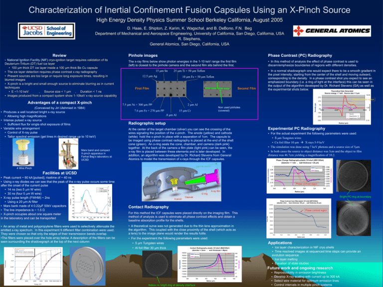
Characterization of Inertial Confinement Fusion Capsules Using an X-Pinch Source High Energy Density Physics Summer School Berkeley California, August 2005 D. Haas, E. Shipton, Z. Karim, K. Wagschal, and B. DeBono, F.N. Beg Department of Mechanical and Aerospace Engineering, University of California, San Diego, California, USA R. Stephens, General Atomics, San Diego, California, USA (Conceived by Jiri Ulshmied in 1984) • Produces a well localized bright x-ray source • Allowing high magnifications • Intense pulsed x-ray source • Sufficient flux for single shot exposure of films • Variable wire arrangement • Control of X-ray pulse • Tailor spectral emission (get lines in desired range up to 10 keV) Marx band and compact X-pinch apparatus in Farhat Beg’s laboratory at UCSD • In this method of analysis the effect of phase contrast is used to discern/emphasize boundaries of regions with different densities. 15 μm Sn 12.5 μm Ag 10 μm Fe + 50 μm Teflon First Film Second Film Source energy = 7 keV , Source size = 5 μm 10.00 270 7.5 μm Ni + 300 μm PP 2 μm Al 7.6 μm Fe + 270 μm PP Non used pinholes (covered) 15 μm Cr 70 -30 4000 4500 5000 -130 1.00 5500 Density Magnification 10 -230 Magnification 28.6 -330 0.10 Radiographic setup Radius (μm) Experimental PC Radiography At the center of the target chamber (silver) you can see the crossing of the wires signaling the position of the x-pinch. The anode (yellow) and cathode (white) hold the x-pinch in place with a separation of 1cm. The capsule to be imaged using phase contrast radiography is placed at the end of the shell cone (green). An o-ring seals the cone, chamber, and camera (dark pink) together. At the back of the camera a film plate (light pink) can be seen, the x-ray film is placed between these elements and is later developed. In addition, an algorithm was developed by Dr. Richard Stevens from General Atomics to model the transmission of x-rays through the ICF capsules. • For the actual experiment the following parameters were used: • 5 μm Tungsten wires • Cu foil filter 10 μm X-rays 5-9 keV • The simulation was done using 7 keV photons and a source size of 5μm • In both cases the source to object distance was 5cm and the object to film distance was 46.7cm yielding a magnification of 10.2. Phase Change Radiography plastic CH shell (#051305s4) (diameter = 1 mm , wall thickness = 20 μm) Facilities at UCSD 1 Experiment 0.95 theory 0.9 0.85 0.8 0.75 Phase contrast region 0.7 444 464 484 X-ray film 504 524 0.995 0.985 0.975 0.965 0.955 0.945 0.935 0.925 0.915 544 Radius (μm) O ring X-pinch Bright PC ring at boundary Capsule Phase Contrast foam filled plastic CH shell (#072105s3) (diamiter 2mm , foam thickness 100 μm , wall thickness 20 μm) 12000 Contact Radiography 11000 For this method the ICF capsules were placed directly on the imaging film. This method of analysis is used to eliminate all phase contrast effects and obtain a baseline absorption profile for the shells. Phase contrast region Pixel Value 10000 • A theoretical curve was not generated due to the thin lens approximation in the algorithm. This coupled with the close proximity of the shell (which acts as a lens) to the image plane would render the results futile. 9000 8000 7000 6000 5000 390 410 430 450 470 490 510 530 Radius (μm) • For the experiment the following parameters were used: Applications • 5 μm Tungsten wires • Al foil filter 30 μm thick Contact Radiography plastic CH shell (#042105s1) (diamiter = 2mm , wall thickness = 49μm) 190000 170000 40μm Intensity Density 295 245 160000 195 150000 145 140000 95 130000 45 120000 -5 110000 100000 300 350 400 450 Radius (μm ) Notice no bright ring at density interface 500 -55 550 Density (g/cc) 180000 Pixel Intensity • An array of metal and polypropylene filters were used to selectively attenuate the emitted x-ray spectrum. In this experiment 9 different filter combination were used. They were chosen so that only the edges of their transmission bands overlap. •The filters were placed over the hole array below. A description of the filters can be seen surrounding the shadowgraph at the top of the next column Phase contrast region 170 .8 μm Al 4 Wire Pinch • Peak current ~ 80 kA [pulsed]; risetime of ~ 40 ns • Using x-ray diodes we can see that the peak of the x-ray pulse occurs some time after the onset of the current pulse • 14 ns (two 5 μm W wire) • 30 ns (four 5 μm W wire) • X-ray pulse length (FWHM) ~ 2ns • Using a 20 μm Al filter • Marx bank made of 4 0.22μF 50kV capacitors • The line impedance is ~ 1.5 Ω • X-pinch occupies about one square meter in the laboratory and can be transported • In a normal shadowgraph one would expect there to be a smooth gradient in the pixel intensity; starting from the center of the shell and moving outward, corresponding to the density. In a phase contrast shot you expect to see an emphasized boundary (i.e. a ring of light at the interface) this can be seen in the output of the algorithm developed by Dr. Richard Stevens (GA) as well as the experimental shots below. Theoredical Data Generated 25 μm Ti + 50 μm Teflon Pixel Intensity Advantages of a compact X-pinch The x-ray films below show photon energies in the 1-10 keV range the first film (left) is closest to the pinhole camera and the second film sits behind the first. Intensity (normalized) Deuterium-Tritium (DT) fuel ice layer • 100 μm thick DT ice layer inside a 100 μm thick Be Cu capsule • The ice layer detection requires phase contrast x-ray radiography • Present sources are too large or require long exposure times, resulting in blurred images • X-pinch is a bright and small enough source to eliminate blurring as in current techniques • E ~1-10 keV , Source size < 1 μm , Duration < 1 ns • Images produced from a compact system show 1-10keV x-ray source capability Phase Contrast (PC) Radiography Density (g/cc) • National Ignition Facility (NIF) cryo-ignition target requires validation of its Pinhole images Intensity (normalized) Review • Ice layer characterization in NIF cryo shells • Time resolved images at sequenced time steps can provide an evolution sequence • Ice layer melting • Equation of state studies Future work and ongoing research • • • • Reproducibility in emission brightness Develop X-ray scaling with current up to 300 kA Select wire material for optimum emission lines Control intervals in multiple pinch systems
