Chapter 07
advertisement
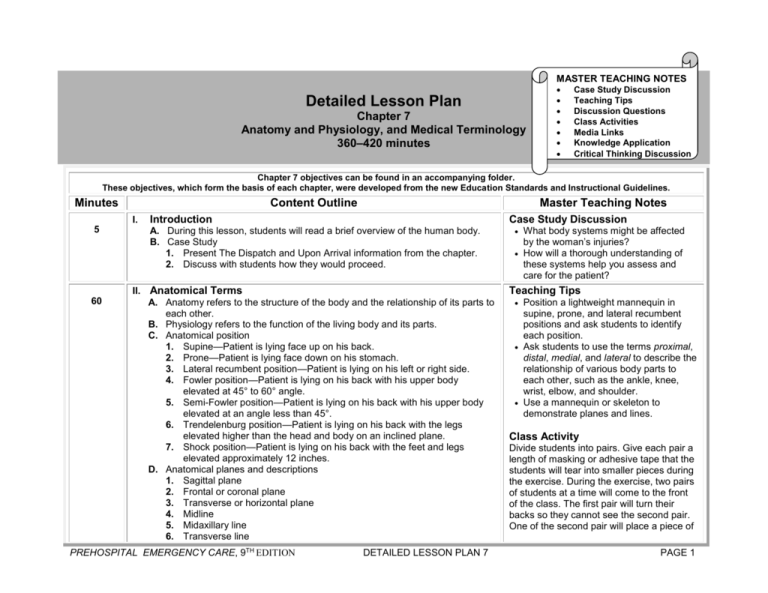
MASTER TEACHING NOTES Detailed Lesson Plan Chapter 7 Anatomy and Physiology, and Medical Terminology 360–420 minutes Case Study Discussion Teaching Tips Discussion Questions Class Activities Media Links Knowledge Application Critical Thinking Discussion Chapter 7 objectives can be found in an accompanying folder. These objectives, which form the basis of each chapter, were developed from the new Education Standards and Instructional Guidelines. Minutes Content Outline I. 5 60 Master Teaching Notes Introduction Case Study Discussion A. During this lesson, students will read a brief overview of the human body. B. Case Study 1. Present The Dispatch and Upon Arrival information from the chapter. 2. Discuss with students how they would proceed. II. Anatomical Terms A. Anatomy refers to the structure of the body and the relationship of its parts to each other. B. Physiology refers to the function of the living body and its parts. C. Anatomical position 1. Supine—Patient is lying face up on his back. 2. Prone—Patient is lying face down on his stomach. 3. Lateral recumbent position—Patient is lying on his left or right side. 4. Fowler position—Patient is lying on his back with his upper body elevated at 45° to 60° angle. 5. Semi-Fowler position—Patient is lying on his back with his upper body elevated at an angle less than 45°. 6. Trendelenburg position—Patient is lying on his back with the legs elevated higher than the head and body on an inclined plane. 7. Shock position—Patient is lying on his back with the feet and legs elevated approximately 12 inches. D. Anatomical planes and descriptions 1. Sagittal plane 2. Frontal or coronal plane 3. Transverse or horizontal plane 4. Midline 5. Midaxillary line 6. Transverse line PREHOSPITAL EMERGENCY CARE, 9TH EDITION DETAILED LESSON PLAN 7 What body systems might be affected by the woman’s injuries? How will a thorough understanding of these systems help you assess and care for the patient? Teaching Tips Position a lightweight mannequin in supine, prone, and lateral recumbent positions and ask students to identify each position. Ask students to use the terms proximal, distal, medial, and lateral to describe the relationship of various body parts to each other, such as the ankle, knee, wrist, elbow, and shoulder. Use a mannequin or skeleton to demonstrate planes and lines. Class Activity Divide students into pairs. Give each pair a length of masking or adhesive tape that the students will tear into smaller pieces during the exercise. During the exercise, two pairs of students at a time will come to the front of the class. The first pair will turn their backs so they cannot see the second pair. One of the second pair will place a piece of PAGE 1 Chapter 7 objectives can be found in an accompanying folder. These objectives, which form the basis of each chapter, were developed from the new Education Standards and Instructional Guidelines. Minutes Content Outline 7. 8. 9. 10. 11. 12. 13. 14. Master Teaching Notes Anterior and posterior Superior and inferior Dorsal and ventral Medial and lateral Proximal and distal Right and left Midclavicular and midaxillary Plantar and palmer tape somewhere on his partner. The class will describe to the first pair, using anatomical and directional terms, the location of the tape. For example, “Two inches inferior to the right elbow, on the posterior aspect.” Without looking, one of the first pair will try to place a piece of tape in the location described by the class. This will assist students in accurately using these terms when communicating with other health care providers. Discussion Questions How are the terms anterior and posterior related to the terms ventral and dorsal? How would you describe a transverse (horizontal) plane? What are Fowler’s and semi-Fowler’s positions? Knowledge Application Point to several locations on your body. Have students describe the locations using anatomical terms. Upon hearing “plain English” descriptions of patient positions, students will substitute the anatomically correct term. Critical Thinking Discussion What are the pros and cons of using anatomical terms of position and direction? Why is it important for EMTs to understand this terminology? Weblinks PREHOSPITAL EMERGENCY CARE, 9TH EDITION DETAILED LESSON PLAN 7 PAGE 2 Chapter 7 objectives can be found in an accompanying folder. These objectives, which form the basis of each chapter, were developed from the new Education Standards and Instructional Guidelines. Minutes Content Outline Master Teaching Notes Go to www.bradybooks.com and click on the mykit link for Prehospital Emergency Care, 9th edition to access web resources on anatomy and physiology. 40 III. Body Systems—The Musculoskeletal System A. The musculoskeletal system consists of the bony framework held together by ligaments, muscles, tendons, and other connective tissues. B. The skeletal system 1. Functions a. Gives body shape b. Protects vital organs c. Allows for movement d. Stores minerals and produces blood cells 2. Components a. Skull houses and protects the brain. i. Cranium forms top, back, and sides of the skull plus the forehead. ii. Face is the area between the brow and chin, which includes the orbits, maxillae, zygomatic bones, and mandible. b. Spinal column, or vertebral column, is the principal support system of the body, which is made up of vertebrae separated by intervertebral disks. i. Cervical spine—First seven vertebrae (neck) ii. Thoracic spine—Twelve thoracic vertebra inferior to the cervical spine (upper back) iii. Lumbar spine—Five vertebrae inferior to thoracic spine that form the lower back (lower back) iv. Sacral spine—Five vertebrae inferior to lumbar spine that are fused together (back wall of pelvis) v. Coccyx—Last four vertebrae that are fused together (tailbone) c. Thorax, or chest, is composed of the sternum and thoracic spine. i. Sternum is a flat, narrow bone in the middle of the anterior chest. ii. Clavicle is attached to the superior portion of the sternum, known as the manubrium. iii. The ribs are attached to the body of the sternum. PREHOSPITAL EMERGENCY CARE, 9TH EDITION DETAILED LESSON PLAN 7 Class Activity Play a game of “Mother May I” using the motions allowed by joints (flexion, extension, abduction, and so on). Teaching Tip Have students come up and point out the bones of different parts of the skeleton on a model skeleton. Discussion Questions What are the functions of the musculoskeletal system? What are examples of each of the three different types of muscle in the body? Animation Go to www.bradybooks.com and click on the mykit link for Prehospital Emergency Care, 9th edition to access an animation labeling the bones of the skeletal system. PAGE 3 Chapter 7 objectives can be found in an accompanying folder. These objectives, which form the basis of each chapter, were developed from the new Education Standards and Instructional Guidelines. Minutes Content Outline Master Teaching Notes iv. The inferior portion of the sternum is the xiphoid process. d. Pelvis is a structure consisting of several bones, including the sacrum and the coccyx. i. Iliac crest is a wing-like structure on either side of the pelvis. ii. Pubis is the anterior and inferior portion of the pelvis. iii. Ischium is the posterior and inferior portion of the pelvis. e. Lower extremities are the legs from the hip to the toes. f. Upper extremities are the shoulders, arms, forearms, wrists, and hands. g. Joints are places where bones connect to one another. i. Types of motion Flexion Extension Abduction Adduction Circumduction Pronation Supination ii. Types of joints Ball-and socket joint Hinged joint Pivot joint Gliding joint Saddle joint Condyloid joint C. Bone injury 1. Fracture breaks continuity in structure. 2. May injure surrounding tissue 3. May result in blood loss D. Muscular system 1. Skeletal muscle, or voluntary muscle, is responsible for all deliberate movement. 2. Smooth muscle, or involuntary muscle, is made up of large fibers that carry out the automatic muscular functions of the body. 3. Cardiac muscle is a special type of involuntary muscle found only in the heart. PREHOSPITAL EMERGENCY CARE, 9TH EDITION DETAILED LESSON PLAN 7 PAGE 4 Chapter 7 objectives can be found in an accompanying folder. These objectives, which form the basis of each chapter, were developed from the new Education Standards and Instructional Guidelines. Minutes 40 Content Outline Master Teaching Notes IV. Body Systems—The Respiratory System A. Functions 1. Respiration, which is the process of moving oxygen and carbon dioxide across membranes 2. Ventilation, which is the mechanical process by which air is move in and out of the lungs 3. Oxygenation, which is the process through which oxygen molecules move across a membrane from an area of high concentration to an area of low concentration, and the removal of carbon dioxide 4. Maintenance of a normal acid-base balance B. Components 1. Nose and mouth 2. Pharynx 3. Trachea and larynx 4. Epiglottis 5. Bronchi 6. Lungs 7. Diaphragm C. Anatomy in infants and children 1. Extra attention is required because mouth and nose are smaller than those of adults and can be more easily obstructed. 2. The tongue can block the pharynx more easily. 3. The trachea is narrower and can be more easily obstructed. 4. Hyperextension can occlude the trachea. 5. The cricoids cartilage is less developed and much less rigid. 6. Excessive movement of the diaphragm is a sign of respiratory distress. D. Mechanics of ventilation 1. Inhalation occurs when the intercostals muscles contract and the diaphragm moves downward, creating negative pressure in the chest. 2. Exhalation occurs when the intercostals muscles relax and the diaphragm moves upward, creating a positive pressure in the chest. 3. Diaphragm receives its stimulation from the phrenic nerve that exits the spinal cord at the cervical spine. E. Physiology of respiration 1. At the alveoli, oxygen enters the bloodstream while carbon dioxide and other wastes leave the bloodstream. 2. At the capillaries, oxygen moves from the blood into the cells while carbon dioxide moves from the cells into the blood. PREHOSPITAL EMERGENCY CARE, 9TH EDITION DETAILED LESSON PLAN 7 Discussion Questions How are respiration, ventilation, and oxygenation different from each other? What is the path of a molecule of oxygen as it moves from the atmosphere to the level of the cell? What are some differences between the respiratory systems of infants and children and those of adults? Critical Thinking Discussion Why might a patient with a respiratory problem feel weak? Discussion Question What are the muscles used in breathing? Teaching Tip Demonstrate increased resistance to airflow by having students breathe through coffee stirrers or drinking straws to simulate reduced diameter of airways. PAGE 5 Chapter 7 objectives can be found in an accompanying folder. These objectives, which form the basis of each chapter, were developed from the new Education Standards and Instructional Guidelines. Minutes Content Outline Master Teaching Notes F. Adequate and inadequate breathing 1. Characteristics of adequate breathing a. Adequate respiratory rate, which is the number of breaths a patient takes in one minute b. Adequate tidal volume, which is the amount of air the patient breathes in and out with one regular breath 2. Characteristics of inadequate breathing a. Rates that are too slow or too fast as compared with what is normal for the patient b. Irregular pattern of breathing c. Diminished or absent breath sounds d. Unequal chest expansion e. Pale or bluish mucous membranes or skin f. Use of accessory muscles g. Nasal flaring h. “Seesaw” breathing i. Heading bobbing j. Agonal respirations k. Grunting 10 V. Body Systems—The Circulatory System A. Functions 1. Provides a medium for perfusion of cells with oxygen and other nutrients and removes carbon dioxide and other wastes 2. Transports blood to cells and alveoli for gas exchange 3. Serves as a reservoir to house blood 4. Serves as a medium for buffering the body’s acid-base balance 5. Provides a mechanism to deliver immune cells and other substances to fight infection 6. Contains substances that promote clotting B. Basic anatomy 1. Heart pumps blood throughout the body. a. Pericardium is a double-walled sac that encloses the heart, gives support, and prevents friction. b. Atria are the upper chambers of the heart, which receive blood from the veins. c. Ventricles are the lower chambers of the heart, which pump blood to the arteries. PREHOSPITAL EMERGENCY CARE, 9TH EDITION DETAILED LESSON PLAN 7 Discussion Question What are signs that breathing is inadequate? Teaching Tip Write “right atrium” on the white board. Have students come up one at a time to write in the next structure through which a drop of blood would pass to complete the circuit. Discussion Questions Where is each of the heart valves located? What is the relationship between hydrostatic pressure and edema? PAGE 6 Chapter 7 objectives can be found in an accompanying folder. These objectives, which form the basis of each chapter, were developed from the new Education Standards and Instructional Guidelines. Minutes Content Outline Master Teaching Notes d. Valves keep blood flowing in one direction. i. Tricuspid valve ii. Pulmonary valve iii. Mitral valve, or bicuspid valve iv. Aortic valve 2. Arteries carry blood away from the heart. a. Aorta is the major artery of the heart, which supplies blood to all other arteries. b. Coronary arteries supply the heart with blood. c. Carotid arteries supply the brain and head with blood. d. Femoral arteries supply the groin and legs with blood. e. Dorsalis pedis arteries extend into the feet. f. Posterior tibial arteries travel from the calf to the feet. g. Brachial arteries are the major arteries of the upper arm. h. Radial arteries are the major arteries of the arm distal to the elbow joint. i. Pulmonary arteries carry oxygen-depleted blood to the lungs. 3. Arterioles are the smallest kinds of arteries, which carry blood from the arteries to the capillaries. 4. Capillaries are tiny blood vessels that connect arterioles to venules and act as sites for the exchange of materials between the blood and the cells. 5. Venules are the smallest branches of veins. 6. Veins carry blood back to the heart. a. Vena cavae carry oxygen-depleted blood back to the right atrium. b. Pulmonary veins carry oxygen-rich blood from the lungs to the left atrium. C. Composition of the blood 1. Red blood cells carry oxygen to the body cells and carry carbon dioxide away from the cells. 2. White blood cells help defend the body against infection. 3. Platelets, along with other clotting factors, are necessary to stop bleeding. 4. Plasma is the liquid part of the blood, which carries blood cells and transports nutrients. D. Physiology of circulation 1. Pulse is a wave of blood propelled thorough the arteries. 2. Blood pressure is the force exerted by the blood on the interior walls of PREHOSPITAL EMERGENCY CARE, 9TH EDITION DETAILED LESSON PLAN 7 Animation Go to www.bradybooks.com and click on the mykit link for Prehospital Emergency Care, 9th edition to access an animation identifying the structures of the heart and the purpose of the cardiovascular system. Teaching Tip Have students locate their carotid, dorsalis pedis, posterior tibial, brachial, and radial pulses. PAGE 7 Chapter 7 objectives can be found in an accompanying folder. These objectives, which form the basis of each chapter, were developed from the new Education Standards and Instructional Guidelines. Minutes Content Outline Master Teaching Notes the arteries. a. Systolic blood pressure is exerted against the walls of the arteries when the left ventricle contracts. b. Diastolic blood pressure is exerted against the walls of the arteries when the left ventricle is at rest. c. Hydrostatic pressure is the force exerted on the inside of the vessel walls as a result of blood pressure and volume. d. Perfusion is the delivery of oxygen, glucose, and other nutrients to the cells, and the elimination of carbon dioxide and other waste products. e. Hypoperfusion is the insufficient supply of oxygen and other nutrients to some of the body’s cells and the inadequate elimination of carbon dioxide and other waste products. E. Transport of gases in the blood 1. Oxygen is attached to hemoglobin and dissolved in plasma. 2. Carbon dioxide is transported in the blood as bicarbonate, attached to hemoglobin, and dissolved in plasma. F. Cell metabolism 1. Aerobic metabolism is the release of energy from glucose in the presence of oxygen. 2. Anaerobic is the release of a small amount of energy from glucose in the absence of oxygen. 25 Discussion Question What is perfusion? Critical Thinking Discussion What are some things that could lead to hypoperfusion? What would happen in the body if the heart rate became very slow? What would happen if the smooth muscle in the blood vessels relaxed and the blood vessels throughout the body dilated? Discussion Question How are carbon dioxide and oxygen carried in the blood? VI. Body Systems—The Nervous System A. Functions 1. Controls and maintains a conscious and aware state 2. Transmits sensory stimuli to the brain 3. Controls motor function and transmits motor impulses to muscles 4. Controls body functions through the autonomic nervous system B. Structural divisions of the nervous system 1. Central nervous system a. Brain is the control center of the nervous system. i. Cerebrum controls specific body functions and initiates and manages motions under conscious control. ii. Cerebellum coordinates muscles activity and maintains balance through impulses from the eyes and ears. iii. Brain stem contains the mesencephalon, the pons, and the medulla oblongata and controls respiration, heart activity, and PREHOSPITAL EMERGENCY CARE, 9TH EDITION DETAILED LESSON PLAN 7 PAGE 8 Chapter 7 objectives can be found in an accompanying folder. These objectives, which form the basis of each chapter, were developed from the new Education Standards and Instructional Guidelines. Minutes Content Outline Master Teaching Notes blood vessels. iv. Pons acts as a bridge to connect the other three parts of the brain. b. Spinal cord is an extension of the brain stem, which conducts nerve impulses. 2. Peripheral nervous system a. Afferent nerves carry sensory information from the body to the spinal cord and brain. b. Efferent nerves carry motor information from the brain and spinal cord to the body. C. Functional divisions of the nervous system 1. Voluntary nervous system influences the activity of skeletal muscles and movement. 2. Autonomic nervous system influences the activities of smooth muscles and glands. a. Sympathetic nervous system b. Parasympathetic nervous system D. Consciousness and unconsciousness 1. Cerebral hemispheres are the large right and left sides of the cerebrum. 2. Reticular activating system is a group of nerves that determine whether a patient remains aware of his surroundings. 25 VII. Body Systems—The Endocrine System A. Produces hormones that regulate the activities of certain organs B. Hormones are secreted by endocrine glands. 1. Thyroid gland 2. Parathyroid gland 3. Adrenal gland 4. Gonads 5. Islets of Langerhans 6. Pituitary gland C. Epinephrine and norepinephrine are the two primary hormones secreted by the sympathetic nervous system. 1. Alpha1 effects cause the vessels to constrict. 2. Alpha2 effects are thought to regulate the release of alpha1. 3. Beta1 effects relate to heart rate, cardiac contraction, and the heart’s electrical conduction system. 4. Beta2 effects cause smooth muscle to dilate. PREHOSPITAL EMERGENCY CARE, 9TH EDITION DETAILED LESSON PLAN 7 Discussion Questions What are examples of voluntary and involuntary functions of the nervous system? What is the reticular activating system? Discussion Question What are some examples of endocrine glands? Critical Thinking Discussion What is the relationship among the nervous, circulatory, and respiratory systems? How does the endocrine system interact with these systems? Animation Go to www.bradybooks.com and click on the mykit link for Prehospital Emergency Care, 9th edition to access an animation identifying the structure and function of the endocrine system. PAGE 9 Chapter 7 objectives can be found in an accompanying folder. These objectives, which form the basis of each chapter, were developed from the new Education Standards and Instructional Guidelines. Minutes Content Outline Master Teaching Notes Discussion Question What are epinephrine’s alpha1, alpha2, beta1, and beta2 effects? 20 VIII. Body Systems—The Integumentary System (Skin) A. Functions 1. Protects the body from the environment 2. Regulates body temperature 3. Serves as a receptor for heat, cold, touch, pain, and pressure 4. Aids in the regulation of water and electrolytes B. Layers 1. Epidermis is the outermost layer of the skin. 2. Dermis is the second layer of the skin. 3. Subcutaneous layer is a layer of fatty tissue below the dermis. C. Accessory structures 1. Nails 2. Hair 3. Sweat glands 4. Oil glands IX. Body Systems—The Digestive System 20 How does loss of skin affect patients who are burned? Discussion Question A. Basic Anatomy 1. Alimentary tract 2. Accessory organs B. Abdominal cavity 1. Stomach is a large, hollow organ in which the majority of digestion takes place. 2. Pancreas secretes pancreatic juices that aid in the digestion of fats, starches, and proteins. 3. Liver produces bile; stores sugars; produces components necessary for immune function, blood clotting, and plasma production; and renders toxic substances produced by digestion harmless. 4. Spleen helps in the filtration of blood and serves as a reservoir of blood. 5. Gallbladder acts as a reservoir for bile, which aids in the digestion of fats. PREHOSPITAL EMERGENCY CARE, 9TH EDITION 10 Critical Thinking Discussion DETAILED LESSON PLAN 7 What are the accessory organs of the digestive system? PAGE Chapter 7 objectives can be found in an accompanying folder. These objectives, which form the basis of each chapter, were developed from the new Education Standards and Instructional Guidelines. Minutes Content Outline Master Teaching Notes 6. Small intestine is the organ in which food is completely broken down into a form that can be used by the body. a. Duodenum b. Jejunum c. Ileum 7. Large intestine, also known as the colon, is the organ that absorbs water from wastes products that cannot be broken down by the small intestine and passes the remains to the rectum. C. Digestive process 1. Mechanical—Includes chewing, swallowing, peristalsis, and defecation 2. Chemical—Occurs when enzymes break down food in components that can be absorbed by the body X. Body Systems—The Urinary or Renal System 15 A. Functions 1. Filters and excretes wastes from the blood 2. Maintains balance of water and chemicals in the body 3. Helps maintain normal acid-base balance in the body B. Components 1. Kidneys filter waste from the bloodstream. 2. Ureters carry wastes from the kidneys to the bladder. 3. Urinary bladder stores urine prior to excretion. 4. Urethra carries urine from the bladder out of the body. XI. Body Systems—The Reproductive System 15 A. Consists of organs that can function to accomplish human reproduction B. Male 1. Sperm 2. Testes 3. Prostate gland 4. Penis C. Female 1. Ovaries 2. Fallopian tubes 3. Uterus 4. Vagina 5. External genitals PREHOSPITAL EMERGENCY CARE, 9TH EDITION 11 DETAILED LESSON PLAN 7 Teaching Tip Ask students tor repeat the correct pronunciation of anatomical structures. Teaching Tip Ask students to explain back to you the physiology of each of the systems. PAGE Chapter 7 objectives can be found in an accompanying folder. These objectives, which form the basis of each chapter, were developed from the new Education Standards and Instructional Guidelines. Minutes Content Outline Master Teaching Notes XII. Medical Terminology—Medical Words and Word Parts 60 A. Refers to specialized language used in all fields of medicine B. Every medical word contains a combining form, which is a root, a combining vowel, and a hyphen. C. Suffix is a word part added to the end of a combining form that modifies or gives specific meaning. D. Prefix is a word part that comes before a combining form or forms, often indicating direction, time, or orientation. Class Activities Have groups of students select medical terms from the glossary in the text or from a medical dictionary and break them down for the class. Have students divide into three groups. One group will be assigned the list of prefixes in the text, one will be assigned combining forms, and one will be assigned suffixes. The first group will call out a prefix, the second will add to it by calling out a combining form, and the third group will complete the term by calling out a suffix. Write each term on the board and discuss it. Be sure to indicate if the term is a legitimate medical term or just a fun term created by the exercise. Teaching Tip Give examples of medical terms using the lists of prefixes, suffixes, and combining forms in the book. Discussion Questions What are the benefits of understanding medical terminology? What are some medical terms you found interesting in your reading? Knowledge Application Given a passage in the text, students should be able to determine the meaning of medical terms. Weblinks PREHOSPITAL EMERGENCY CARE, 9TH EDITION 12 DETAILED LESSON PLAN 7 PAGE Chapter 7 objectives can be found in an accompanying folder. These objectives, which form the basis of each chapter, were developed from the new Education Standards and Instructional Guidelines. Minutes Content Outline Master Teaching Notes Go to www.bradybooks.com and click on the mykit link for Prehospital Emergency Care, 9th edition to access web resources on medical terminology. XI. Follow-Up 10 Case Study Follow-Up Discussion A. Answer student questions. B. Case Study Follow-Up 1. Review the case study from the beginning of the chapter. 2. Remind students of some of the answers that were given to the discussion questions. 3. Ask students if they would respond the same way after discussing the chapter material. Follow up with questions to determine why students would or would not change their answers. C. Follow-Up Assignments 1. Review Chapter 7 Summary. 2. Complete Chapter 7 In Review questions. 3. Complete Chapter 7 Critical Thinking. D. Assessments 1. Handouts 2. Chapter 7 quiz PREHOSPITAL EMERGENCY CARE, 9TH EDITION 13 DETAILED LESSON PLAN 7 Why is assessment of the face and mouth important in this patient? What are some explanations for the patient’s increased pulse and blood pressure? Class Activity Alternatively, assign each question to a group of students and give them several minutes to generate answers to present to the rest of the class for discussion. Teaching Tips Answers to In Review and Critical Thinking questions are in the appendix to the Instructor’s Wraparound Edition. Advise students to review the questions again as they study the chapter. The Instructor’s Resource Package contains handouts that assess student learning and reinforce important information in each chapter. This can be found under mykit at www.bradybooks.com. PAGE
