Bio.17.02.2012
advertisement
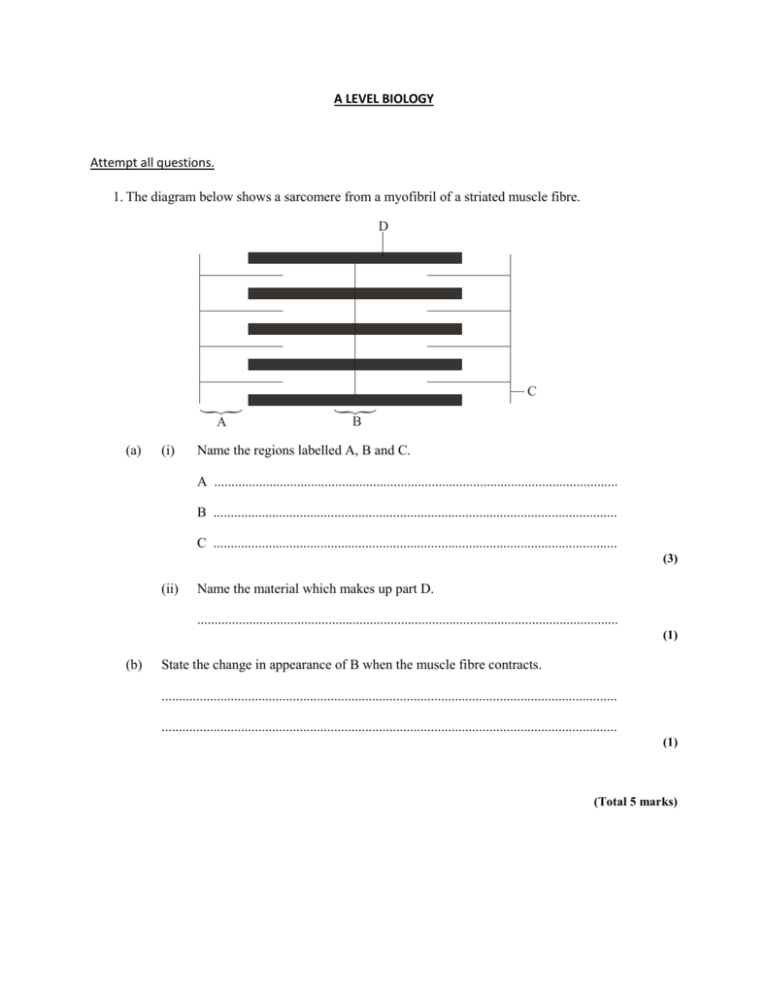
A LEVEL BIOLOGY Attempt all questions. 1. The diagram below shows a sarcomere from a myofibril of a striated muscle fibre. D C A (a) (i) B Name the regions labelled A, B and C. A ..................................................................................................................... B ..................................................................................................................... C ..................................................................................................................... (3) (ii) Name the material which makes up part D. .......................................................................................................................... (1) (b) State the change in appearance of B when the muscle fibre contracts. .................................................................................................................................... .................................................................................................................................... (1) (Total 5 marks) 2. The diagrams below, (labelled 1, 2, 3, 4 and 5), show five possible states of a sarcomere from striated muscle. The relative positions of some actin and myosin filaments are shown. Z disk Myosin Z disk Actin 1 2 3 4 5 The graph below shows the relationship between sarcomere length and the amount of tension generated during the contraction. The letters A, B, C, D and E on the graph correspond to the different positions of the actin and myosin filaments, shown on the diagrams above, during the contraction. A C B D E 100 80 Tension 60 as % of maximum 40 20 0 1.0 1.5 2.0 2.5 3.0 Sarcomere length / m 3.5 4.0 (a) (i) State the letter of the stage on the graph which corresponds to the greatest length of the sarcomere shown in the diagrams. (1) (ii) State the length of the sarcomere when the maximum tension is generated during contraction (1) (iii) Which diagram of the sarcomere shows the position of the actin and myosin filaments when the maximum tension is generated (1) (b) (i) Describe two ways in which the positions of structures in the sarcomere you have identified in (a) (ii) differ from those in the sarcomere at its greatest length 1 ….…………………………………………………………………………. 2 ….…………………………………………………………………………. (2) (ii) Explain how a sarcomere in a contracting muscle would change between the state shown in diagram 1 and that in diagram 3 (4) (c) Suggest why the maximum tension is not achieved when the sarcomere length is at its shortest. (2) (Total 11 marks) 3The diagrams below illustrate part of a sarcomere to show the sequence of events during muscle contraction. A B Diagram 1 Diagram 2 Diagram 3 (a) Name the proteins labelled A and B. A .............................................................................................................................. B .............................................................................................................................. (1) (b) With reference to the diagrams, describe how shortening of the sarcomere is brought about. ................................................................................................................................. ................................................................................................................................. ................................................................................................................................. ................................................................................................................................. ................................................................................................................................. (4) (Total 5 marks)
