The Tissue Level of Organization
advertisement
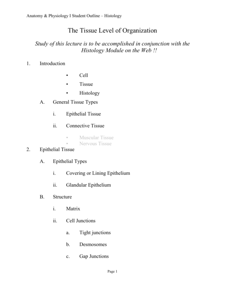
Anatomy & Physiology I Student Outline – Histology The Tissue Level of Organization Study of this lecture is to be accomplished in conjunction with the Histology Module on the Web !! 1. Introduction A. 2. • Cell • Tissue • Histology General Tissue Types i. Epithelial Tissue ii. Connective Tissue • Muscular Tissue • Nervous Tissue Epithelial Tissue A. B. Epithelial Types i. Covering or Lining Epithelium ii. Glandular Epithelium Structure i. Matrix ii. Cell Junctions a. Tight junctions b. Desmosomes c. Gap Junctions Page 1 Anatomy & Physiology I Student Outline – Histology • C. Intercellular Communication iii. Stem Cells iv. Avascular v. Basement Membrane vi. a. Basal Lamina b. Reticular Lamina Orientation a. Apical b. Basal Covering or lining Epithelium i. Simple Epithelium a. b. b. Simple Squamous Epithelium (Study images on web carefully) • Endothelium • Mesothelium Simple Cuboidal Epithelium (Study images on web carefully) • Functions • Locations Simple Columnar Epithelium (Study images on web carefully) • Functions • Locations Page 2 Anatomy & Physiology I Student Outline – Histology ii. Stratified Epithelium a. Stratified Squamous Epithelium (Study images on web carefully) • Keratinized Stratified Squamous Epithelium • b. c. d. Keratin * Functions * Location Nonkeratinized Stratified Squamous Epithelium * Functions * Location Stratified Cuboidal Epithelium (Study images on web carefully) • Functions • Locations Stratified Columnar Epithelium (Study images on web carefully) • Function • Locations Transitional Epithelium (Study images on web carefully) • Functions • Location Page 3 Anatomy & Physiology I Student Outline – Histology e. Ciliated Pseudostratified Columnar Epithelium (Study images on web carefully) 3. • Functions • Locations Connective Tissue Embryonic Connective Tissue i. Mesenchyme ii. Mucous Connective Tissue A. Adult Connective Tissue i. Loose (Areolar) Connective Tissue (Study images on web carefully) a. Matrix b. Fiber Components • Collagenous Fibers * • Elastic Fibers • Reticular Fibers * c. ii. Fibril Reticulum Cellular Component • Fibroblasts • Macrophages • Plasma Cells • Mast Cells Adipose Tissue (Study images on web carefully) Page 4 Anatomy & Physiology I Student Outline – Histology a. Adipocytes • b. iii. Location / Function Relationships Dense (Collagenous) Connective Tissue (Study images on web carefully) a. b. iv. Regular Dense Connective Tissue • Function • Locations * Tendons * Ligaments * Aponeuroses Irregular Dense Connective Tissue • Location • Function Elastic Connective Tissue a. Function • b. v. Inclusion Body of Lipid Elastic Laminae (Arteries) Locations Reticular Connective Tissue Page 5 Anatomy & Physiology I Student Outline – Histology * B. a. Function b. Locations Reticulum Cartilage i. ii. General Characteristics of all Cartilages a. Chondrocytes b. Lacunae c. Perichondrium d. Avascular Types a. b. c. Hyaline Cartilage (Study images on web carefully) • Characteristics and Functions • Locations * Costal Cartilage * Articular Cartilage Elastic Cartilage (Study images on web carefully) • Function • Locations Fibrocartilage (Study images on web carefully) Page 6 Anatomy & Physiology I Student Outline – Histology iii. Function • Locations Growth of Cartilage (Study images on web carefully) a. 4. • Interstitial Growth Appositional Growth Glandular Epithelium A. Exocrine Glands i. Morphology a. b. Unicellular glands Multicellular Glands * Tubular Gland * ii. Holocrine Glands * • • Examples Merocrine (Eccrine) Glands * Example Apocrine glands * B. Alveolar (Acinar) Gland * Tubuloacinar gland * Simple Gland * Compound Gland Functional Types • 5. b. Example Endocrine Glands Membranes - (Pull out your handout on Membranes), you may also want to pull out the handout on Body Cavities from the first lecture. Page 7 Anatomy & Physiology I Student Outline – Histology A. B. Serous Membranes (Serosa) i. Epithelia = Mesothelium ii. Visceral Portion iii. Parietal Portion iv. Serous Fluid v. Examples a. Visceral and Parietal Pericardium b. Visceral and Parietal Pleura c. Visceral and Parietal Peritoneum Mucous Membrane i. Epithelia ii. Lamina Propria iii. Muscularis Mucosa C. Cutaneous Membranes – will be covered during the lecture on the Integumentary System. D. Synovial Membranes– will be covered during the lecture on Articulations. The subjects below will be covered in subsequent lectures! 5. Osseous Tissue (Bone) 6. Vascular Tissue (Blood) 7. Muscle Tissue A. Skeletal Muscle Tissue Page 8 Anatomy & Physiology I Student Outline – Histology B. C. 8. Cardiac Muscle Tissue Smooth Muscle Tissue Nervous Tissue Page 9


