Cell Fractionation Kit - Cell Signaling Technology, Inc.
advertisement
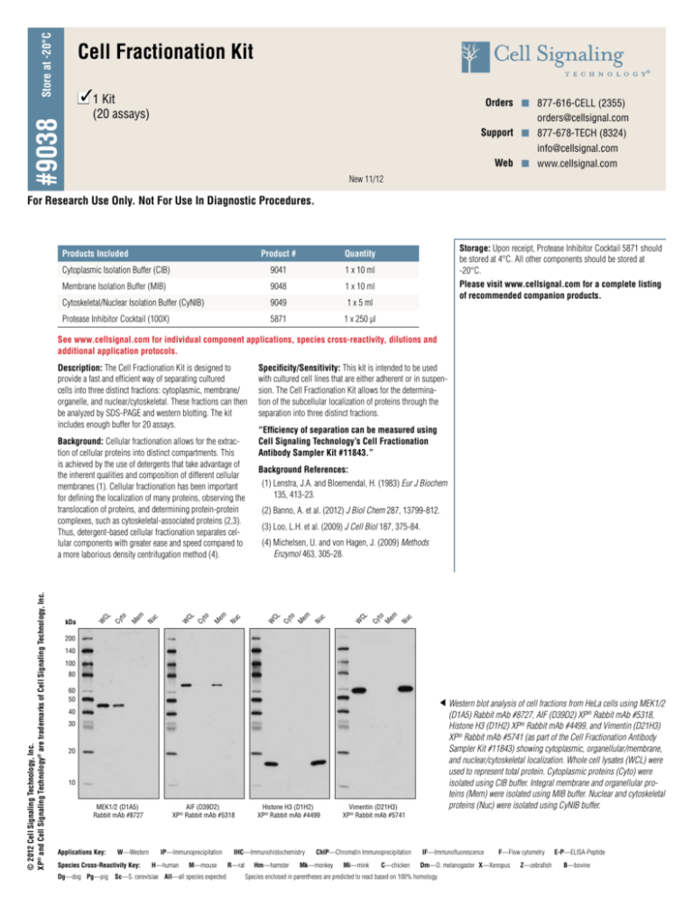
Store at -20°C Cell Fractionation Kit 31 Kit n Orders n 877-616-CELL (2355) orders@cellsignal.com Support n 877-678-TECH (8324) info@cellsignal.com Web n www.cellsignal.com #9038 (20 assays) New 11/12 For Research Use Only. Not For Use In Diagnostic Procedures. Products Included Product # Quantity Cytoplasmic Isolation Buffer (CIB) 9041 1 x 10 ml Membrane Isolation Buffer (MIB) 9048 1 x 10 ml Cytoskeletal/Nuclear Isolation Buffer (CyNIB) 9049 1 x 5 ml Protease Inhibitor Cocktail (100X) 5871 1 x 250 µl Storage: Upon receipt, Protease Inhibitor Cocktail 5871 should be stored at 4°C. All other components should be stored at -20°C. Please visit www.cellsignal.com for a complete listing of recommended companion products. See www.cellsignal.com for individual component applications, species cross-reactivity, dilutions and additional application protocols. Description: The Cell Fractionation Kit is designed to provide a fast and efficient way of separating cultured cells into three distinct fractions: cytoplasmic, membrane/ organelle, and nuclear/cytoskeletal. These fractions can then be analyzed by SDS-PAGE and western blotting. The kit includes enough buffer for 20 assays. “Efficiency of separation can be measured using Cell Signaling Technology’s Cell Fractionation Antibody Sampler Kit #11843.” Background References: (1) Lenstra, J.A. and Bloemendal, H. (1983) Eur J Biochem 135, 413-23. (2) Banno, A. et al. (2012) J Biol Chem 287, 13799-812. (3) Loo, L.H. et al. (2009) J Cell Biol 187, 375-84. c Nu to em M CL Cy W c Nu to em M Cy CL (4) Michelsen, U. and von Hagen, J. (2009) Methods Enzymol 463, 305-28. W c Nu to em M CL W Cy c Nu to em M CL Cy kDa W © 2012 Cell Signaling Technology, Inc. XP® and Cell Signaling Technology® are trademarks of Cell Signaling Technology, Inc. Background: Cellular fractionation allows for the extraction of cellular proteins into distinct compartments. This is achieved by the use of detergents that take advantage of the inherent qualities and composition of different cellular membranes (1). Cellular fractionation has been important for defining the localization of many proteins, observing the translocation of proteins, and determining protein-protein complexes, such as cytoskeletal-associated proteins (2,3). Thus, detergent-based cellular fractionation separates cellular components with greater ease and speed compared to a more laborious density centrifugation method (4). Specificity/Sensitivity: This kit is intended to be used with cultured cell lines that are either adherent or in suspension. The Cell Fractionation Kit allows for the determination of the subcellular localization of proteins through the separation into three distinct fractions. 200 140 100 80 60 50 Western blot analysis of cell fractions from HeLa cells using MEK1/2 (D1A5) Rabbit mAb #8727, AIF (D39D2) XP® Rabbit mAb #5318, Histone H3 (D1H2) XP® Rabbit mAb #4499, and Vimentin (D21H3) XP® Rabbit mAb #5741 (as part of the Cell Fractionation Antibody Sampler Kit #11843) showing cytoplasmic, organellular/membrane, and nuclear/cytoskeletal localization. Whole cell lysates (WCL) were used to represent total protein. Cytoplasmic proteins (Cyto) were isolated using CIB buffer. Integral membrane and organellular proteins (Mem) were isolated using MIB buffer. Nuclear and cytoskeletal proteins (Nuc) were isolated using CyNIB buffer. 40 30 20 10 MEK1/2 (D1A5) Rabbit mAb #8727 Applications Key: W—Western Species Cross-Reactivity Key: AIF (D39D2) XP® Rabbit mAb #5318 IP—Immunoprecipitation H—human M—mouse Dg—dog Pg—pig Sc—S. cerevisiae All—all species expected Histone H3 (D1H2) XP® Rabbit mAb #4499 IHC—Immunohistochemistry R—rat Hm—hamster Vimentin (D21H3) XP® Rabbit mAb #5741 ChIP—Chromatin Immunoprecipitation Mk—monkey Mi—mink C—chicken IF—Immunofluorescence F—Flow cytometry Dm—D. melanogaster X—Xenopus Species enclosed in parentheses are predicted to react based on 100% homology. Z—zebrafish E-P—ELISA-Peptide B—bovine #9038 Cell Fractionation Protocol ABuffers E Cell Fractionation Cytoplasm Isolation Buffer (CIB) – 10 ml, Store at -20°C. Membrane Isolation Buffer (MIB) – 10 ml, Store at -20°C. Cytoskeleton/Nucleus Isolation Buffer (CyNIB) – 5 ml, Store at -20°C. Protease Inhibitor Cocktail (100X) (#5871) – 250 μl, Store at 4°C. 1. Aliquot the remaining 400 μl into a 1.5 ml tube. 2. Centrifuge for 5 min at 500 x g at 4°C. 3. Aspirate the supernatant. 4. Resuspend pellet in 500 μl of CIB. 5. Vortex for 5 sec. 6. Incubate on ice for 5 min. 7. Centrifuge for 5 min at 500 x g. 8. Save the supernatant. This is the Cytoplasmic Fraction. 9. Resuspend pellet in 500 μl of MIB. 10. Vortex for 15 sec. 11. Incubate on ice for 5 min. 12. Centrifuge for 5 min at 8,000 x g. 13. Save the supernatant. This is the Membrane and Organelle Fraction. 14. Resuspend pellet in 250 μl of CyNIB. 15. Sonicate for 5 sec at 20% power 3 times. This is the Cytoskeletal and Nuclear Fraction. BNotes • • • • • • • • • A ll steps, except for the addition and sonication of the CyNIB buffer, should be done on ice or at 4°C. Adherent or suspension cultured cells can be used for this assay. 1X protease inhibitors (#5871) (5 μl of 100X per 500 μl buffer, included in kit) and 1 mM fresh PMSF (#8553) (2.5 μl of 200 mM PMSF per 500 μl buffer, not included in kit) should be added to each buffer immediately before use. Phosphatase inhibitors are already included in the buffers. There is no need to add them. Please refer to Table 1 for the appropriate volumes for your specific cell concentration. The volumes given in Sections D and E are based on cell counts obtained from HeLa cells at ~90% confluency in a 10 cm cell culture dish (5x10⁶ cells). If the CyNIB buffer is cloudy after thawing, please warm the solution in a 37°C water bath until the solution is clear. Be cautious when saving fractions so that you do not get any contamination from the resulting pellet. All lysates should be stored at -20°C for short term storage (less than 1 month) or -80°C for long term storage (greater than 1 month). F Western Blot 1. A dd 60 μl of 3X SDS Loading Buffer with DTT (#7722) for every 100 μl of supernatant. 2. Boil each sample for 5 min at 95°C and centrifuge for 3 min at 15,000 x g. 3. Load 15 μl of each fraction along with 15 μl of WCL. Table 1: Volumes in μl for WCL or buffer at indicated cell numbers. Cell Count C Isolating Cell Population 2.5 x 10 cells 5 x 10 cells WCL 50 100 150 200 CIB 250 500 750 1000 MIB 250 500 750 1000 CyNIB 125 250 375 500 6 For adherent cells 1. Wash plate with cold 1X PBS. 2. Trypsinize the plate. 3. Add cold growth media to deactivate trypsin. For both adherent and suspension cells 1. Spin down cells at 350 x g for 5 min. 2. Aspirate media. 3. Wash cell pellet with cold 1X PBS. 4. Resuspend pellet in 0.5 ml of cold 1X PBS. 5. Count live cells using Trypan Blue and a hemacytometer. 6 7.5 x 10⁶ cells 1 x 10⁷ cells D Detection of Proteins © 2012 Cell Signaling Technology, Inc. 1. A liquot 100 μl of cell suspension into a 1.5 ml tube for the whole cell lysate (WCL). 2. Add 60 μl of 3X SDS Loading Buffer with DTT (#7722) to make a final volume of 160 μl of WCL. 3. Sonicate WCL tube for 15 sec at 20% power 3 times, heat for 5 min at 95°C, and centrifuge for 3 min at 15,000 x g. Orders n 877-616-CELL (2355) orders@cellsignal.com Support n 877-678-TECH (8324) info@cellsignal.com Web n www.cellsignal.com


