Study on Chemical Constituents and Cytotoxic Activities of Salacia
advertisement

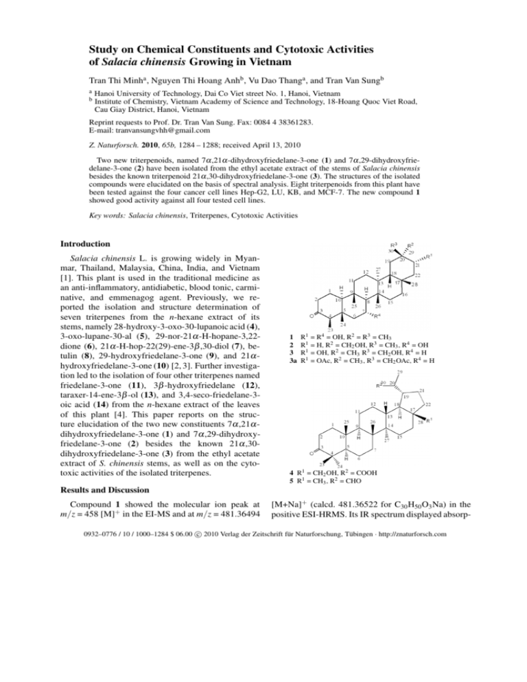
Study on Chemical Constituents and Cytotoxic Activities of Salacia chinensis Growing in Vietnam Tran Thi Minha , Nguyen Thi Hoang Anhb , Vu Dao Thanga , and Tran Van Sungb a b Hanoi University of Technology, Dai Co Viet street No. 1, Hanoi, Vietnam Institute of Chemistry, Vietnam Academy of Science and Technology, 18-Hoang Quoc Viet Road, Cau Giay District, Hanoi, Vietnam Reprint requests to Prof. Dr. Tran Van Sung. Fax: 0084 4 38361283. E-mail: tranvansungvhh@gmail.com Z. Naturforsch. 2010, 65b, 1284 – 1288; received April 13, 2010 Two new triterpenoids, named 7α ,21α -dihydroxyfriedelane-3-one (1) and 7α ,29-dihydroxyfriedelane-3-one (2) have been isolated from the ethyl acetate extract of the stems of Salacia chinensis besides the known triterpenoid 21α ,30-dihydroxyfriedelane-3-one (3). The structures of the isolated compounds were elucidated on the basis of spectral analysis. Eight triterpenoids from this plant have been tested against the four cancer cell lines Hep-G2, LU, KB, and MCF-7. The new compound 1 showed good activity against all four tested cell lines. Key words: Salacia chinensis, Triterpenes, Cytotoxic Activities Introduction Salacia chinensis L. is growing widely in Myanmar, Thailand, Malaysia, China, India, and Vietnam [1]. This plant is used in the traditional medicine as an anti-inflammatory, antidiabetic, blood tonic, carminative, and emmenagog agent. Previously, we reported the isolation and structure determination of seven triterpenes from the n-hexane extract of its stems, namely 28-hydroxy-3-oxo-30-lupanoic acid (4), 3-oxo-lupane-30-al (5), 29-nor-21α -H-hopane-3,22dione (6), 21α -H-hop-22(29)-ene-3β ,30-diol (7), betulin (8), 29-hydroxyfriedelane-3-one (9), and 21α hydroxyfriedelane-3-one (10) [2, 3]. Further investigation led to the isolation of four other triterpenes named friedelane-3-one (11), 3β -hydroxyfriedelane (12), taraxer-14-ene-3β -ol (13), and 3,4-seco-friedelane-3oic acid (14) from the n-hexane extract of the leaves of this plant [4]. This paper reports on the structure elucidation of the two new constituents 7α ,21α dihydroxyfriedelane-3-one (1) and 7α ,29-dihydroxyfriedelane-3-one (2) besides the known 21α ,30dihydroxyfriedelane-3-one (3) from the ethyl acetate extract of S. chinensis stems, as well as on the cytotoxic activities of the isolated triterpenes. 1 2 3 3a R1 R1 R1 R1 = R4 = OH, R2 = R3 = CH3 = H, R2 = CH2 OH, R3 = CH3 , R4 = OH = OH, R2 = CH3 R3 = CH2 OH, R4 = H = OAc, R2 = CH3 , R3 = CH2 OAc, R4 = H 4 R1 = CH2 OH, R2 = COOH 5 R1 = CH3 , R2 = CHO Results and Discussion Compound 1 showed the molecular ion peak at m/z = 458 [M]+ in the EI-MS and at m/z = 481.36494 [M+Na]+ (calcd. 481.36522 for C30 H50 O3 Na) in the positive ESI-HRMS. Its IR spectrum displayed absorp- c 2010 Verlag der Zeitschrift für Naturforschung, Tübingen · http://znaturforsch.com 0932–0776 / 10 / 1000–1284 $ 06.00 T. T. Minh et al. · Chemical Constituents and Cytotoxic Activities of Salacia chinensis 1285 6 13 7 14 8 9 10 11 12 R1 R1 R1 R1 = O, R2 = H, R3 = CH2 OH = O, R2 = OH, R3 = CH3 = O, R2 = H, R3 = CH3 = OH, R2 = H, R3 = CH3 tion bands at 3446 (OH) and 1716 cm−1 (>C=O). The 1 H NMR spectrum revealed 8 methyl signals, one of them being a doublet at δH = 0.91 (J = 7.0 Hz); the others are singlets at δH = 0.80, 0.93, 0.99, 1.08, 1.12, 1.17, and 1.23. Furthermore, the signals of two >CH-OH groups were observed at δH = 4.08 (1H, ddd, 3.5, 10.5, 10.5 Hz) and 3.70 (1H, dd, 4.5, 12.1 Hz). The 13 C NMR spectrum showed signals of 30 carbon atoms including one carbonyl (δC = 212.3), two hydroxymethines (δC = 68.8; 73.8), eight methyls, nine methylenes, four methines, and six quaternary carbon atoms. The analysis of the spectroscopic data and a comparison of the chemical shift of the tertiary methyl groups with those of other friedelane derivatives [5 – 7] suggested that compound 1 is a friedelanone with two hydroxyl substituents. The locations of the carbonyl as well as of the two hydroxyl groups have been determined by HMBC and NOESY experiments. In the HMBC spectrum the observed correlations between δC = 68.8 (C-7) and δH = 1.49 (H-8), 1.40 and 2.01 (H2 -6) suggested one hydroxyl group at C-7, which was confirmed by the correlations of δH = 4.08 (H-7) and δH = 2.01, 1.40 (H2 -6) and 1.49 (H-8) in the 1 H-1 H COSY spectrum. The position of the second hydroxyl group at C-21 was confirmed by correlations between δC = 73.8 (C-21) and δH = 0.99 (H3 -29), 1.08 (H3 30), 1.23 (H3 -28), 1.61, and 1.35 (H2 -22) in the HMBC spectrum as well as by the correlations between δH = 3.70 (H-21) and δH = 1.61 and 1.35 (H2 -22) in the 1 H1 H COSY spectrum. The 3-oxo group was deduced from the 3 JCH correlations between C-3 (δC = 212.3) and H3 -23 (δH = 0.91), H2 -1 (δH = 1.99 and 1.68), and H2 -2 (δH = 2.41 and 2.30) in the HMBC spectrum. The configuration of both hydroxyl groups was established as α by the NOESY experiment, which showed NOE correlations between δH = 4.08 (H-7) and δH = 0.8 (H3 24), 0.93 (H3 -25) and 1.17 (H3 -26), and between δH = 3.70 (H-21) and δH = 1.23 (H3 -28) and 1.08 (H3 -30). Consequently, the structure of 1 was determined as the new triterpene 7α ,21α -dihydroxyfriedelane-3-one. Compound 2 was obtained as colorless crystals. Its IR spectrum showed absorptions of hydroxyl and T. T. Minh et al. · Chemical Constituents and Cytotoxic Activities of Salacia chinensis 1286 Table 1. 13 C NMR spectral data (125 MHz, CD3 OD) of compounds 1 – 3 and 3a (δ in ppm). Position 1 2 3 4 5 6 7 8 9 10 11 12 13 14 15 16 17 18 19 20 21 22 23 24 25 26 27 28 29 30 CH3 CO CH3 CO CH3CO CH3CO 1 21.9 41.2 212.3 58.2 42.6 52.6 68.8 57.6 39.1 58.9 35.7 30.2 40.3 39.7 33.6 36.5 31.8 43.5 36.2 34.3 73.8 47.6 6.9 15.9 19.2 19.0 19.7 32.8 25.4 31.2 – – – – 2 21.9 41.2 212.3 58.3 42.6 52.3 68.8 58.7 39.1 58.9 35.9 30.4 40.1 40.4 35.4 36.1 29.7 42.4 29.5 33.5 28.0 38.3 6.9 15.9 18.9 20.8 19.0 31.9 71.4 29.1 – – – – 3 22.2 41.3 214.4 58.1 42.0 41.0 18.1 50.8 37.4 59.3 35.0 29.7 39.0 38.7 29.8 36.0 32.2 44.1 29.5 38.3 71.2 44.6 6.5 14.4 19.1 19.4 19.4 32.9 16.5 73.3 – – – – 3a 22.3 41.5 212.7 58.3 42.1 41.3 18.3 52.2 37.2 59.6 35.4 30.3 39.4 38.7 31.3 35.8 32.1 43.3 31.1 37.5 71.3 42.7 6.8 14.7 18.2 18.8 18.5 32.5 21.8 71.0 20.9 22.1 170.5 171.1 ketone groups at 3532 and 1714 cm−1 , respectively. The molecular formula of C30 H50 O3 and the molecular weight of m/z = 458 were obtained from the positive high-resolution ESI-MS through the peak at m/z = 497.33922 [M+K]+ (calcd. 497.33915). The 1 H and 13 C NMR spectra of 2 were very similar to those of 1 with two exceptions. Instead of signals for 8 methyl and two oxygenated methine groups in the spectra of 1, compound 2 exhibited 7 methyls, one oxygenated methine (δH = 4.09, δC = 68.8) and one oxygenated methylene (δH = 3.37, 3.46; δC = 71.4) groups. These spectral data suggested that compound 2 has also a dihydroxyfriedelane-3-one structure like 1 but with a different localization of the hydroxyl groups. The first hydroxyl group was located at C-7 as in 1, due to the correlations between δC = 68.8 (C-7) and δH = 1.5 (H8), 2.02 and 1.39 (H2 -6) in the HMBC spectra as well as the correlations between δH = 4.09 (H-7) and 1.5 (H-8), 2.02 and 1.39 (H2 -6) in the COSY spectrum. The second OH group was connected at C-29, due to the cross peaks between: δC = 71.4 (C-29) and δH = 1.0 (H3 -30), 1.19 (H-19); between δH = 3.37 (H-29B) and δC = 29.5 (C-19), 33.5 (C-20); between δH = 3.46 (H29A) and δC = 29.1 (C-30), 33.5 (C-20) in the HMBC experiments as well as the correlation between H3 -30 (1.0 ppm) and H3 -28 (1.23 ppm) in the NOESY spectrum. The configuration of the 7α -hydroxyl group in 2 was determined by NOE effects of H-7 (4.09 ppm) and H3 -24 (0.8 ppm), H3 -25 (0.92 ppm) and H3 -26 (1.22 ppm). The structure of 2 was thus established as the new triterpene 7α ,29-dihydroxyfriedelane-3-one. The NMR spectral data assigned to carbon signals for 1 and 2 are listed in Table 1. Compound 3 was isolated as colorless needles. It showed the molecular ion peak at m/z = 458 [M]+ in the EIMS, the same as that of 1 and 2. Its IR spectrum indicated the presence of hydroxyl and carbonyl functions at 3421 and 1714 cm−1 , respectively. The 1 H and 13 C NMR spectra showed similar signals to those of compound 2 with a slight difference in the chemical shifts of the oxygenated methine [δH = 3.92 (1H, dd, 4.0, 12.0 Hz), δC = 71.2] and the oxygenated methylene group [δH = 3.35 (2H, s), δC = 73.3]. From a detailed spectral analysis and by a comparison with the data of 21α ,30-dihydroxyfriedelane3-one as well as with those of its diacetate (3a), the structure of compound 3 was determined as 21α ,30dihydroxyfriedelane-3-one. This compound has been previously isolated from Salacia reticulata (Celastraceae) [8]. Compounds 1, 2, 3, 4, 6, 11, 12, and 14 were tested for cytotoxicity with four cancer cell lines: liver cancer (Hep-G2), lung cancer (LU), mouth cancer (KB), and breast cancer (MCF-7). Compound 1 was active against all four cancer cell lines tested, with the IC50 values 16.22, 19.27, 16.86, and 27.35 µ g mL−1 , respectively (Table 2). Compounds 4 and 14 were also found to be cytotoxic against all four cell lines with IC50 higher than that of 1. Compound 6 was active against human lung carcinoma cells (LU) (IC50 = 117.33 µ g mL−1 ), whereas compounds 3, 11 and 12 were inactive (IC50 > 128 µ g mL−1 ) (Table 2). Experimental Section General Melting points were determined on a Botius melting point apparatus (Germany). Optical rotation values: Polarimeter T. T. Minh et al. · Chemical Constituents and Cytotoxic Activities of Salacia chinensis 1287 In vitro cytotoxicity IC50 (µ g mL−1 ) MCF7 a LUb HepG2c KBd 27.35 19.27 16.22 16.86 > 128 > 128 > 128 > 128 > 128 > 128 > 128 > 128 69.48 25.77 61.79 62.90 > 128 117.33 > 128 > 128 > 128 > 128 > 128 > 128 > 128 > 128 > 128 > 128 77.12 67.07 88.14 83.61 0.31 – 0.62 0.31 – 0.62 0.31 – 0.62 0.62 – 1.25 Table 2. Cytotoxic activity of triterpenoids from Salacia chinensis L. Compounds 7α ,21α -dihydroxyfriedelane-3-one (1) 7α ,29-dihydroxyfriedelane-3-one (2) 21α ,30-dihydroxyfriedelane-3-one (3) 28-hydroxy-3-oxo-30-lupanoic acid (4) 29-nor-21α -H-hopane-3,22-dione (6) friedelane-3-one (11) 3β -hydroxyfriedelane (12) 3,4-seco-friedelane-3-oic acid (14) Ellipticine POLAX-2L (Japan). FT-IR: Nicolet IMPACT 410. EI-MS: HP5989B. ESI-MS: AGILENT 1100 LC-MSD Trap spectrometer. HR-ESI-MS: Qstar pulsar (Applied Bioystems). NMR: Bruker Avance 500 MHz. Column chromatography (CC): silica gel (70 – 230 and 230 – 400 mesh, Merck). Thin layer chromatography (TLC): DC-Alufolien 60 F254 (Merck). Plant material The leaves of Salacia chinensis L. were collected in Quang Binh province, Vietnam, in April 2007. The species was identified by Dr. Ngo Van Trai, Institute of Materia Medica, Hanoi. A voucher specimen (No. SC-02) is deposited in the Hanoi University of Technology, Vietnam. The dried and powdered leaves of Salacia chinensis L. (1.8 kg) were extracted with 80 % aqueous MeOH at r. t. After MeOH was evaporated in vacuo, the residue was partitioned with n-hexane followed by EtOAc and n-BuOH. The EtOAc extract (6.5 g) was chromatographed on silica gel with solvents of increasing polarity (0 – 100 % MeOH in dichlomethane) to give 7 fractions. The fractions were further purified to afford compounds 1, 2 and 3. Extraction and isolation 7α ,21α -Dihydroxyfriedelane-3-one (1) Fraction 3 (0.34 g), eluated with dichloromethane : MeOH = 95 : 5, was rechromatographed over a silica gel column with a mixture of CH2 Cl2 : MeOH = 95 : 5 followed by crystallization (CHCl3 : MeOH = 9 : 1), afforded 80 mg of compound 1 (0.0044 %) as colorless plates. – M. p. 272 – 273 ◦C. – [α ]25 D = +219.8 (c = 0.2, CH3 OH : CHCl3 = 10 : 1). – IR (KBr): ν = 3446, 2928, 1716, 1453, 1386, 1038 cm−1 . – EI-MS: m/z (%) = 458 (0.7) [M]+ , 440 (8) [M–H2 O]+ , 422 (5) [M–2H2 O]+ , 231 (10), 203 (22), 177 (19), 161 (24), 133 (29), 123 (73), 95 (70), 69 (86), 55 (100). – HRMS ((+)-ESI): m/z = 481.36494 (calcd. 481.36522 for C30 H50 O3 Na, [M+Na]+ ). – 1 H NMR (500 MHz, CDCl3 ): δ = 4.08 (1H, ddd, J = 3.5, 10.5, 10.5 Hz, H-7), 3.70 (1H, dd, J = 4.5, 12.1 Hz, H-21), 1.23 (s, H3 -28), 1.17 (s, H3 -26), 1.12 (s, H3 -27), 1.08 (s, H3 -30), a Human breast carcinoma; b human lung carcinoma; c human epatocellular carcinoma; d human epidermic carcinoma. 0.99 (s, H3 -29), 0.93 (s, H3 -25), 0.91 (d, J = 7.0 Hz, H3 -23), 0.80 (s, H3 -24). – 13 C NMR: see Table 1. 7α ,29-Dihydroxyfriedelane-3-one (2) Fraction 2 (0.18 g) (dichloromethane : MeOH = 98 : 2) was crystallized from a mixture of CHCl3 : MeOH (9 : 1) to give 90 mg (0.005 %) of compound 2 as colorless crystals. – M. p. 325 – 327 ◦C. – [α ]25 D = −152.4 (c = 0.2, CH3 OH : CHCl3 = 10 : 1). – IR (KBr): ν = 3532, 2944, 2874, 1714, 1458, 1386, 1197, 1037 cm−1 . – HRMS ((+)-ESI): m/z = 497.33922 (calcd. 497.33915 for C30 H50 O3 K, [M+K]+ ). – 1 H NMR (500 MHz, CDCl3 ): δ = 4.09 (1H, m, H-7), 3.46 (1H, d, J = 10.5 Hz, H-29A), 3.37 (1H, d, J = 10.5 Hz, H-29B), 1.22 (s, H3 -26), 1.18 (s, H3 -28), 1.09 (s, H3 -27), 1.00 (s, H3 -30), 0.92 (s, H3 -25), 0.91 (d, J = 6.5 Hz, H3 -23), 0.80 (s, H3 -24). – 13 C NMR: see Table 1. 21α ,30-Dihydroxyfriedelane-3-one (3) Fraction 3 (0.34 g) was rechromatographed over a silica gel column with a mixture of CH2 Cl2 : MeOH = 95 : 5. Crystallization (CHCl3 : MeOH = 9 : 1) afforded 180 mg (0.01 %) of compound 3 as colorless crystals. – M. p. 285 – 286 ◦C. – IR (KBr): ν = 3421 – 3300, 2929, 2879, 1714, 1460, 1383, 1039 cm−1 . – EI-MS: m/z (%) = 458 (1) [M]+ , 440 (10), 410 (2), 302 (4), 273 (12), 231 (10), 175 (12), 161 (15), 121 (41), 95 (51), 81 (61), 55 (100). – 1 H NMR (500 MHz, CDCl3 ): δ = 3.92 (1H, dd, J = 4.0, 12.0 Hz, H-21), 3.35 (2H, s, H2 30), 1.16 (6H, s), 1.04 (3H, s), 0.88 (6H, s), 0.87 (3H, d, J = 7.0 Hz), 0.72 (3H, s). – 13 C NMR: see Table 1. Acetylation of diol 3 with Ac2 O-pyridine (1 : 1) at r. t. for 24 h gave 21α ,30-dihydroxy-3-friedelanone diacetate (3a). 21α ,30-Dihydroxyfriedelane-3-one diacetate (3a) Colorless crystals (n-hexane : CHCl3 = 9 : 1). – M. p. 175 – 178 ◦C. – IR (KBr): ν = 2936, 1733, 1465, 1385, 1239, 1022, 759 cm−1 . – EI-MS: m/z = 542 [M]+ (C34 H54 O5 ). – 1 H NMR (500 MHz, CDCl ): δ = 5.09 (1H, dd, J = 4.5, 3 12.5 Hz, H-21), 3.95 (1H, d, J = 10.9 Hz, H-30A), 3.86 (1H, d, J = 10.9 Hz, H-30B), 2.07 and 2.01 (each 3H, s, OAc), 1288 T. T. Minh et al. · Chemical Constituents and Cytotoxic Activities of Salacia chinensis 1.28, 1.12, 1.02, 0.94, 0.87, 0.73 (each 3H, s, Me) and 0.88 (3H, d, J = 6.5 Hz, H-23). – 13 C NMR: see Table 1. Assay of cytotoxic activities using Hep-G2, LU, KB and MCF-7 cell lines The human cancer cell lines supplied by the American Type Culture Collection (ATCC) were maintained in a suitable medium adding FBS and were incubated at 37 ◦C in a humidified atmosphere of 5 % CO2 . The Hep-G2 (the human epatocellular carcinoma), KB (the human mouth epidermal carcinoma) and MCF7 (the human breast carcinoma) cell lines were maintained in RPMI1640 culture medium with 10 % fetal bovine serum (FBS). The LU (the human lung carcinoma) cell line was maintained in DMEM culture medium with 10 % fetal bovine serum (FBS). The cell line was cultured at 37 ◦C in an atmosphere of 5 % CO2 in air (100 % humidity). The cells were treated in [1] T. Morikawa, A. Kishi, Y. Pongpiriyadacha, H. Matsuda, M. Yoshikawa, J. Nat. Prod. 2003, 66, 1191 – 1196. [2] T. T. Minh, N. T. H. Anh, V. D. Thang, T. V. Sung, Z. Naturforsch. 2008, 63b, 1411 – 1414. [3] T. T. Minh, N. T. H. Anh, V. D. Thang, T. V. Sung, J. Chem. Vietnam 2009, 47, 469 – 473. [4] T. T. Minh, N. T. H. Anh, V. D. Thang, T. V. Sung, J. Chem. Vietnam 2009, 47, 192 – 196. triplicate at various concentration of the natural compounds (1 and 10 µ g mL−1 ) and incubated for 72 h at 37 ◦C in an atmosphere of 5 % CO2 . The cell growth inhibition was determined by the MTT assay. After incubation for 72 h, the media was removed, and the cells were incubated with 10 µ L of media containing 5 mg/mL stock solution of MTT in 40 µ L RPMI-1640 (for Hep-G2, KB, MCF7 ) or in 40 µ L DMEM (for LU). After incubation for 4 h at 37 ◦C in an atmosphere of 5 % CO2 , the formazan crystals formed were dissolved by adding 150 µ L of DMSO per well. The optical density was measured at 570 nm. The number of viable cells was proportional to the extent of formazan production. % CI = [1–(OD570 treated/OD570 control)] × 10018 . Acknowledgements We thank Mr. D. V. Luong, Institute of Chemistry, Hanoi, Vietnam for NMR measurement, Dr. J. Schmidt, IPB, Halle (Saale), Germany, for HRESIMS measurement and Dr. N. V. Trai, Hanoi, for identification of the plant material. [5] A. Kishi, T. Morikawa, H. Matsuda, M. Yoshikawa, Chem. Pharm. Bull. 2003, 51, 1051 – 1055. [6] W.-H. Hui, M.-M. Li, K.-M. Wong, Phytochemistry 1975, 15, 797 – 798. [7] A. Patra, S. K. Chaudhuri, Magn. Reson. Chem. 1987, 25, 95 – 100. [8] V. Kumar, D. B. T. Wijeratne, C. Abeygunawardena, Phytochemistry 1990, 29, 333 – 335.


