REVIEW: Hyaluronan and synovial joint: function, distribution and
advertisement
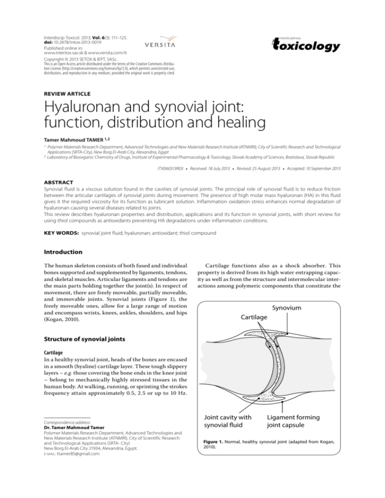
Interdiscip Toxicol. 2013; Vol. 6(3): 111–125. doi: 10.2478/intox-2013-0019 Published online in: www.intertox.sav.sk & www.versita.com/it Copyright © 2013 SETOX & IEPT, SASc. This is an Open Access article distributed under the terms of the Creative Commons Attribution License (http://creativecommons.org/licenses/by/2.0), which permits unrestricted use, distribution, and reproduction in any medium, provided the original work is properly cited. interdisciplinary REVIEW ARTICLE Hyaluronan and synovial joint: function, distribution and healing Tamer Mahmoud TAMER 1,2 1 Polymer Materials Research Department, Advanced Technologies and New Materials Research Institute (ATNMRI), City of Scientific Research and Technological Applications (SRTA-City), New Borg El-Arab City, Alexandria, Egypt 2 Laboratory of Bioorganic Chemistry of Drugs, Institute of Experimental Pharmacology & Toxicology, Slovak Academy of Sciences, Bratislava, Slovak Republic ITX060313R03 • Received: 18 July 2013 • Revised: 25 August 2013 • Accepted: 10 September 2013 ABSTRACT Synovial fluid is a viscous solution found in the cavities of synovial joints. The principal role of synovial fluid is to reduce friction between the articular cartilages of synovial joints during movement. The presence of high molar mass hyaluronan (HA) in this fluid gives it the required viscosity for its function as lubricant solution. Inflammation oxidation stress enhances normal degradation of hyaluronan causing several diseases related to joints. This review describes hyaluronan properties and distribution, applications and its function in synovial joints, with short review for using thiol compounds as antioxidants preventing HA degradations under inflammation conditions. KEY WORDS: synovial joint fluid; hyaluronan; antioxidant; thiol compound Introduction The human skeleton consists of both fused and individual bones supported and supplemented by ligaments, tendons, and skeletal muscles. Articular ligaments and tendons are the main parts holding together the joint(s). In respect of movement, there are freely moveable, partially moveable, and immovable joints. Synovial joints (Figure 1), the freely moveable ones, allow for a large range of motion and encompass wrists, knees, ankles, shoulders, and hips (Kogan, 2010). Cartilage functions also as a shock absorber. This property is derived from its high water entrapping capacity as well as from the structure and intermolecular interactions among polymeric components that constitute the Synovium Cartilage Structure of synovial joints Cartilage In a healthy synovial joint, heads of the bones are encased in a smooth (hyaline) cartilage layer. These tough slippery layers – e.g. those covering the bone ends in the knee joint – belong to mechanically highly stressed tissues in the human body. At walking, running, or sprinting the strokes frequency attain approximately 0.5, 2.5 or up to 10 Hz. Correspondence address: Dr. Tamer Mahmoud Tamer Polymer Materials Research Department, Advanced Technologies and New Materials Research Institute (ATNMRI), City of Scientific Research and Technological Applications (SRTA- City) New Borg El-Arab City 21934, Alexandria, Egypt. E-MAIL: ttamer85@gmail.com Joint cavity with synovial fluid Ligament forming joint capsule Figure 1. Normal, healthy synovial joint (adapted from Kogan, 2010). 112 Hyaluronan and synovial joint Tamer Mahmoud Tamer cartilage tissue (Servaty et al., 2000). Figure 2 sketches a section of the cartilage – a chondrocyte cell that permanently restructures/rebuilds its extracellular matrix. Three classes of proteins exist in articular cartilage: collagens (mostly type II collagen); proteoglycans (primarily aggrecan); and other noncollagenous proteins (including link protein, fibronectin, COMP – cartilage oligomeric matrix protein) and the smaller proteoglycans (biglycan, decorin, and fibromodulin). The interaction between highly negatively charged cartilage proteoglycans and type II collagen fibrils is responsible for the compressive and tensile strength of the tissue, which resists applied load in vivo. Synovium/synovial membrane Each synovial joint is surrounded by a fibrous, highly vascular capsule/envelope called synovium, whose internal surface layer is lined with a synovial membrane. Inside this membrane, type B synoviocytes (fibroblast-like cell lines) are localized/embedded. Their primary function is to continuously extrude high-molar-mass hyaluronans (HAs) into synovial fluid. Synovial fluid The synovial fluid (SF) of natural joints normally functions as a biological lubricant as well as a biochemical pool through which nutrients and regulatory cytokines traverse. SF contains molecules that provide low-friction and low-wear properties to articulating cartilage surfaces. Molecules postulated to play a key role in lubrication alone or in combination, are proteoglycan 4 (PRG4) (Swann et al., 1985) present in SF at a concentration of 0.05–0.35 mg/ml (Schmid et al., 2001), hyaluronan (HA) (Ogston & Stanier, 1953) at 1–4 mg/ml (Mazzucco et al., 2004), and surface-active phospholipids (SAPL) (Schwarz & Hills, 1998) at 0.1 mg/ml (Mazzucco et al., 2004). Synoviocytes secrete PRG4 (Jay et al., 2000; Schumacher et al., 1999) and are the major source of SAPL (Dobbie et al., 1995; Hills & Crawford, 2003; Schwarz & Hills, 1996), as well as HA (Haubeck et al., 1995; Momberger et al., 2005) in SF. Other cells also secrete PRG4, including chondrocytes in the superficial layer of articular cartilage (Schmid et al., 2001b; Schumacher et al., 1994) and, to a much lesser extent, cells in the meniscus (Schumacher et al., 2005). As a biochemical depot, SF is an ultra filtrate of blood plasma that is concentrated by virtue of its filtration through the synovial membrane. The synovium is a thin lining (~50 μm in humans) comprised of tissue macrophage A cells, fibroblast-like B cells (Athanasou & Quinn, 1991; Revell, 1989; Wilkinson et al., 1992), and fenestrated capillaries (Knight & Levick, 1984). It is backed Link protein Aggrecan Hyaluronan Fibronectin S–S Integrin COMP Biglycan Chondrocyte Decorin Type IX collagen Fibromodulin Type II collagen Figure 2. Articular cartilage main components and structure (adapted from Chen et al., 2006). ISSN: 1337-6853 (print version) | 1337-9569 (electronic version) Interdisciplinary Toxicology. 2013; Vol. 6(3): 111–125 Also available online on PubMed Central by a thicker layer (~100 μm) of loose connective tissue called the subsynovium (SUB) that includes an extensive system of lymphatics for clearance of transported molecules. The cells in the synovium form a discontinuous layer separated by intercellular gaps of several microns in width (Knight & Levick, 1984; McDonald & Levick, 1988). The extracellular matrix in these gaps contains collagen types I, III, and V (Ashhurst et al., 1991; Rittig et al., 1992), hyaluronan (Worrall et al., 1991), chondroitin sulphate (Price et al., 1996; Worrall et al., 1994), biglycan and decorin proteoglycans (Coleman et al., 1998), and fibronectin (Poli et al., 2004). The synovial matrix provides the permeable pathway through which exchange of molecules occurs (Levick, 1994), but also offers sufficient outflow resistance (Coleman et al., 1998; Scott et al., 1998) to retain large solutes of SF within the joint cavity. Together, the appropriate reflection of secreted lubricants by the synovial membrane and the appropriate lubricant secretion by cells are necessary for development of a mechanically functional SF (Blewis et al., 2007). In the joint, HA plays an important role in the protection of articular cartilage and the transport of nutrients to cartilage. In patients with rheumatoid arthritis (RA), (Figure 3) it has been reported that HA acts as an anti inflammatory substance by inhibiting the adherence of immune complexes to neutrophils through the Fc receptor (Brandt, 1970), or by protecting the synovial tissues from the attachment of inflammatory mediators (Miyazaki et al., 1983, Mendichi & Soltes, 2002). Reactive oxygen species (ROS) (O2•–, H2O2 , •OH) are generated in abundance by synovial neutrophils from RA patients, as compared with synovial neutrophils of osteoarthritis (OA) patients and peripheral neutrophils of both RA and OA patients (Niwa et al., 1983). McCord (1973) demonstrated that HA was susceptible to degradation by ROS in vitro, and that this could be protected by superoxide dismutase (SOD) and/or catalase, which suggests the possibility that there is pathologic oxidative damage to synovial fluid components in RA patients. Dahl et al. (1985) reported that there are reduced HA concentrations in synovial fluids from RA patients. It has also been reported that ROS scavengers inhibit the degradation of HA by ROS (Soltes, 2010; Blake et al., 1981; Betts & Cleland, 1982; Soltes et al., 2004). These findings appear to support the hypothesis that ROS are responsible for the accelerated degradation of HA in the rheumatoid joint. In the study of Juranek and Soltes (2012) the oxygen radical scavenging activities of synovial fluids from both RA and OA patients were assessed, and the antioxidant activities of these synovial fluids were analyzed by separately examining HA, d-glucuronic acid, and N-acetyl-d-glucosamine. Hyaluronan In 1934, Karl Meyer and his colleague John Palmer isolated a previously unknown chemical substance from the vitreous body of cows’ eyes. They found that the substance NORMAL JOINT Cartilage Muscle Tendon Synovium Bone Joint Capsule Synovial Fluid Bone JOINT AFFECTED BY RHEUMATOID ARTHRITIS Bone Loss/Erosion Cartilage Loss Bone Loss (Generalized) Inflamed Synovium Swollen Joint Capsule Figure 3. Normal, (healthy) and rheumatoid arthritis synovial joint. contained two sugar molecules, one of which was uronic acid. For convenience, therefore, they proposed the name “hyaluronic acid”. The popular name is derived from “hyalos”, which is the Greek word for glass + uronic acid (Meyer & Palmer, 1934). At the time, they did not know that the substance which they had discovered would prove to be one of the most interesting and useful natural macromolecules. HA was first used commercially in 1942 Copyright © 2013 SETOX & Institute of Experimental Pharmacology and Toxicology, SASc. 113 114 Hyaluronan and synovial joint Tamer Mahmoud Tamer when Endre Balazs applied for a patent to use it as a substitute for egg white in bakery products (Necas et al., 2008). The term “hyaluronan” was introduced in 1986 to conform to the international nomenclature of polysaccharides and is attributed to Endre Balazs (Balazs et al., 1986) who coined it to encompass the different forms the molecule can take, e.g, the acid form, hyaluronic acid, and the salts, such as sodium hyaluronate, which forms at physiological pH (Laurent, 1989). HA was subsequently isolated from many other sources and the physicochemical structure properties and biological role of this polysaccharide were studied in numerous laboratories (Kreil, 1995). This work has been summarized in a Ciba Foundation Symposium (Laurent, 1989) and a recent review (Laurent & Fraser, 1992; Chabrecek et al., 1990; Orvisky et al., 1992). Hyaluronan (Figure 4) is a unique biopolymer composed of repeating disaccharide units formed by N-acetyld-glucosamine and d-glucuronic acid. Both sugars are spatially related to glucose which in the β-configuration allows all of its bulky groups (the hydroxyls, the carboxylate moiety, and the anomeric carbon on the adjacent sugar) to be in sterically favorable equatorial positions while all of the small hydrogen atoms occupy the less sterically favorable axial positions. Thus, the structure of the disaccharide is energetically very stable. HA is also unique in its size, reaching up to several million Daltons and is synthesized at the plasma membrane rather than in the Golgi, where sulfated glycosaminoglycans are added to protein cores (Itano & Kimata, 2002; Weigel et al., 1997; Kogan et al., 2007a). In a physiological solution, the backbone of a HA molecule is stiffened by a combination of the chemical structure of the disaccha ride, internal hydrogen bonds, and interactions with the solvent. The axial hydrogen atoms form a non-polar, relatively hydrophobic face while the equatorial side chains form a more polar, hydrophilic face, thereby creating a twisting ribbon structure. Solutions of hyaluronan manifest very unusual rheological properties and are exceedingly lubricious and very hydrophilic. In solution, the hyaluronan polymer chain takes on the form of an expanded, random coil. These chains entangle with each other at very low concentrations, which may contribute to the unusual rheological proper ties. At higher concentrations, solutions have an extremely high but shear-dependent viscosity. A 1% solution is like jelly, but when it is put under pressure it moves easily and can be administered through a small-bore needle. It has therefore been called a “pseudo-plastic” material. The extraordinary rheological properties of hyaluronan solutions make them ideal as lubricants. There is evidence OH HO OH O O C H 3OC HO O NH OC C H 3OC O O HO OH NH O OC O HO OH OH O OH Figure 4. Structural formula of hyaluronan – the acid form. ISSN: 1337-6853 (print version) | 1337-9569 (electronic version) n that hyaluronan separates most tissue surfaces that slide along each other. The extremely lubricious properties of hyaluronan have been shown to reduce postoperative adhesion formation following abdominal and orthopedic surgery. As mentioned, the polymer in solution assumes a stiffened helical configuration, which can be at tributed to hydrogen bonding between the hydroxyl groups along the chain. As a result, a coil structure is formed that traps approximately 1000 times its weight in water (Chabrecek et al., 1990; Cowman & Matsuoka, 2005; Schiller et al., 2011) Properties of hyaluronan Hyaluronan networks The physico-chemical properties of hyaluronan were studied in detail from 1950 onwards (Comper & Laurent, 1978). The molecules behave in solution as highly hydrated randomly kinked coils, which start to entangle at concentrations of less than 1 mg/mL. The entanglement point can be seen both by sedimentation analysis (Laurent et al., 1960) and viscosity (Morris et al., 1980). More recently Scott and his group have given evidence that the chains when entangling also interact with each other and form stretches of double helices so that the network becomes mechanically more firm (Scott et al., 1991). Rheological properties Solutions of hyaluronan are viscoelastic and the viscosity is markedly shearing dependent (Morris et al., 1980; Gibbs et al., 1968). Above the entanglement point the viscosity increases rapidly and exponentially with concentration (~c3.3) (Morris et al., 1980) and a solution of 10 g/l may have a viscosity at low shear of ~106 times the viscosity of the solvent. At high shear the viscosity may drop as much as ~103 times (Gibbs et al., 1968). The elasticity of the system increases with increasing molecular weight and concentration of hyaluronan as expected for a molecular network. The rheological properties of hyaluronan have been connected with lubrication of joints and tissues and hyaluronan is commonly found in the body between surfaces that move along each other, for example cartilage surfaces and muscle bundles (Bothner & Wik, 1987). Water homeostasis A fixed polysaccharide network offers a high resistance to bulk flow of solvent (Comper & Laurent, 1978). This was demonstrated by Day (1950) who showed that hyaluronidase treatment removes a strong hindrance to water flow through a fascia. Thus HA and other polysaccharides prevent excessive fluid fluxes through tissue compartments. Furthermore, the osmotic pressure of a hyaluronan solution is non-ideal and increases exponentially with the concentration. In spite of the high molecular weight of the polymer the osmotic pressure of a 10 g/l hyaluronan solution is of the same order as an l0 g/l albumin solution. The exponential relationship makes hyaluronan and other polysaccharides excellent osmotic buffering substances – moderate changes in concentration lead Interdisciplinary Toxicology. 2013; Vol. 6(3): 111–125 Also available online on PubMed Central to marked changes in osmotic pressure. Flow resistance together with osmotic buffering makes hyaluronan an ideal regulator of the water homeostasis in the body. Network interactions with other macromolecules The hyaluronan network retards the diffusion of other molecules (Comper & Laurent, 1978; Simkovic et al., 2000). It can be shown that it is the steric hindrance which restricts the movements and not the viscosity of the solution. The larger the molecule the more it will be hindered. In vivo hyaluronan will therefore act as a diffusion barrier and regulate the transport of other substances through the intercellular spaces. Furthermore, the network will exclude a certain volume of solvent for other molecules; the larger the molecule the less space will be available to it (Comper & Laurent, 1978). A solution of 10 g/l of hyaluronan will exclude about half of the solvent to serum albumin. Hyaluronan and other polysaccharides therefore take part in the partition of plasma proteins between the vascular and extravascular spaces. The excluded volume phenomenon will also affect the solubility of other macromolecules in the interstitium, change chemical equilibria and stabilize the structure of, for example, collagen fibers. Medical applications of hyaluronic acid The viscoelastic matrix of HA can act as a strong biocompatible support material and is therefore commonly used as growth scaffold in surgery, wound healing and embryology. In addition, administration of purified high molecular weight HA into orthopaedic joints can restore the desirable rheological properties and alleviate some of the symptoms of osteoarthritis (Balazs & Denlinger, 1993; Balazs & Denlinger, 1989; Kogan et al., 2007). The success of the medical applications of HA has led to the production of several successful commercial products, which have been extensively reviewed previously. Table 1 summarizes both the medical applications and the commonly used commercial preparations containing HA used within this field. HA has also been extensively studied in ophthalmic, nasal and parenteral drug delivery. In addition, more novel applications including pulmonary, implantation and gene delivery have also been suggested. Generally, HA is thought to act as either a mucoadhesive and retain the drug at its site of action/absorption or to modify the in vivo release/absorption rate of the therapeutic agent. A summary of the drug delivery applications of HA is shown in Table 2. Table 1. Summary of the medical applications of hyaluronic acid (Brown & Jones, 2005). Disease state Applications Commercial products Osteoarthritis Lubrication and mechanical support for the joints Hyalgan® (Fidia, Italy) Artz® (Seikagaku, Japan) ORTHOVISC® (Anika, USA) Healon®, Opegan® and Opelead® Surgery and wound healing Implantation of artificial intraocular lens, viscoelastic gel Bionect®, Connettivina® and Jossalind® Culture media for the use of in vitro fertilization EmbryoGlue® (Vitrolife, USA) Embryo implantation Publications Hochburg, 2000; Altman, 2000; Dougados, 2000; Guidolin et al., 2001; Maheu et al., 2002; Barrett & Siviero, 2002; Miltner et al., 2002;Tascioglu and Oner, 2003; Uthman et al., 2003; Kelly et al., 2003; Hamburger et al., 2003; Kirwan, 2001; Ghosh & Guidolin, 2002; Mabuchi et al., 1999; Balazs, 2003; Fraser et al., 1993; Zhu & Granick, 2003. Ghosh & Jassal, 2002; Risbert, 1997; Inoue & Katakami, 1993; Miyazaki et al., 1996; Stiebel-Kalish et al., 1998; Tani et al., 2002; Vazquez et al., 2003; Soldati et al., 1999; Ortonne, 1996; Cantor et al., 1998; Turino & Cantor, 2003. Simon et al., 2003; Gardner et al., 1999; Vanos et al., 1991; Kemmann, 1998; Suchanek et al., 1994; Joly et al., 1992; Gardner, 2003; Lane et al., 2003; Figueiredo et al., 2002, Miyano et al., 1994; Kano et al., 1998; Abeydeera, 2002; Jaakma et al., 1997; Furnus et al., 1998;Jang et al., 2003. Table 2. Summary of the drug delivery applications of hyaluronic acid. Route Ophthalmic Nasal Pulmonary Parenteral Justification Therapeutic agents Publications Jarvinen et al., 1995; Sasaki et al., 1996; Gurny et al., 1987; Camber et al., 1987; Camber & Edman, 1989; Increased ocular residence of drug, Pilocarpine, tropicamide, timolol, genSaettone et al., 1994; Saettone et al., 1991; Bucolo et al., 1998; which can lead to increased timycin, tobramycin, Bucolo & Mangiafico, 1999; Herrero-Vanrell et al., 2000; Moreira bioavailability arecaidine polyester, (S) aceclidine et al., 1991; Bernatchez et al., 1993; Gandolfi et al., 1992; Langer et al., 1997. Bioadhesion resulting in increased Xylometazoline, vasopressin, Morimoto et al., 1991; Lim et al., 2002. bioavailability gentamycin Absorption enhancer Insulin Morimoto et al., 2001; Surendrakumar et al., 2003. and dissolution rate modification Drobnik, 1991; Sakurai et al., 1997; Luo and Prestwich, 1999; Luo Taxol, superoxide dismutase, Drug carrier and facilitator of liposoet al., 2000; Prisell et al., 1992; Yerushalmi et al., 1994; Yerushalmi human recombinant insulin-like mal entrapment & Margalit, 1998; Peer & Margalit, 2000; growth factor, doxorubicin Eliaz & Szoka, 2001; Peer et al., 2003. Implant Dissolution rate modification Insulin Surini et al., 2003; Takayama et al., 1990. Gene Dissolution rate modification and protection Plasmid DNA/monoclonal antibodies Yun et al., 2004; Kim et al., 2003. Copyright © 2013 SETOX & Institute of Experimental Pharmacology and Toxicology, SASc. 115 116 Hyaluronan and synovial joint Tamer Mahmoud Tamer Cosmetic uses of hyaluronic acid HA has been extensively utilized in cosmetic products because of its viscoelastic properties and excellent biocompatibility. Application of HA containing cosmetic products to the skin is reported to moisturize and restore elasticity, thereby achieving an antiwrinkle effect, albeit so far no rigorous scientific proof exists to substantiate this claim. HA-based cosmetic formulations or sunscreens may also be capable of protecting the skin against ultraviolet irradiation due to the free radical scavenging properties of HA (Manuskiatti & Maibach, 1996). HA, either in a stabilized form or in combination with other polymers, is used as a component of commercial dermal fillers (e.g. Hylaform®, Restylane® and Dermalive®) in cosmetic surgery. It is reported that injection of such products into the dermis, can reduce facial lines and wrinkles in the long term with fewer side-effects and better tolerability compared with the use of collagen (Duranti et al., 1998; Bergeret-Galley et al., 2001; Leyden et al., 2003). The main side-effect may be an allergic reaction, possibly due to impurities present in HA (Schartz, 1997; Glogau, 2000). Biological function of hyaluronan Naturally, hyaluronan has essential roles in body functions according to organ type in which it is distributed (Laurent et al., 1996). Space filler The specific functions of hyaluronan in joints are still essentially unknown. The simplest explanation for its presence would be that a flow of hyaluronan through the joint is needed to keep the joint cavity open and thereby allow extended movements of the joint. Hyaluronan is constantly secreted into the joint and removed by the synovium. The total amount of hyaluronan in the joint cavity is determined by these two processes. The half-life of the polysaccharide at steady-state is in the order of 0.5–1 day in rabbit and sheep (Brown et al., 1991; Fraser et al., 1993). The volume of the cavity is determined by the pressure conditions (hydrostatic and osmotic) in the cavity and its surroundings. Hyaluronan could, by its osmotic contributions and its formation of flow barriers in the limiting layers, be a regulator of the pressure and flow rate (McDonald & Leviek, 1995). It is interesting that in fetal development the formation of joint cavities is parallel with a local increase in hyaluronan (Edwards et al., 1994). Lubrication Hyaluronan has been regarded as an ideal lubricant in the joints due to its shear-dependent viscosity (Ogston & Stanier, 1953) but its role in lubrication has been refuted by others (Radin et al., 1970). However, there are now reasons to believe that the function of hyaluronan is to form a film between the cartilage surfaces. The load on the joints may press out water and low-molecular solutes from the hyaluronan layer into the cartilage matrix. As a ISSN: 1337-6853 (print version) | 1337-9569 (electronic version) result, the concentration of hyaluronan increases and a gel structure of micrometric thickness is formed which protects the cartilage surfaces from frictional damage (Hlavacek, 1993). This mechanism to form a protective layer is much less effective in arthritis when the synovial hyaluronan has both a lower concentration and a lower molecular weight than normal. Another change in the arthritic joint is the protein composition of the synovial fluid. Fraser et al. (1972) showed more than 40 years ago that addition of various serum proteins to hyaluronan substantially increased the viscosity and this has received a renewed interest in view of recently discovered hyaladherins (see above). TSG-6 and inter-α-trypsin inhibitor and other acute phase reactants such as haptoglobin are concentrated to arthritic synovial fluid (Hutadilok et al., 1988). It is not known to what extent these are affecting the rheology and lubricating properties. Scavenger functions Hyaluronan has also been assigned scavenger functions in the joints. It has been known since the 1940s that hyaluronan is degraded by various oxidizing systems and ionizing irradiation and we know today that the common denominator is a chain cleavage induced by free radicals, essentially hydroxy radicals (Myint et al., 1987). Through this reaction hyaluronan acts as a very efficient scavenger of free radicals. Whether this has any biological importance in protecting the joint against free radicals is unknown. The rapid turnover of hyaluronan in the joints has led to the suggestion that it also acts as a scavenger for cellular debris (Laurent et al., 1995). Cellular material could be caught in the hyaluronan network and removed at the same rate as the polysaccharide (Stankovska et al., 2007; Rapta, et al., 2009). Regulation of cellular activities As discussed above, more recently proposed functions of hyaluronan are based on its specific interactions with hyaladherins. One interesting aspect is the fact that hyaluronan influences angiogenesis but the effect is different depending on its concentration and molecular weight (Sattar et al., 1992). High molecular weight and high concentrations of the polymer inhibit the formation of capillaries, while oligosaccharides can induce angiogenesis. There are also reports of hyaluronan receptors on vascular endothelial cells by which hyaluronan could act on the cells (Edwards et al., 1995). The avascularity of the joint cavity could be a result of hyaluronan inhibition of angiogenesis. Another interaction of some interest in the joint is the binding of hyaluronan to cell surface proteins. Lymphocytes and other cells may find their way to joints through this interaction. Injection of high doses of hyaluronan intra-articularly could attract cells expressing these proteins. Cells can also change their expression of hyaluronan-binding proteins in states of disease, whereby hyaluronan may influence immunological reactions and cellular traffic in the path of physiological processes in cells (Edwards et al., 1995). The observation often Interdisciplinary Toxicology. 2013; Vol. 6(3): 111–125 Also available online on PubMed Central reported that intra-articular injections of hyaluronan alleviate pain in joint disease (Adams, 1993) may indicate a direct or indirect interaction with pain receptors. Hyaluronan and synovial fluid In normal/healthy joint, the synovial fluid, which consists of an ultrafiltrate of blood plasma and glycoproteins contains HA macromolecules of molar mass ranging between 6–10 mega Daltons (Praest et al., 1997). SF serves also as a lubricating and shock absorbing boundary layer between moving parts of synovial joints. SF reduces friction and wear and tear of the synovial joint playing thus a vital role in the lubrication and protection of the joint tissues from damage during motion (Oates et al., 2002). As SF of healthy humans exhibits no activity of hyaluronidase, it has been inferred that oxygen-derived free radicals are involved in a self-perpetuating process of HA catabolism within the joint (Grootveld et al., 1991; Stankovska et al., 2006; Rychly et al., 2006). This radical-mediated process is considered to account for ca. twelve-hour half-life of native HA macromolecules in SF. Acceleration of degradation of high-molecular-weight HA occurring under inflammation and/or oxidative stress is accompanied by impairment and loss of its viscoelastic properties (Parsons et al., 2002; Soltes et al., 2005; Stankovska et al., 2005; Lath et al., 2005; Hrabarova et al., 2007; Valachova & Soltes, 2010; Valachova et al., 2013a). Low-molecular weight HA was found to exert different biological activities compared to the native high-molecular-weight biopolymer. HA chains of 25–50 disaccharide units are inflammatory, immune-stimulatory, and highly angiogenic. HA fragments of this size appear to function as endogenous danger signals, reflecting tissues under stress (Noble, 2002; West et al., 1985; Soltes et al., 2007; Stern et al., 2007; Soltes & Kogan, 2009). Figure 5 describes the fragmentation mechanism of HA under free radical stress. a. b. c. d. e. Initiation phase: the intact hyaluronan macromolecule entering the reaction with the HO• radical formed via the Fenton-like reaction: Cu+ + H2O2 Cu2+ + HO• + OH– H2O2 has its origin due to the oxidative action of the Weissberger system (see Figure 6) Formation of an alkyl radical (C-centered hyaluronan macroradical) initiated by the HO• radical attack. Propagation phase: formation of a peroxy-type C-macroradical of hyaluronan in a process of oxygenation after entrapping a molecule of O2. Formation of a hyaluronan-derived hydroperoxide via the reaction with another hyaluronan macromolecule. Formation of highly unstable alkoxy-type C-macroradical of hyaluronan on undergoing a redox reaction with a transition metal ion in a reduced state. f. Termination phase: quick formation of alkoxytype C-fragments and the fragments with a terminal C=O group due to the glycosidic bond scission of hyaluronan. Alkoxy-type C fragments may continue the propagation phase of the free-radical hyaluronan degradation reaction. Both fragments are represented by reduced molar masses (Kogan, 2011; Rychly et al., 2006; Hrabarova et al., 2012; Surovcikova et al., 2012; Valachova et al., 2013b; Banasova et al., 2012). Several thiol compounds have attracted much attention from pharmacologists because of their reactivity toward endobiotics such as hydroxyl radical-derived species. Thiols play an important role as biological reductants (antioxidants) preserving the redox status of cells and protecting tissues against damage caused by the elevated reactive oxygen/nitrogen species (ROS/RNS) levels, by which oxidative stress might be indicated. Soltes and his coworkers examined the effect of several thiol compounds on inhibition of the degradation kinetics of a high-molecular-weight HA in vitro. High molecular weight hyaluronan samples were exposed to free-radical chain degradation reactions induced by ascorbate in the presence of Cu(II) ions, the so called HO O HOOC HO O HOOC HO O HOOC Ac OH C O C O HO NH C Ac O H O C O HO H2O NH C H Ac O O HO O O OH C COOH O O HO O C OH HO OH HO O O OH O O HO O OH O C COOH O OH HO O O OH NH Ac HA A NH Ac O2 H NH C NH Ac OH OH OH C C COOH O HO O OH OH C H HO HO O HOOC HO O HOOC HO O HOOC Ac OH C O C O HO H NH C O OH Ac OH C O O C O HO O O C COOH O HO O Ac O C O HO H NH C O OH O O O C COOH H2O NH Ac OH HO O O OH OH OH C OH CuI CuII OH- H NH C O C COOH O HO O HO OH Ac NH OH O HO OH O O Ac NH Figure 5. Schematic degradation of HA under free radical stress (Hrabarova et al., 2012). Copyright © 2013 SETOX & Institute of Experimental Pharmacology and Toxicology, SASc. 117 Hyaluronan and synovial joint Tamer Mahmoud Tamer Weissberger’s oxidative system. The concentrations of both reactants [ascorbate, Cu(II)] were comparable to those that may occur during an early stage of the acute phase of joint inflammation (see Figure 6) (Banasova et al., 2011; Valachova et al., 2011; Soltes et al., 2006a; Soltes et al., 2006b; Stankovska et al., 2004; Soltes et al., 2006c; Soltes et al., 2007; Valachova et al., 2008; 2009; 2010; 2011; 2013; Hrabarova et al., 2009, 2011; Rapta et al., 2009; 2010; Surovcikova-Machova et al., 2012; Banasova et al., 2011; Drafi et al., 2010; Fisher & Naughton, 2005). Figure 7 illustrates the dynamic viscosity of hyaluronan solution in the presence and absence of bucillamine, d-penicillamine and l-cysteine as inhibitors for free radical degradation of HA. The study showed that bucillamine to be both a preventive and chain-breaking antioxidant. On the other hand, d-penicillamine and l-cysteine dose dependently act as scavenger of •OH radicals within the first 60 min. Then, however, the inhibition activity is lost and degradation of hyaluronan takes place (Valachova et al., 2011; Valachova et al., 2009; 2010; Hrabarova et al., 2009). O O O H O + Cu(II) + O2 O H CH OH 2 CH2OH O H O H CH OH 2 CH2OH O O + H+ + Cu(II) + H2 O2 O O O O O O H Cu (I) O O H CH OH 2 CH2OH l-Glutathione (GSH; l-γ-glutamyl-l-cysteinyl-glycine; a ubiquitous endogenous thiol, maintains the intracellular reduction-oxidation (redox) balance and regulates signaling pathways during oxidative stress/conditions. GSH is mainly cytosolic in the concentration range of ca. 1–10 mM; however, in the plasma as well as in SF, the range is only 1–3 μM (Haddad & Harb, 2005). This unique thiol plays a crucial role in antioxidant defense, nutrient metabolism, and in regulation of pathways essential for the whole body homeostasis. Depletion of GSH results in an increased vulnerability of the cells to oxidative stress (Hultberg & Hultberg, 2006). It was found that l-glutathione exhibited the most significant protective and chain-breaking antioxidative effect against hyaluronan degradation. Thiol antioxidative activity, in general, can be influenced by many factors such as various molecule geometry, type of functional groups, radical attack accessibility, redox potential, thiol concentration and pKa, pH, ionic strength of solution, as well as different ability to interact with transition metals (Hrabarova et al., 2012). Figure 8 shows the dynamic viscosity versus time profiles of HA solution stressed to degradation with Weissberger’s oxidative system. As evident, addition of different concentrations of GSH resulted in a marked protection of the HA macromolecules against degradation. The greater the GSH concentration used, the longer was the observed stationary interval in the sample viscosity values. At the lowest GSH concentration used, i.e. 1.0 μM (Figure 8), the time-dependent course of the HA degradation was more rapid than that of the reference experiment with the zero thiol concentration. Thus, one could classify GSH traces as functioning as a pro-oxidant. The effectiveness of antioxidant activity of 1,4-dithioerythritol expressed as the radical scavenging capacity was studied by a rotational viscometry method (Hrabarova et al., 2010). 1,4-dithioerythritol, widely accepted and used as an effective antioxidant in the field of enzyme and protein oxidation, is a new potential antioxidant standard exhibiting very good solubility in a variety of solvents. Figure 9 describes the effect of 1,4-dithioerythritol on Cu (I) O − O H CH OH 2 CH2OH O Figure 6. Scheme. Generation of H2O2 by Weissberger’s system from ascorbate and Cu(II) ions under aerobic conditions (Valachova et al., 2011) 10 Dynamic viscosity [mPa·s] 118 10 10 100 50 100 8 8 8 0 6 6 A 4 0 50 B 50 60 120 180 240 300 Time [min] 0 0 100 6 4 0 C 4 60 120 180 Time [min] 240 300 0 60 120 180 240 300 Time [min] Figure 7. Effect of A) L-penicillamine, B) L-cysteine and C) bucillamine with different concentrations (50, 100 μM) on HA degradation induced by the oxidative system containing 1.0 μM CuCl2 + 100 μM ascorbic acid (Valachova et al., 2011). ISSN: 1337-6853 (print version) | 1337-9569 (electronic version) Interdisciplinary Toxicology. 2013; Vol. 6(3): 111–125 Also available online on PubMed Central 11 10 3 4 5 9 Dynamic viscosity [mPa·s] Dynamic viscosity [mPa·s] 11 2 8 7 0 1 6 5 10 1 9 8 OH 7 HS 5 OH 4 4 0 60 120 180 240 0 300 60 120 180 240 300 Time [min] Time [min] Figure 8. Comparison of the effect of L-glutathione on HA degradation induced by the system containing 1.0 μM CuCl2 plus 100 μM L-ascorbic acid. Concentration of L-glutathione in μM: 1–1.0; 2–10; 3, 4, 5–50, 100, and 200. Concentration of reference experiment: 0–nil thiol concentration (Hrabarova et al., 2009; Valachova et al., 2010a). Figure 9. Effect of 1,4-dithioerythritol (1) on HA degradation induced by Weissberger’s oxidative system (0) (Hrabarova et al., 2010). 11 Dynamic viscosity [mPa·s] 0 SH 6 10 10 100 9 100 50 6 0 60 120 180 Time [min] 240 25 7 0 25 A 50 8 8 7 9 300 6 0 B 0 60 120 180 Time [min] 240 300 Figure 10. Evaluation of antioxidative effects of N-acetyl-L-cysteine against high-molar-mass hyaluronan degradation in vitro induced by Weissberger´s oxidative system. Reference sample (black): 1 μM Cu(II) ions plus 100 μM ascorbic acid; nil thiol concentration. N-Acetyl-Lcysteine addition at the onset of the reaction (A) and after 1 h (B) (25, 50,100 μM). (Hrabarova et al., 2012). degradation of HA solution under free radical stress (Hrabarova et al., 2010). N-Acetyl-l-cysteine (NAC), another significant precursor of the GSH biosynthesis, has broadly been used as effective antioxidant in a form of nutritional supplement (Soloveva et al., 2007; Thibodeau et al., 2001). At low concentrations, it is a powerful protector of α1-antiproteinase against the enzyme inactivation by HOCl. NAC reacts with HO• radicals and slowly with H 2O2; however, no reaction of this endobiotic with superoxide anion radical was detected (Aruoma et al., 1989). Investigation of the antioxidative effect of N-Acetyll-cysteine. Unlike l-glutathione, N-acetyl-l-cysteine was found to have preferential tendency to reduce Cu(II) ions to Cu(I), forming N-acetyl-l-cysteinyl radical that may subsequently react with molecular O2 to give O2•– (Soloveva et al., 2007; Thibodeau et al., 2001). Contrary to l-cysteine, NAC (25 and 50 μM), when added at the beginning of the reaction, exhibited a clear antioxidative effect within ca. 60 and 80 min, respectively (Figure 10A). Subsequently, NAC exerted a modest pro-oxidative effect, more profound at 25-μM than at 100-μM concentration (Figure 10A). Copyright © 2013 SETOX & Institute of Experimental Pharmacology and Toxicology, SASc. 119 Hyaluronan and synovial joint Tamer Mahmoud Tamer Dynamic viscosity [mPa·s] 120 10 100 50 50 9 B 10 100 9 25 8 8 25 7 A 0 7 0 60 120 180 Time [min] 240 300 0 B 0 60 120 180 Time [min] 240 300 Figure 11. Evaluation of antioxidative effects of cysteamine against high-molar-mass hyaluronan degradation in vitro induced by Weissberger´s oxidative system. Reference sample (black): 1 mM CuII ions plus 100 μM ascorbic acid; nil thiol concentration. Cysteamine addition at the onset of the reaction (a) and after 1 h (b) (25, 50,100 μM). (Hrabarova et al., 2012). Application of NAC 1 h after the onset of the reaction (Figure 10B) revealed its partial inhibitory effect against formation of the peroxy-type radicals, independently from the concentration applied (Hrabarova et al., 2012). An endogenous amine, cysteamine (CAM) is a cystinedepleting compound with antioxidative and anti-inflammatory properties; it is used for treatment of cystinosis – a metabolic disorder caused by deficiency of the lysosomal cystine carrier. CAM is widely distributed in organisms and considered to be a key regulator of essential metabolic pathways (Kessler et al., 2008). Investigation of the antioxidative effect of cysteamine. Cysteamine (100 μM), when added before the onset of the reaction, exhibited an antioxidative effect very similar to that of GSH (Figure 8A and Figure 11A). Moreover, the same may be concluded when applied 1 h after the onset of the reaction (Figure 11B) at the two concentrations (50 and 100 μM), suggesting that CAM may be an excellent scavenger of peroxy radicals generated during the peroxidative degradation of HA (Hrabarova et al., 2012). Acknowledgements The author would like to thank the Institute of Experimental Pharmacology & Toxicology for having invited him and oriented him in the field of medical research. He would also like to thank Slovak Academic Information Agency (SAIA) for funding him during his work in the Institute. Adams ME. (1993). Viseosupplementation: A treatment for osteoarthritis. J Rheumatol 20: Suppl. 39: 1–24. Altman RD. (2000). Intra-articular sodium hyaluronate in osteoarthritis of the knee. Semin Arthritis Rheum 30: 11–18. Aruoma OI, Halliwell B, Hoey BM, Butler J. (1989). The antioxidant action of N-acetylcysteine: its reaction with hydrogen peroxide, hydroxyl radical, superoxide, and hypochlorous acid. Free Radic Biol Med 6: 593. Ashhurst DE, Bland YS, Levick JR. (1991). An immunohistochemical study of the collagens of rabbit synovial interstitium. J Rheumatol 18: 1669–1672. Athanasou NA, Quinn J. (1991). Immunocytochemical analysis of human synovial lining cells: phenotypic relation to other marrow derived cells. Ann Rheum Dis 50: 311–315. Balazs EA, Denlinger JL. (1989). Clinical uses of hyaluronan. Ciba Found Symp 143: 265–280. Balazs EA, Laurent TC, Jeanloz RW. (1986). Nomenclature of hyaluronic acid. Biochemical Journal 235: 903. Balazs EA. (2003). Analgesic effect of elastoviscous hyaluronan solutions and the treatment of arthritic pain. Cells Tissues Organs 174: 49–62. Balazs EA, Denlinger JL. (1993). Viscosupplementation: a new concept in the treatment of osteoarthritis. J Rheumatol 20: 3–9. Banasova M, Valachova K, Juranek I, Soltes L. (2012). Effect of thiol compounds on oxidative degradation of high molar hyaluronan in vitro. Interdiscip Toxicol 5(Suppl. 1): 25–26. Banasova M, Valachova K, Juranek I, Soltes L. (2013b). Aloevera and methylsulfonylmethane as dietary supplements: Their potential benefits for arthritic patients with diabetic complications. Journal of Information Intelligence and Knowledge 5: 51–68. Banasova M, Valachova K, Rychly J, Priesolova E, Nagy M, Juranek I, Soltes L. (2011). Scavenging and chain breaking activity of bucillamine on free-radical mediated degradation of high molar mass hyaluronan. ChemZi 7: 205– 206. Baňasová M, Valachová K, Hrabárová E, Priesolová E, Nagy M, Juránek I, Šoltés L. (2011). Early stage of the acute phase of joint inflammation. In vitro testing of bucillamine and its oxidized metabolite SA981 in the function of antioxidants. 16th Interdisciplinary Czech-Slovak Toxicological Conference in Prague. Interdiscip Toxicol 4(2): 22. Barrett J P, Siviero P. (2002). Retrospective study of outcomes in Hyalgan(R)treated patients with osteoarthritis of the knee. Clin Drug Invest 22: 87–97. REFERENCES Abeydeera LR. (2002). In vitro production of embryos in swine. Theriogenology 57: 257–273. ISSN: 1337-6853 (print version) | 1337-9569 (electronic version) Bergeret-Galley C, Latouche X, Illouz Y G.(2001). The value of a new filler material in corrective and cosmetic surgery: DermaLive and DermaDeep. Aesthetic Plast Surg 25: 249–255. Interdisciplinary Toxicology. 2013; Vol. 6(3): 111–125 Also available online on PubMed Central Bernatchez SF, Tabatabay C, Gurny R. (1993). Sodium hyaluronate 0.25-percent used as a vehicle increases the bioavailability of topically administered gentamicin. Graefes Arch Clin Exp Ophthalmol 231: 157–161. Betts WH, Cleland LG. (1982): Effect of metal chelators and antiinflammatory drugs on the degradation of hyaluronic acid. Arthritis Rheum 25: 1469–1476. Blake DR, Hall ND, Treby DA. (1981). Protection against superoxide and hydrogen peroxide in synovial fluid from rheumatoid patients. Clin Sci 61: 483–486. Blewis ME, Nugent-Derfus GE, Schmidt TA, Schumacher BL, Sah RL. (2007). A model of synovial fluid lubricant composition in normal and injured. European cells and materials 13: 26–39. Bothner H, Wik O. (1987). Rheology of hyaluronate. Acta Otolaryngol Suppl 442: 25–30. Brandt K. (1970). Modification of chemotaxis by synovial fluid hyaluronate. Arthritis Rheum 13: 308–309. Brown MB, Jones SA. (2005). Hyaluronic acid: a unique topical vehicle for the localized delivery of drugs to the skin. J Eur Acad Dermatol Venereol 19: 308–318. Brown TJ, Laurent UBG, Fraser JRE. (1991). Turnover of hyaluronan in synovial joints: elimination of labelled hyaluronan from the knee joints of the rabbit. Exp Physiol 76: 125–34. Bucolo C, Mangiafico P. (1999). Pharmacological profile of a new topical pilocarpine formulation. J Ocul Pharmacol Ther 15: 567–573. Bucolo C, Spadaro A, Mangiafico S. (1998). Pharmacological evaluation of a new timolol/pilocarpine formulation. Ophthalmic Res 30: 101–106. Camber O, Edman P, Gurny R. (1987). Influence of sodium hyaluronate on the meiotic effect of pilocarpine in rabbits. Curr Eye Res 6: 779–784. Camber O, Edman P. (1989). Sodium hyaluronate as an ophthalmic vehicle – some factors governing its effect on the ocular absorption of pilocarpine. Curr Eye Res 8: 563–567. Cantor JO, Cerreta JM, Armand G, Turino GM. (1998). Aerosolized hyaluronic acid decreases alveolar injury induced by human neutrophil elastase. Proc Soc Exp Biol Med 217: 471–475. Chabrecek P, Soltes L, Kallay Z, Fugedi A. (1990). Isolation and characterization of high molecular weight (3H) hyaluronic acid. J Label Compd Radiopharm 28: 1121–1125. Chabrecek P, Soltes L, Kallay Z, Novak I. (1990). Gel permeation chromatographic characterization of sodium hyaluronate and its reactions prepared by ultrasonic degradation. Chromatographia 30: 201–204. Chen FH, Rousche KT, Tuan RS. (2006). Technology Insight: adult stem cells in cartilage regeneration and tissue engineering. Nat Clin Pract Rheumatol 2(7): 373–82. Coleman P, Kavanagh E, Mason RM, Levick JR, Ashhurst DE. (1998). The proteoglycans and glycosaminoglycan chains of rabbit synovium. Histochem J 30: 519–524. Comper WD, Laurent TC. (1978). Physiological function of connective tissue polysaccharidcs. Physiol Rev 58: 255–315. Cowman MK, Matsuoka S. (2005). Experimental approaches to hyaluronan structure. Carbohydrate Research 340: 791–809. Dahl LB, Dahl IM, Engstrom-Laurent A, Granath K. (1985). Concentration and molecular weight of sodium hyaluronate in synovial fluid from patients with rheumatoid arthritis and other arthropathies. Ann Rheum Dis 44: 817–822. Dobbie JW, Hind C, Meijers P, Bodart C, Tasiaux N, Perret J, Anderson JD. (1995). Lamellar body secretion: ultrastructural analysis of an unexplored function of synoviocytes. Br J Rheumatol 34: 13–23. Dougados M. (2000). Sodium hyaluronate therapy in osteoarthritis: arguments for a potential beneficial structural effect. Semin Arthritis Rheum 30: 19–25. Dráfi F, Valachová K, Hrabárová E, Juránek I, Bauerová K, Šoltés L. (2010). Study of methotrexate and β-alanyl-L-histidine in comparison with L-glutathione on high-molar-mass hyaluronan degradation induced by ascorbate plus Cu (II) ions via rotational viscometry. 60th Pharmacological Days in Hradec Králové. Acta Medica 53(3): 170. Drobnik J. (1991). Hyaluronan in drug delivery. Adv Drug Dev Rev 7: 295–308. Duranti F, Salti G, Bovani B, Calandra M, Rosati ML. (1998). Injectable hyaluronic acid gel for soft tissue augmentation – a clinical and histological study. Dermatol Surg 24: 1317–1325. Edwards JCW, Wilkinson LS, Jones HM. (1994). The formation of human synovial cavities: a possible role for hyaluronan and CD44 in altered interzone cohesion. J Anat 185: 355–67. Edwards JCW (1995). Consensus statement. Second international meeting on synovium. Cell biology, physiology and pathology. Ann Rheum Dis 54: 389–91. Eliaz RE, Szoka FC. (2001). Liposome-encapsulated doxorubicin targeted to CD44: a strategy to kill CD44-overexpressing tumor cells. Cancer Res 61: 2592–2601. Figueiredo F, Jones GM, Thouas GA, Trounson AO. (2002). The effect of extracellular matrix molecules on mouse preimplantation embryo development in vitro. Reprod Fertil Dev 14: 443–451. Fisher AE, Naughton ODP. (2005). Therapeutic chelators for the twenty first century: new treatments for iron and copper mediated inflammatory and neurological disorders. Curr Drug Delivery 2: 261–268. Fraser JRE, Foo WK, Maritz JS. (1972). Viscous interactions of hyaluronic acid with some proteins and neutral saccharides. Ann Rheum Dis 31: 513–20. Fraser JRE, Kimpton WG, Pierscionek BK, Cahill RNP. (1993). The kinetics of hyaluronan in normal and acutely inflamed synovial joints – observations with experimental arthritis in sheep. Semin Arthritis Rheum 22: 9–17. Furnus CC, de Matos DG, Martinez AG. (1998). Effect of hyaluronic acid on development of in vitro produced bovine embryos. Theriogenology 49: 1489–99. Gandolfi SA, Massari A, Orsoni JG. (1992). Low-molecular-weight sodium hyaluronate in the treatment of bacterial corneal ulcers. Graefes Arch Clin Exp Ophthalmol 230: 20–23. Gardner DK, Lane M, Stevens J, Schoolcraft WB. (2003). Changing the start temperature and cooling rate in a slow-freezing protocol increases human blastocyst viability. Fertil Steril 79: 407–410. Gardner DK, Rodriegez-Martinez H, Lane M. (1999). Fetal development after transfer is increased by replacing protein with the glycosaminoglycan hyaluronan for mouse embryo culture and transfer. Hum Reprod 14: 2575–2580. Ghosh P, Guidolin D. (2002). Potential mechanism of action of intraarticular hyaluronan therapy in osteoarthritis: are the effects molecular weight dependent? Semin Arthritis Rheum 32: 10–37. Ghosh S, Jassal M. (2002). Use of polysaccharide fibres for modem wound dressings. Indian J Fibre Textile Res 27: 434–450. Gibbs DA, Merrill EW, Smith KA, Balazs EA. (1968). Rheology of hyaluronic acid. Biopolymers 6: 777–91. Glogau RG. (2000). The risk of progression to invasive disease. J Am Acad Dermatol 42: S23–S24. Grootveld M, Henderson EB, Farrell A, Blake DR, Parkes HG, Haycock P. (1991). Oxidative damage to hyaluronate and glucose in synovial fluid during exercise of the inflamed rheumatoid joint. Detection of abnormal low-molecular-mass metabolites by proton-N.M.R. spectroscopy. Biochem J 273: 459–467. Guidolin DD, Ronchetti IP, Lini E. (2001). Morphological analysis of articular cartilage biopsies from a randomized. clinical study comparing the effects of 500–730 kDa sodium hyaluronate Hyalgan(R) and methylprednisolone acetate on primary osteoarthritis of the knee. Osteoarthritis Cartilage 9: 371–381. Gurny R, Ibrahim H, Aebi A. (1987). Design and evaluation of controlled release systems for the eye. J Control Release 6: 367–373. Haddad JJ, Harb HL. (2005). L-gamma-Glutamyl-L-cysteinyl-glycine (glutathione; GSH) and GSH-related enzymes in the regulation of pro- and anti-inflammatory cytokines: a signaling transcriptional scenario for redox(y) immunologic sensor(s). Mol Immunol 42: 987–1014. Hamburger MI, Lakhanpal S, Mooar PA, Oster D. (2003). Intra-articular hyaluronans: a review of product-specific safety profiles. Semin Arthritis Rheum 32: 296–309. Haubeck HD, Kock R, Fischer DC, van de Leur E, Hoffmeister K, Greiling H. (1995). Transforming growth factor ß1, a major stimulator of hyaluronan synthesis in human synovial lining cells. Arthritis Rheum 38: 669–677. Herrero-Vanrell R, Fernandez-Carballido A, Frutos G, Cadorniga R. (2000). Enhancement of the mydriatic response to tropicamide by bioadhesive polymers. J Ocul Pharmacol Ther 16: 419–428. Hills BA, Crawford RW. (2003) Normal and prosthetic synovial joints are lubricated by surface-active phospholipid: a hypothesis. J Arthroplasty 18: 499–505. Hlavacek M. (1993). The role of synovial fluid filtration by cartilage in lubrication of synovial joints. J Biomech 26(10): 1145–50. Hochberg MC. (2000). Role of intra-articular hyaluronic acid preparations in medical management of osteoarthritis of the knee. Semin Arthritis Rheum 30: 2–10. Copyright © 2013 SETOX & Institute of Experimental Pharmacology and Toxicology, SASc. 121 122 Hyaluronan and synovial joint Tamer Mahmoud Tamer Hrabarova E, Valachova K, Rapta P, Soltes L. (2010). An alternative standard for trolox-equivalent antioxidant-capacity estimation based on thiol antioxidants. Comparative 2,2’-azinobis[3-ethylbenzothiazoline-6-sulfonic acid] decolorization and rotational viscometry study regarding hyaluronan degradation. Chemistry & Biodiversity 7(9): 2191–2200. Hrabarova E, Valachova K, Rychly J, Rapta P, Sasinkova V, Malikova M, Soltes L. (2009). High-molar-mass hyaluronan degradation by Weissberger’s system: Pro- and anti-oxidative effects of some thiol compounds. Polymer Degradation and Stability 94: 1867–1875. Hrabarova E, Valachova K, Juranek I, Soltes L. (2012). Free-radical degradation of high-molar-mass hyaluronan induced by ascorbate plus cupric ions: evaluation of antioxidative effect of cysteine-derived compounds. Chemistry & Biodiversity 9: 309–317. Hrabarova E, Gemeiner P, Soltes L. (2007). Peroxynitrite: In vivo and in vitro synthesis and oxidant degradative action on biological systems regarding biomolecular injury and inflammatory processes. Chem Pap 61: 417–437. Hrabárová E, Valachová K, Juránek I, Šoltés L. (2011). Free-radical degradation of high-molar-mass hyaluronan induced by ascorbate plus cupric ions. Antioxidative properties of the Piešťany-spa curative waters from healing peloid and maturation pool. In: “Kinetics, Catalysis and Mechanism of Chemical Reactions” G. E. Zaikov (eds), Nova Science Publishers, New York, pp. 29–36. Hrabárová E, Valachová K, Rychlý J, Rapta P, Sasinková V, Gemeiner P, Šoltés L. (2009). High-molar-mass hyaluronan degradation by the Weissberger´s system: pro- and antioxidative effects of some thiol compounds. Polym Degrad Stab 94: 1867–1875. Hultberg M, Hultberg B. (2006). The effect of different antioxidants on glutathione turnover in human cell lines and their interaction with hydrogen peroxide. Chem Biol Interact 163(3): 192–198. Hutadilok N. Ghosh P, Brooks PM. (1988). Binding of haptoglobin. inter-αtrypsin inhibitor, and l proteinase inhibitor to synovial fluid hyaluronate and the influence of these proteins on its degradation byoxygen derived free radicals. Ann Rheum Dis 47: 377–85. Inoue M, Katakami C. (1993). The effect of hyaluronic-acid on corneal epithelial-cell proliferation. Invest Ophthalmol Vis Sci 34: 2313–2315. Itano N, Kimata K. (2002). Mammalian hyaluronan synthases. IUBMB Life 54: 195–199. Jaakma U, Zhang B R, Larsson B. (1997). Effects of sperm treatments on the in vitro development of bovine oocytes in semidefined and defined media. Theriogenology 48: 711–720. Jang G, Lee BC, Kang SK, Hwang WS. (2003). Effect of glycosaminoglycans on the preimplantation development of embryos derived from in vitro fertilization and somatic cell nuclear transfer. Reprod Fertil Dev 15: 179–185. Jarvinen K, Jarvinen T, Urtti A. (1995). Ocular absorption following topical delivery. Adv Drug Dev Rev 16: 3–19. Jay GD, Britt DE, Cha DJ. (2000). Lubricin is a product of megakaryocyte stimulating factor gene expression by human synovial fibroblasts. J Rheumatol 27: 594–600. Joly T, Nibart M, Thibier M. (1992). Hyaluronic-acid as a substitute for proteins in the deep-freezing of embryos from mice and sheep – an in vitro investigation. Theriogenology 37: 473–480. Juranek I, Soltes L. (2012). Reactive oxygen species in joint physiology: Possible mechanism of maintaining hypoxia to protect chondrocytes from oxygen excess via synovial fluid hyaluronan peroxidation. In: “Kinetics, Catalysis and Mechanism of Chemical Reactions: From Pure to Applied Science. Volume 2 – Tomorrow and Perspectives” R.M. Islamova, S.V. Kolesov, G.E. Zaikov (eds), Nova Science Publishers, New York pp. 1–10 Kano K, Miyano T, Kato S. (1998). Effects of glycosaminoglycans on the development of in vitro matured and fertilized porcine oocytes to the blastocyst stage in vitro. Biol Reprod 58: 1226–1232. Kelly MA, Goldberg VM, Healy WL. (2003). Osteoarthritis and beyond: a consensus on the past, present, and future of hyaluronans in orthopedics. Orthopedics 26: 1064–1079. Kemmann E. (1998). Creutzfeldt-Jakob disease (CJD) and assisted reproductive technology (ART) – quantification of risks as part of informed consent. Hum Reprod 13: 1777. Kessler A, Biasibetti M, da Silva Melo DA, Wajner M, Dutra-Filho CS, de Souza Wyse AT, Wannmacher CMD. (2008). Antioxidant effect of cysteamine in brain cortex of young rats. Neurochem Res 33: 737–44. Kim A, Checkla DM, Dehazya P, Chen WL. (2003). Characterization of DNAhyaluronan matrix for sustained gene transfer. J Control Release 90: 81–95. Kirwan J. (2001). Is there a place for intra-articular hyaluronate in osteoarthritis of the knee? Knee 8: 93–101. ISSN: 1337-6853 (print version) | 1337-9569 (electronic version) Knight AD, Levick JR. (1984). Morphometry of the ultrastructure of the bloodjoint barrier in the rabbit knee. Q J Exp Physiol 69: 271–288. Kogan G. (2010). Hyaluronan – A High Molar mass messenger reporting on the status of synovial joints: part 1. Physiological status In: New Steps in Chemical and Biochemical Physics. ISBN: 97 8-1-61668-923 -0. pp. 121–133. Kogan G, Soltes L, Stern R, Mendichi R. (2007a). Hyaluronic acid: A biopolymer with versatile physico-chemical and biological properties. Chapter 31 – in: Handbook of Polymer Research: Monomers, Oligomers, Polymers and Composites. Pethrick R. A, Ballada A, Zaikov G. E. (eds.), Nova Science Publishers, New York, pp. 393–439. Kogan G, Soltes L, Stern R, Gemeiner P. (2007). Hyaluronic acid: A natural biopolymer with a broad range of biomedical and industrial applications. Biotechnol Lett 29: 17–25. Kreil G. (1995). Hyaluronidases-A group of neglected enzymes. Protein Sciences 4: 1666–1669. Lane M, Maybach JM, Hooper K. (2003). Cryo-survival and development of bovine blastocysts are enhanced by culture with recombinant albumin and hyaluronan. Mol Reprod Dev 64: 70–78. Langer K, Mutschler E, Lambrecht G. (1997). Methylmethacrylate sulfopropylmethacrylate copolymer nanoparticles for drug delivery – Part III. Evaluation as drug delivery system for ophthalmic applications. Int J Pharm 158: 219–231. Lath D, Csomorova K, Kollarikova G, Stankovska M, Soltes L. (2005). Molar mass-intrinsic viscosity relationship of high-molar-mass yaluronans: Involvement of shear rate. Chem Pap 59: 291–293. Laurent TC, Laurent UBG, Fraser JRE. (1996). The structure and function of hyaluronan: An over view. Immunology and Cell Biology 74: A1–A7. Laurent TC. (1989). The biology of hyaluronan. In: Ciba Foundation Symposium. John Wiley and Sons, New York. 143: 1–298. Laurent TC, Fraser JRE. (1992). Hyaluronan. FASEB J 6: 2397–2404. Laurent TC. Laurent UBG, Fraser JRE. (1995). Functions of hyaluronan. Ann Rheum Dis 54: 429–32. Laurent TC, Ryan M, Pictruszkiewicz A. (1960). Fractionation of hyaluronic acid. The polydispersity of hyaluronic acid from the vitreous body. Biochim Biophys Acta 42: 476–85. Levick JR. (1994). An analysis of the interaction between interstitial plasma protein, interstitial flow, and fenestral filtration and its application to synovium. Microvasc Res 47: 90–125. Leyden J, Narins RS, Brandt F. (2003). A randomized, double-blind, multicenter comparison of the efficacy and tolerability of Restylane versus Zyplast for the correction of nasolabial folds. Dermatol Surg 29: 588–595. Lim ST, Forbes B, Berry DJ, Martin GP, Brown MB. (2002). In vivo evaluation of novel hyaluronan/chitosan microparticulate delivery systems for the nasal delivery of gentamicin in rabbits. Int J Pharm 231: 73–82. Luo Y, Prestwich GD. (1999). Synthesis and selective cytotoxicity of a hyaluronic acid-antitumor bioconjugate. Bioconjug Chem 10: 755–763. Luo Y, Ziebell MR, Prestwich GD. (2000). A hyaluronic acid-taxol antitumor bioconjugate targeted to cancer cells. Biomacromolecules 1: 208–218. Maheu E, Ayral X, Dougados M. (2002). A hyaluronan preparation (500– 730 kDa) in the treatment of osteoarthritis: a review of clinical trials with Hyalgan(R). Int J Clin Pract 56: 804–813. Manuskiatti W, Maibach HI. (1996). Hyaluronic acid and skin: wound healing and aging. Int J Dermatol 35: 539–544. Mazzucco D, Scott R, Spector M. (2004). Composition of joint fluid in patients undergoing total knee replacement and revision arthroplasty: correlation with flow properties. Biomaterials 25: 4433–4445. McCord JM. (1974). Free radicals and inflammation: protection of synovial fluid by superoxide dismutase. Science 185: 529–531. McDonald JN, Levick JR. (1988). Morphology of surface synoviocytes in situ at normal and raised joint pressure, studied by scanning electron microscopy. Ann Rheum Dis 47: 232–240. McDonald JN, Leviek JR. (1995). Effect of intra-articular hyaluronan on pressure-flow relation across synovium in anaesthetized rabbits. J Physiol 485(Pt.1): 179–93. Mendichi R, Soltes L. (2002). Hyaluronan molecular weight and polydispersity in some commercial intra-articular injectable preparations and in synovial fluid Inflamm Res 51: 115–116. Meyer K, Palmer JW. (1934). The polysaccharide of the vitreous humor. Journal of Biology and Chemistry 107: 629–634. Interdisciplinary Toxicology. 2013; Vol. 6(3): 111–125 Also available online on PubMed Central Miltner O, Schneider U, Siebert CH. (2002). Efficacy of intraarticular hyaluronic acid in patients with osteoarthritis–a prospective clinical trial. Osteoarthritis Cartilage 10: 680–686. Miyano T, Hirooka RE, Kano K. (1994). Effects of hyaluronic-acid on the development of 1-cell and 2-cell porcine embryos to the blastocyst stage invitro. Theriogenology 41: 1299–1305. Miyazaki M, Sato S, Yamaguchi T. (1983). Analgesic and antiinflammatory action of hyaluronic sodium, Japan Pharmacological Conference. Tokyo, April 4, 1983. Miyazaki T, Miyauchi S, Nakamura T. (1996). The effect of sodium hyaluronate on the growth of rabbit cornea epithelial cells in vitro. J Ocul Pharmacol Ther 12: 409–415. Momberger TS, Levick JR, Mason RM. (2005). Hyaluronan secretion by synoviocytes is mechanosensitive. Matrix Biol 24: 510–519. Moreira CA, Armstrong DK, Jelliffe RW. (1991). Sodium hyaluronate as a carrier for intravitreal gentamicin – an experimental study. Acta Ophthalmol (Copenh) 69: 45–49. Moreira CA, Moreira AT, Armstrong DK. (1991). In vitro and in vivo studies with sodium hyaluronate as a carrier for intraocular gentamicin. Acta Ophthalmol (Copenh) 69: 50–56. Morimoto K, Metsugi K, Katsumata H. (2001). Effects of lowviscosity sodium hyaluronate preparation on the pulmonary absorption of rh-insulin in rats. Drug Dev Ind Pharm 27: 365–371. Morimoto K, Yamaguchi H, Iwakura Y. (1991). Effects of viscous hyaluronatesodium solutions on the nasal absorption of vasopressin and an analog. Pharmacol Res 8: 471–474. Morris ER, Rees DA, Welsh EJ. (1980). Conformation and dynamic interactions in hyaluronate solutions. J Mol Biol 138: 383–400. Myint P. (1987). The reactivity of various free radicals with hyaluronic acid steady-state and pulse radiolysis studies. Biochim Biophys-Aeta 925: 194–202. Necas J, Bartosikova L, Brauner P, Kolar J. (2008). Hyaluronic acid (hyaluronan): a review. Veterinarni Medicina 53(8): 397–411. Niwa Y, Sakane T, Shingu M, Yokoyama MM. (1983). Effect of stimulated neutrophils from the synovial fluid of patients with rheumatoid arthritis on lymphocytes: a possible role of increased oxygen radicals generated by the neutrophils. J Clin Immunol 3: 228–240. Noble PW. (2002). Hyaluronan and its catabolic products in tissue injury and repair. Matrix Biol 21: 25–29. Oates KMN, Krause WE, Colby RH. (2002). Using rheology to probe the mechanism of joint lubrication: polyelectrolyte/protein interactions in synovial fluid. Mat Res Soc Syrnp Proc 711: 53–58. Ogston AG, Stanier JE. (1953). The physiological function of hyaluronic acid in synovial fluid viscous, elastic and lubricant properties. J Physiol 199: 244– 52. Ortonne JP. (1996). A controlled study of the activity of hyaluronic acid in the treatment of venous leg ulcers. J Dermatol Treatment 7: 75–81. Orvisky E, Soltes L, Chabrecek P, Novak I, Kery V, Stancikova M, Vins I. (1992). The determination of hyaluronan molecular weight distribution by means of high perfeormance size exclusion chromatography. J Liq Chromatogr 15: 3203–3218. Parsons BJ, Al-Assaf S, Navaratnam S, Phillips GO. (2002). Comparison of the reactivity of different oxidative species (ROS) towards hyaluronan, in: Kennedy JF, Phillips GO, Williams PA, Hascall VC (Eds.), Hyaluronan: Chemical, Biochemical and Biological Aspects, Woodhead, Publishing Ltd, Cambridge, MA, pp. 141–150. Peer D, Florentin A, Margalit R. (2003). Hyaluronan is a key component in cryoprotection and formulation of targeted unilamellar liposomes. Biochim Biophys Acta-Biomembranes 1612: 76–82. Peer D, Margalit R. (2000). Physicochemical evaluation of a stability-driven approach to drug entrapment in regular and in surface-modified liposomes. Arch Biochem Biophys 383: 185–190. Poli A, Mason RM, Levick JR. (2004). Effects of Arg- Gly-Asp sequence peptide and hyperosmolarity on the permeability of interstitial matrix and fenestrated endothelium in joints. Microcirculation 11: 463–476. Praest BM, Greiling H, Kock R. (1997). Effects of oxygen-derived free radicals on the molecular weight and the polydispersity of hyaluronan solutions. Carbohydr Res 303 :153–157 . Price FM, Levick JR, Mason RM. (1996). Glycosaminoglycan concentration in synovium and other tissues of rabbit knee in relation to synovial hydraulic resistance. J Physiol (Lond) 495: 803–820. Prisell PT, Camber O, Hiselius J, Norstedt G. (1992). Evaluation of hyaluronan as a vehicle for peptide growth factors. Int J Pharm 85: 51–56. Radin EL, Swann DA, Weisser PA. (1970). Separation of a hyaluronate-frec lubricating fraction from synovial fluid. Nature 228: 377–8. Rapta P, Valachova K, Gemeiner P, Soltes L. (2009). High-molar-mass hyaluronan behavior during testing its radical scavenging capacity in organic and aqueous media: Effects of the presence of Manganese (II) ions. Chem Biodivers 6: 162–169. Rapta P, Valachová K, Gemeiner P, Šoltés L. (2009). High-molar-mass hyaluronan behavior during testing its antioxidant properties in organic and aqueous media: effects of the presence of Mn(II) ions. Chem Biodivers 6: 162–169. Rapta P, Valachová K, Zalibera M, Šnirc V, Šoltés L. (2010). Hyaluronan degradation by reactive oxygen species: scavenging eggect of the hexapyridoindole stobadine and two of its derivatives. In Monomers, Oligomers, Polymers, Composites, and Nanocomposites, Ed: R. A. Pethrick P. Petkov, A. Zlatarov G. E. Zaikov, S. K. Rakovsky, Nova Science Publishers, N.Y, Chapter 7, pp. 113–126. Rees MD, Kennett EC, Whitelock JM, Davies MJ. (2008). Oxidative damage to extracellular matrix and its role in human pathologies. Free Radical Biol. Med 44: 1973–2001. Revell PA. (1989). Synovial lining cells. Rheumatol Int 9: 49–51. Risberg B. (1997). Adhesions: preventive strategies. Eur J Surg 163: 32–39. Rittig M, Tittor F, Lutjen-Drecoll E, Mollenhauer J, Rauterberg J. (1992). Immunohistochemical study of extracellular material in the aged human synovial membrane. Mech Ageing Dev 64: 219–234. Rychly J, Soltes L, Stankovska M, Janigova I, Csomorova K, Sasinkova V, Kogan G, Gemeiner P. (2006). Unexplored capabilities of chemiluminescence and thermoanalytical methods in characterization of intact and degraded hyaluronans. Polym Degrad Stab 91(12): 3174–3184. Saettone MF, Giannaccini B, Chetoni P, et al. (1991). Evaluation of highmolecular-weight and low-molecular-weight fractions of sodium hyaluronate and an ionic complex as adjuvants for topical ophthalmic vehicles containing pilocarpine. Int J Pharm 72: 131–139. Saettone MF, Monti D, Torracca MT, Chetoni P. (1994). Mucoadhesive ophthalmic vehicles – evaluation polymeric low-viscosity formulations. J Ocul Pharmacol 10: 83–92. Sakurai K, Miyazaki K, Kodera Y. (1997). Anti-inflammatory activity of superoxide dismutase conjugated with sodium hyaluronate. Glycoconj J 14: 723–728. Sasaki H, Yamamura K, Nishida K. (1996). Delivery of drugs to the eye by topical application. Prog Retinal Eye Res 15: 583–620. Sattar A, Kumar S, West DC. (1992). Does hyaluronan have a role in endothelial cell proliferation ofthe synovium. Semin. Arthritis Rheum 22: 37–43. Schartz RA. (1997). The actinic keratoses. A perspective and update. Dermatol Surg 23: 1009–1019. Schiller J, Volpi N, Hrabarova E, Soltes L. (2011). Hyaluronic acid: a natural biopolymer In: “Handbook of Biopolymers and Their Applications” S. Kalia and L. Averous (eds), Wiley & Scrivener Publishing, USA pp. 3–34. Schmid T, Lindley K, Su J, Soloveychik V, Block J, Kuettner K, Schumacher B. (2001a). Superficial zone protein (SZP) is an abundant glycoprotein in human synovial fluid and serum. Trans Orthop Res Soc 26: 82. Schmid T, Soloveychik V, Kuettner K, Schumacher B. (2001b). Superficial zone protein (SZP) from human cartilage has lubrication activity. Trans Orthop Res Soc 26: 178. Schumacher BL, Block JA, Schmid TM, Aydelotte MB, Kuettner KE. (1994). A novel proteoglycan synthesized and secreted by chondrocytes of the superficial zone of articular cartilage. Arch Biochem Biophys 311: 144–152. Schumacher BL, Hughes CE, Kuettner KE, Caterson B, Aydelotte MB. (1999). Immunodetection and partial c DNA sequence of the proteoglycan, superficial zone protein, synthesized by cells lining synovial joints. J Orthop Res 17: 110–120. Schumacher BL, Schmidt TA, Voegtline MS, Chen AC, Sah RL. (2005). Proteoglycan 4 (PRG4) synthesis and immunolocalization in bovine meniscus. J Orthop Res 23: 562–568. Schwarz IM, Hills BA. (1996). Synovial surfactant: lamellar bodies in type B synoviocytes and proteolipid in synovial fluid and the articular lining. Br J Rheumatol 35: 821–827. Schwarz IM, Hills BA. (1998). Surface-active phospholipids as the lubricating component of lubricin. Br J Rheumatol 37: 21–26. Scott DL, Shipley M, Dawson A, Edwards S, Symmons DP, Woolf AD. (1998). The clinical management of rheumatoid arthritis and osteoarthritis: strategies for improving clinical effectiveness. Br J Rheumatol 37: 546–554. Copyright © 2013 SETOX & Institute of Experimental Pharmacology and Toxicology, SASc. 123 124 Hyaluronan and synovial joint Tamer Mahmoud Tamer Scott JE, Cummings C, Brass A, Chen Y. (1991). Secondary and tertiary structures of hyaluronan in aqueous solution, investigated by rotary shadowingelectron microscopy and computer simulation. Biochem J 274: 600–705. Servaty R, Schiller J, Binder H, Arnold K. (2000). Hydration of polymeric components of the cartilage – An infrared spectroscopic study on hyaluronic acid and chondroitin sulfate. Int J Biol Macromol 28: 123–129. Simkovic I, Hricovini M, Soltes L, Mendichi R, Cosentino C. (2000). Preparation of water soluble/insoluble derivatives of Hyaluronic acid by cross linking with epichlorohydrin in aqueous NaOH/NH4OH solution. Carbohydr Polym 41: 9–14. Simon A, Safran A, Revel A. (2003). Hyaluronic acid can successfully replace albumin as the sole macromolecule in a human embryo transfer medium. Fertil Steril 79: 1434–1438. Soldati D, Rahm F, Pasche P. (1999). Mucosal wound healing after nasal surgery. A controlled clinical trial on the efficacy of hyaluronic acid containing cream. Drugs Exp Clin Res 25: 253–261. Soloveva ME, Solovev VV, Faskhutdinova AA, Kudryavtsev AA, Akatov VS. (2007). Prooxidant and cytotoxic action of N-acetylcysteine and glutathione in combinations with vitamin B12b. Cell Tissue Biol 1: 40–49. Soltes L, Kogan G. (2009). Impact of transition metals in the free-radical degradation of hyaluronan biopolymer In: “Kinetics & Thermodynamics for Chemistry & Biochemistry: Vol. 2” E. M. Pearce, G. E. Zaikov, G. Kirshenbaum (eds), Nova Science Publishers, New York (181–199). Soltes L, Mendichi R, Kogan G, Mach M. (2004). Associating Hyaluronan Derivatives: A Novel Horizon in Viscosupplementation of Osteoarthritic Joints. Chem Biodivers 1: 468–472. Soltes L, Brezova V, Stankovska M, Kogan G, Gemeiner P. (2006a). Degradation of high-molecular-weight hyaluronan by hydrogen peroxide in the presence of cupric ions. Carbohydr Res 341: 639–644. Soltes L, Mendichi R, Kogan G, Schiller J, Stankovska M, Arnhold J. (2006b) Degradative action of reactive oxygen species on hyaluronan. Biomacromolecules 7: 659–668. Soltes L, Stankovska M, Brezova V, Schiller J, Arnhold J, Kogan G, Gemeiner P. (2006c). Hyaluronan degradation by copper (II) chloride and ascorbate: rotational viscometric, EPR spin-trapping, and MALDI-TOF mass spectrometric investigations Carbohydr Res 341: 2826–2834. Soltes L, Stankovska M, Kogan G, Germeiner P, Stern R. (2005). Contribution of oxidative reductive reations to high molecular weight hyaluronan catabolism. Chem Biodivers 2: 1242–1245. Soltes L, Valachova K, Mendichi R, Kogan G, Arnhold J, Gemeiner P. (2007). Solution properties of high-molar-mass hyaluronans: the biopolymer degradation by ascorbate. Carbohydr Res 342: 1071–1077. Soltes L. (2010). Hyaluronan – A High-Molar-Mass Messenger Reporting on the Status of Synovial Joints: Part II. Pathophysiological Status In: “New Steps in Chemical and Biochemical Physics. Pure and Applied Science” E. M. Pearce, G. Kirshenbaum, G. E. Zaikov (eds), Nova Science Publishers, New York pp. 137–152. Stankovska M, Arnhold J, Rychly J, Spalteholz H, Gemeiner P, Soltes L. (2007). In vitro screening of the action of non-steroidal anti-inflammatory drugs on hypochlorous acid-induced hyaluronan degradation. Polym Degrad Stabil 92: 644–652. Stankovska M, Soltes L, Vikartovska A, Mendichi r, Lath D, Molnarova M, Gemeiner P. (2004). Study of hyaluronan degradation by means of rotational Viscometry: Contribution of the material of viscometer. Chem Pap 58: 348–352. Stankovska M, Hrabarova E, Valachova K, Molnarova M, Gemeiner P, Soltes L. (2006). The degradative action of peroxynitrite on high-molecular-weight hyaluronan . Neuroendocrinol Lett 27(Suppl. 2): 31–34. Stankovska M, Soltes L, Vikartovska A, Gemeiner P, Kogan G, Bakos D. (2005). Degradation of high-molecular-weight hyaluronan: a rotational viscometry study. Biologia 60(Suppl. 17): 149–152. Stern R, Kogan G, Jedrzejas M. J, Soltes L. (2007). The many ways to cleave hyaluronan. Biotechnol Adv 25: 537–557. Stiebel-Kalish H, Gaton DD, Weinberger D. (1998). A comparison of the effect of hyaluronic acid versus gentamicin on corneal epithelial healing. Eye 12: 829–833. Suchanek E, Simunic V, Juretic D, Grizelj V. (1994). Follicular-fluid contents of hyaluronic-acid, follicle-stimulating-hormone and steroids relative to the success of in-vitro fertilization of human oocytes. Fertil Steril 62: 347–352. Surendrakumar K, Martyn GP, Hodgers ECM. (2003). Sustained release of insulin from sodium hyaluronate based dry powder formulations after pulmonary delivery to beagle dogs. J Control Release 91: 385–394. ISSN: 1337-6853 (print version) | 1337-9569 (electronic version) Surini S, Akiyama H, Morishita M. (2003). Polyion complex of chitosan and sodium hyaluronate as an implant device for insulin delivery. STP Pharm Sci 13: 265–268. Surovcikova L, Valachova K, Banasova M, Snirc V, Priesolova E, Nagy M, Juranek I, Soltes L. (2012). Free-radical degradation of high-molar-mass hyaluronan induced by ascorbate plus cupric ions: Testing of stobadine and its two derivatives in function as antioxidants. General Physiol Biophys 31: 57–64. Swann DA, Silver FH, Slayter HS, Stafford W, Shore E. (1985). The molecular structure and lubricating activity of lubricin isolated from bovine and human synovial fluids. Biochem J 225: 195–201. Takayama K, Hirata M, Machida Y. (1990). Effect of interpolymer complex-formation on bioadhesive property and drug release phenomenon of compressed tablet consisting of chitosan and sodium hyaluronate. Chem Pharmaceut Bull 38: 1993–1997. Tani E, Katakami C, Negi A (2002). Effects of various eye drops on corneal wound healing after superficial keratectomy in rabbits. Jpn J Ophthalmol 46: 488–495. Tascioglu F, Oner C. (2003). Efficacy of intra-articular sodium hyaluronate in the treatment of knee osteoarthritis. Clin Rheumatol 22: 112–117. Thibodeau PA, Kocsis-Bedard S, Courteau J, Niyonsenga T, Paquette B. (2001). Thiols can either enhance or suppress DNA damage induction by catecholestrogens. Free Radic Biol Med 30: 62–73. Turino GM, Cantor JO. (2003). Hyaluronan in respiratory injury and repair. Am J Respir Crit Care Med 167: 1169–1175. Uthman I, Raynauld JP, Haraoui B. (2003). Intra-articular therapy in osteoarthritis. Postgrad Med J 79: 449–453. Valachova K, Vargova A, Rapta P, Hrabarova E, Drafi F, Bauerova K, Juranek I, Soltes L. (2011). Aurothiomalate as preventive and chain-breaking antioxidant in radical degradation of high-molar-mass hyaluronan. Chemistry & Biodiversity 8: 1274–1283. Valachova K, Banasova M, Machova L, Juranek I, Bezek S, Soltes L. (2013b). Antioxidant activity of various hexahydropyridoindoles. Journal of Information Intelligence and Knowledge 5: 15–32. Valachova K, Hrabarova E, Priesolova E, Nagy M, Banasova M, Juranek I, Soltes L. (2011). Free-radical degradation of high-molecular-weight hyaluronan induced by ascorbate plus cupric ions. Testing of bucillamine and its SA981metabolite as antioxidants. J Pharma & Biomedical Analysis 56: 664–670. Valachová K, Hrabárová E, Dráfi F, Juránek I, Bauerová K, Priesolová E, Nagy M, Šoltés L. (2010a). Ascorbate and Cu(II) induced oxidative degradation of high-molar-mass hyaluronan. Pro- and antioxidative effects of some thiols. Neuroendocrinol Lett 31(2): 101–104. Valachová K, Hrabárová E, Gemeiner P, Šoltés L. (2008). Study of pro- and anti-oxidative properties of d-penicillamine in a system comprising highmolar-mass hyaluronan, ascorbate, and cupric ions. Neuroendocrinol Lett 29: 697–701. Valachová K, Hrabárová E, Juránek I, Šoltés L. (2011b). Radical degradation of high-molar-mass hyaluronan induced by Weissberger oxidative system. Testing of thiol compounds in the function of antioxidants. 16th Interdisciplinary Slovak-Czech Toxicological Conference in Prague. Interdiscip Toxicol 4(2): 65. Valachová K, Kogan G, Gemeiner P, Šoltés L. (2008b). Hyaluronan degradation by ascorbate: Protective effects of manganese (II). Cellulose Chem. Technol 42(9–10): 473−483. Valachová K, Kogan G, Gemeiner P, Šoltés L. (2009b). Hyaluronan degradation by ascorbate: protective effects of manganese (II) chloride. In: Progress in Chemistry and Biochemistry. Kinetics, Thermodynamics, Synthesis, Properties and Application, Nova Science Publishers, N.Y, Chapter 20, pp. 201–215. Valachová K, Mendichi R, Šoltés L. (2010c). Effect of L-glutathione on high-molar-mass hyaluronan degradation by oxidative system Cu(II) plus ascorbate. In: Monomers, Oligomers, Polymers, Composites, and Nanocomposites, Ed: R. A. Pethrick P. Petkov, A. Zlatarov G. E. Zaikov, S. K. Rakovsky, Nova Science Publishers, N.Y, Chapter 6, pp. 101–111. Valachová K, Rapta P, Kogan G, Hrabárová E, Gemeiner P, Šoltés L. (2009a). Degradation of high-molar-mass hyaluronan by ascorbate plus cupric ions: effects of D -penicillamine addition. Chem Biodivers 6: 389–395. Valachová K, Rapta P, Slováková M, Priesolová E, Nagy M, Mislovičová D, Dráfi F, Bauerová K, Šoltés L. (2013a). Radical degradation of high-molar-mass hyaluronan induced by ascorbate plus cupric ions. Testing of arbutin in the function of antioxidant. In: Advances in Kinetics and Mechanism of Chemical Reactions, G. E. Zaikov, A. J. M. Valente, A. L. Iordanskii (eds), Apple Academic Press, Waretown, NJ, USA, pp. 1–19. Interdisciplinary Toxicology. 2013; Vol. 6(3): 111–125 Also available online on PubMed Central Valachová K, Šoltés L. (2010b). Effects of biogenic transition metal ions Zn(II) and Mn(II) on hyaluronan degradation by action of ascorbate plus Cu(II) ions. In: New Steps in Chemical and Biochemical Physics. Pure and Applied Science, Nova Science Publishers, Ed: E. M. Pearce, G. Kirshenbaum, G.E. Zaikov, Nova Science Publishers, N.Y, Chapter 10, pp. 153–160. Valachová K, Vargová A, Rapta P, Hrabárová E, Dráfi F, Bauerová K, Juránek I, Šoltés L. (2011a). Aurothiomalate in function of preventive and chainbreaking antioxidant at radical degradation of high-molar-mass hyaluronan. Chem Biodivers 8: 1274–1283. Vanos HC, Drogendijk AC, Fetter WPF. (1991). The influence of contamination of culture-medium with hepatitis-B virus on the outcome of in vitro fertilization pregnancies. Am J Obstet Gynecol 165: 152–159. Vazquez JR, Short B, Findlow AH. (2003). Outcomes of hyaluronan therapy in diabetic foot wounds. Diabetes Res Clin Pract 59: 123–127. Weigel PH, Hascall VC, Tammi M. (1997). Hyaluronan synthases. J Biol Chem 272: 13997–14000. West DC, Hampson IN, Arnold F, Kumar S. (1985). Angiogenesis induced by degradation products of hyaluronic acid. Science 228: 1324–1326. Wilkinson LS, Pitsillides AA, Worrall JG, Edwards JC. (1992). Light microscopic characterization of the fibroblastlike synovial intimal cell (synoviocyte). Arthritis Rheum 35: 1179–1184. Worrall JG, Bayliss MT, Edwards JC. (1991). Morphological localization of hyaluronan in normal and diseased synovium. J Rheumatol 18: 1466–1472. Worrall JG, Wilkinson LS, Bayliss MT, Edwards JC. (1994). Zonal distribution of chondroitin-4-sulphate/ dermatan sulphate and chondroitin-6-sulphate in normal and diseased human synovium. Ann Rheum Dis 53: 35–38. Yerushalmi N, Arad A, Margalit R. (1994). Molecular and cellular studies of hyaluronic acid-modified liposomes as bioadhesive carriers for topical drugdelivery in wound-healing. Arch Biochem Biophys 313: 267–273. Yerushalmi N, Margalit R. (1998). Hyaluronic acid-modified bioadhesive liposomes as local drug depots: effects of cellular and fluid dynamics on liposome retention at target sites. Arch Biochem Biophys 349: 21–26. Yun YH, Goetz DJ, Yellen P, Chen W. (2004). Hyaluronan microspheres for sustained gene delivery and site-specific targetting. Biomaterials 25: 147–157. Zhu YX, Granick S. (2003). Biolubrication: hyaluronic acid and the influence on its interfacial viscosity of an antiinflammatory drug. Macromolecules 36: 973–976. Copyright © 2013 SETOX & Institute of Experimental Pharmacology and Toxicology, SASc. 125
