Short-latency somatosensory evoked potentials in dystrophia
advertisement
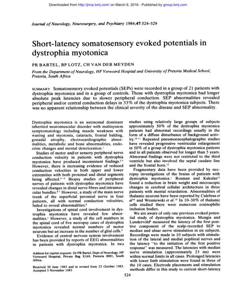
Downloaded from http://jnnp.bmj.com/ on March 6, 2016 - Published by group.bmj.com Journal of Neurology, Neurosurgery, and Psychiatry 1984;47:524-529 Short-latency somatosensory evoked potentials in dystrophia myotonica PR BARTEL, BP LOTZ, CH VAN DER MEYDEN From the Department of Neurology, HF Verwoerd Hospital and University of Pretoria Medical School, Pretoria, South Africa Somatosensory evoked potentials (SEPs) were recorded in a group of 21 patients with dystrophia myotonica and in a group of controls. Those with dystrophia myotonica had longer absolute peak latencies due to slower peripheral conduction. SEP abnormalities revealed peripheral and/or central conduction delays in 33% of the dystrophia myotonica subjects. There was no apparent relationship between the clinical severity of the disease and SEP abnormality. SUMMARY studies using relatively large groups of subjects approximately 50% of the dystrophia myotonica patients had abnormal recordings usually in the form of a diffuse disturbance of background activity.'01' Repeated pneumoencephalographic studies have revealed progressive ventricular enlargement in 50% of a group of dystrophia myotonica patients and in all patients observed for longer than 5 years. Abnormal findings were not restricted to the third ventricle but also involved the septal caudate line and the frontal horn.'2 Fragmentary data have been provided by necropsy investigations of the brains of patients with dystrophia myotonica.' Rosman and Kakulas'3 found a reduction in brain weight and microscopic changes in cerebral cellular architecture in three patients with mental retardation. Abnormalities of thalamic neurons have been reported by Culebras et al'4 and Wesniewski et al.'5 In 10-30% of thalamic cells studied there were numerous eosinophilic inclusion bodies. We are aware of only one previous evoked potential study of dystrophia myotonica. Mongia and Lundervold4 measured the latency of the first positive component of the scalp-recorded SEP to median and ulnar nerve stimulation in six subjects. Recordings were made in 10 subjects with stimulation of the lateral and medial popliteal nerves and the latency "to the initiation of the first positive response" was measured. The latencies with median nerve stimulation (approximately 23 ms) were Address for reprint requests: Dr PR Bartel, Dept of Neurology, HF within normal limits in all cases. Prolonged latencies Verwoerd Hospital, Private Bag X169, Pretoria 0001, South Africa. with lower limb stimulation were found in three of the 10 cases. Electrode placements and stimulation 1983. 23 October form in revised and 1983 Received 30 June methods differ in this study to current short-latency 5 1983 November Accepted 524 Dystrophia myotonica is an autosomal dominant inherited neuromuscular disorder with multisystem symptomatology including muscle weakness with wasting and myotonia, cataracts, frontal balding, gonadal atrophy, electrocardiographic abnormalities, metabolic and bone abnormalities, endocrine changes and mental deterioration.' Studies of motor and/or sensory peripheral nerve conduction velocity in patients with dystrophia myotonica have produced inconsistent findings.2 3 However, there is increasing evidence of reduced conduction velocities in both upper and lower extremities with both proximal and distal segments being affected.3-5 Biopsy studies of peripheral nerves of patients with dystrophia myotonica have revealed changes in distal nerve fibres and intramuscular bundles.6' However, a study of the main nerve trunk of the superficial peroneal nerve in four patients, all with normal conduction velocities, failed to reveal abnormalities.8 Investigations of spinal cord involvement in dystrophia myotonica have revealed few abnormalities.' However, a study of the cell numbers in the spinal cord of five necropsy cases of dystrophia myotonica revealed normal numbers of motor neurons but an increase in the number of glial cells.9 Evidence of central nervous system involvement has been provided by reports of EEG abnormalities in patients with dystrophia myotonica. In two Downloaded from http://jnnp.bmj.com/ on March 6, 2016 - Published by group.bmj.com Short-latency somatosensory evoked potentials in dystrophia myotonica LS SEP techniques. Furthermore, with median nerve stimulation, abnormal interpeak conduction times have been reported for the pa!thway segment Erb's point to cervical spine (N9-N 13) and for thepathway segment cervical spine to cortex (N13-N20) in cases with normal absolute latencies for the first cortical response, that is, peak N20.'617 The purpose of the present study was to assess SEP absolute latencies and interpeak conduction times with both right and left median nerve stimulation in a relatively large group of dystrophia myotonica patients and matched controls. 525 C4'- Fpz N20 Il25uV Cv2-Fpz N13 Patients and methods The dystrophia myotonica group consisted of 21 subjects. There were 13 females and eight males with a mean age of 34.4 years (SD 11*79, range 13-54 years). Each of the dystrophia myotonica subjects fitted into a known family tree with the disease and only those subjects who showed at least percussion myotonica of the thenar muscles were included in the study. Each subject was clinically examined by one of us (BL) and there was no incidence of sensory symptoms or signs. Absent deep tendon arm reflexes were found in 10 of our patients, five of whom had intact deep tendon reflexes in the legs. The control group consisted of 21 age- and sex-matched subjects. Informed consent was obtained from all subjects prior to testing. A total of 23 dystrophia myotonica patients were tested, but two raised objections during the testing procedure which was then discontinued and these two subjects were excluded from the group. The recordings were made in a sound-attenuated, airconditioned room. Amplification of recorded signals, stimulation, averaging and latency measurements were achieved by means of Nicolet Pathfinder II system. Recording electrodes were silver-plated cup electrodes secured by means of collodion and filled with conductive paste. Electrodes were placed over Erb's point, over the second cervical vertebra and at C3' and C4' (2 cm behind C3 and C4 of the 10-20 system). The reference electrode was placed on the mid-forehead (Fpz). A large metal disc ground electrode was placed on each forearm. Electrode impedances were below 3 kohm. Signals were passed into high-gain differential amplifiers with a bandpass of 30 Hz-1-5 kHz (12 dB/octave roll-off) and with sensitivities individually set for each channel. Automatic artefact rejection was used. Rectangular stimulating pulses with a duration of 0-1 ms and a rate of 5 1/s were applied to stimulating electrodes 9 mm in diameter mounted in a plastic holder 3 cm apart. The cathode was placed proximally to the anode over the median nerve at the wrist. Stimulus intensity was adjusted to produce a consistent moderate twitch of the thenar muscle. The right median nerve was stimulated first. The stimulus was well-tolerated by all control subjects. However, two dystrophia myotonica patients objected to the stimulation and had to be excluded from the group while others had to be encouraged to complete the recording. Subjects were supine on a hospital bed and were encouraged to minimise muscle activity during recordings and Erb-Fpz I250uV N9 L I 7ms Fig 1 Somatosensory evoked potentials with left-sided stimulation in a dystrophia myotonica patient. Electrodes were located over Erb's point, over the second cervical vertebra (CV2) and over the somatosensory cortex (C4'). The latency ofjy9 is delayed (12.88 ms) but the interpeak latencies N9-N13 (3.22 ms) and N13-N20 (5.18 ms) are within normal limits indicating peripheral but not central conduction delay. they were instructed to maintain their heads in an upright position (900 to the bed). For each recording 500 stimuli were averaged. The analysis time was 35 ms and the sampling interval 0-07 ms. Recordings were replicated at least once to ascertain stability of the responses. In all recordings and tracings a relative negativity at grid 1 of the amplifier produced a downwards deflection. Absolute latencies were measured from stimulus onset to the major negative peak at Erb's point (N9), at the second cervical vertebra (N13) and at the contralateral somatosensory cortex (N20) as shown in fig 1. Measurements were made by means of interactive cursors and software-based routines on the displascreen. Interpeak conduction times are designated N9-N13 and N13-N20. The nomenclature is that of Jones.'8 Peak N9 is considered to originate in the region of the brachial plexus, N13 in the spinal grey matter or dorsal column nuclei and N20 probably represents the first response of the somatosensory cortex contralateral to the side of stimulation." '8 The influence of skin temperature on peripheral nerve conduction velocity has been elucidated'9 and the effects on SEP absolute latencies should be taken into account.20 While facilities were not available to maintain a specific skin temperature in the present study, the skin temperature of the forearm of each subject was measured. Jones'8 showed that absolute SEP latencies are directly related to arm length. The arm lengths of each subject were measured from the distal flexor fold of the wrist to the head of the humerus in the axilla with the arm fully stretched. Downloaded from http://jnnp.bmj.com/ on March 6, 2016 - Published by group.bmj.com Bartel, Lotz, Van der Meyden 526 HF b C4 -Fpz C3-Fpz- Cv2-Fpz N3 Cv2-Fpz. I125UV C4-Fpz 125 uV i C3 -Fpz 720 Cv2-Fpz Cv2-Fpz I I I 1 -L I 1, I I I 7ms Fig 2 Unilaterally abnormal N13-N20 interpeak latency of the somatosensory evoked potential in a dystrophia myotonica patient. Electrodes were located over the second cervical vertebra (CV2), over the somatosensory cortex (C4' and C3') and at a mid-pj rontal position (FpZ). The lower 2 tracings show a N13-N20 interpeak latency of 7-44 ms with lIf-sided stimulation and the upper 2 tracings show an N13-N20 interpeak latency of 5.40 ms with right-sided stimulation. The right-left interpeak latency difference of 2-04 ms is abnormal. Fig 3 Unilaterally abnormal N13-N20 interpeak latency of the somatosensory evoked potential in a dystrophia myotonica patient. Electrodes were located over the second cervical vertebra (CV2), over the somatosensory cortex (C4' and C3') and at a mid-p frontal position (FpZ). The lower 2 tracings show a N13-N20 interpeak latency of 7.84 ms with right-sided stimulation and the upper 2 tracings show a N13-N20 interpeak latency of 574 ms with left-sided stimulation. The right-left interpeak latency difference of 2 10 ms is abnormal. Group mean scores for absolute latencies and interpeak latencies were compared using t tests. Individual scores were regarded as abnormal if an interpeak latency score and/or the right-left differences in interpeak latencies exceeded the control group mean plus 3 SD. Absent N9, N13 or N20 peaks were also regarded as abnormal.20 Each subject was classified according to the clinical severity of the disease as follows: 1 = minimally affected with subject showing only myotonia upon thenar percussion; 2 = moderately affected with the presence of muscular weakness, but without producing functional impairment; 3 = severely affected with muscle weakness leading to some degree of functional impairment and in some cases resulting in the subject being bound to a wheel-chair; 4 = bedridden. INTERPEAK LATENCIES Results ABSOLUTE PEAK LATENCIES The dystrophia myotonica group mean values for N9 and N13 for right median nerve stimulation and N9, N13 and N20 for left median nerve stimulation were statistically significantly longer than control group means (table 1). The two groups' means for interpeak latencies and for right-left interpeak latency differences were not statistically significantly different (table 1). ABNORMAL FINDINGS In the dystrophia myotonica group seven subjects (33%) had one or more abnormal findings. There were 12 such findings in these subjects. The criteria used and the type of abnormality found are shown in table 2. Of these seven subjects, six had abnormal interpeak latencies: four had a single abnormal value with right or left stimulation; one had abnormal conduction times for the same interpeak latency (N-N13) with both right and left stimulation; one had two abnormal conduction times involving different interpeak latencies (N13-N20, wrist-N9), the former with right stimulation and the latter with left stimulation. Abnormal right-left differences in interpeak latencies were found for two of the three subjects with abnormal N13-N20 conduction times. Absent responses (N13) were found in one subject with both right and left stimulation, precluding the Downloaded from http://jnnp.bmj.com/ on March 6, 2016 - Published by group.bmj.com Short-latency somatosensory evoked potentials in dystrophia myotonica 527 Table 1 Absolute peak latencies, interpeak latencies for right (R) and left (L) median nerve stimulation, interpeak right-left (R-L) difference scores, and T-test results SEP peak(s) N29 R NL3 R N20 R N9 L N13 L N2 L Dystrophia myotonica group Control group Mean SD Mean SD 10-704 14-143 19-580 10-591 14-059 0-907 1-186 1-529 0-983 1-169 1-380 0-688 0-715 0-609 0-815 0-398 0-658 10-039 13-170 18-913 9-855 13-079 18-698 3-131 5-747 0-698 0-982 1-012 0-730 2-66* 2-90t 1-67 1-008 2-91t 2-18* 1-55 1-32 1-16 0-85 0-78 0-41 19-525 3-421 N29-N1 R N13-N20 R N9-N13 L N13-N20 L 5-511 3-425 5-449 0-004 0062 Ng-Ni3 R-L N13-N20 R-L 3-230 5-619 0-100 0-128 t 1-051 0-509 0-398 0-470 0-423 0-395 0-322 2.75t *p = 0-05 tp= 0-01 Table 2 Criteria for abnormal scores (control group means + 3 SD) and number and percentage of dystrophia myotonica subjects exceeding criteria. W = wrist and other abbreviations are the same as in table I Criteria Control group No. of mean (rn) + 3 SD subjects W-N9R N9-N13 R N13-N20 R 12-40 4-66 6-94 12-08 4-64 6-89 1-34 1-28 1-09 W-N9L N_-N1J L N13-N20 L W-N9 R-L W-MNT R-L N13-N2QR-L Absent N9 Absent N13 Absent N20 0 2 (10%) 1 (5%) 2 (10%) 1 (5%) 2 (10%) 0 0 2 (10%) 0 1 (5%) 0 Table 3 Distribution ofabnormal findings and clinical ratings in dystrophia myotonica group. Abbreviations as in table I Subject Age (yr) Sex Abnormality Clinical rating CO MP 46 20 F F N-N13 R 4 1 FP 54 M WbN2..L N13.N20 R 2 LS HF 22 19 F M MJ IE 34 36 F F N9-N13 R N9-N13 L N13-N20 R-L W-N9 L N13-N20 L NfL-R R-L N13-N20 L Absent N13 R Absent N13 L CLINICAL RATINGS The criteria for the 4-point clinical rating scale have been described previously. The 21 dystrophia myotonica subjects were classified as follows: 1-7, 2-10, 3-2, 4-2. The clinical ratings of subjects with SEP abnormalities are shown in table 3. SKIN TEMPERATURE The mean skin temperature for the dystrophia myotonica group was 33-62°C (SD = 1-61, range = 30°-35°) and the mean for the control group was 33 40°C (SD = 1-49, range = 30°-35°). The means did not differ significantly (t = 0-46, p = 34.7). ARM LENGTHS The mean right arm length for the dystrophia myotonica group was 50-48 cm (SD = 2.56) and for the control group 51*57 cm (SD = 3.58). The means did not differ statistically significantly (t = 1 13, p = 26-20). Left arm mean length was 50-00 cm (SD = 2.72) for the dystrophia myotonica group and 51*13 cm (SD = 3.80) for the control group. The means were not significantly different (t = 1-11, p = 27.40). Discussion 2 2 2 2 calculation of the N9-N13 and N13-N20 interpeak latencies (table 3). Abnormal findings occurred in five females (38%) and in two males (25%). The age-range of subjects with abnormalities varied widely (19-54 years). No abnormal interpeak latency values or absent components were encountered in the control group. Peripheral sensory conduction delays in the median nerves of the dystrophia myotonica group are revealed by the prolonged Erb's point responses (N9). The inter-group N9 mean differences of approximately 0-8 ms are further reflected in the same magnitude in the longer approximately N13 and N20 mean latencies for the dystrophia myotonica group. These findings cannot be accounted for by differences between the groups in arm lengths or in skin temperature at the time of testing. These results are in accordance with previous findings of prolonged peripheral sensory nerve Downloaded from http://jnnp.bmj.com/ on March 6, 2016 - Published by group.bmj.com 528 conduction velocity in patients with dystrophia myotonica.2'23 Glucose intolerance was not controlled for in the present study as a previous investigation has shown this not to be a significant factor in peripheral nerve conduction delays in dystrophia myotonica.23 Intergroup comparisons of interpeak latencies failed to reveal_significant differences in conduction times for N9-N13 and N13-N20. However, 33% (7/21) of the dystrophia myotonica group had at least one SEP abnormality. There appeared to be no clear pattern in these findings with evidence of peripheral (wrist-N9) and/or proximal (N-N13) and/or central (ND13-N20) segments of the somatosensory pathway being involved. Unilateral abnormalities (with either right- or left-sided stimulation) predominated with only one subject showing bilateral abnormalities (with both right- and leftsided stimulation) for the same interpeak-latency (N-N 13). Abnormal right-left difference scores were encountered in two of the three subjects with unilaterally abnormal central conduction times. With the exclusion of the abnormal peripheral conduction times, all the other forms of SEP abnormalities found in this study have been reported in patients with demyelinating disorders of the central nervous system.24 Delays in central conduction along the somatosensory pathway is not an altogether surpris- ing finding in the light of previous reports of EEG and pneumoencephalographic changes in dystrophia myotonica.'0-'2 Furthermore, changes in thalamic neurons in patients with this disease may directly impinge upon the somatosensory pathway.'4 i5 Mongia and Lundervold4 reported abnormal SEP onset times with lower limb stimulation in the pres- of normal antidromic peripheral sensory conduction velocities. The present findings confirm this earlier indication of possible spinal and/or central conduction delays in dystrophia myotonica. Furthermore, Mongia and Lundervold4 found abnormal SEP onset latencies in almost every case showing abnormal peripheral conduction velocities, as would be expected using this particular SEP lateny measure. We were able to show normal N9-N13 and N13-N20 conduction times in subjects with abnormal peripheral (wrist-N9) conduction times. Using the control group's means for N20 + 3 SDs as criteria, three of our dystrophia myotonica patients had abnormal values, that is, values exceeding 21-95 ms and 21-85 ms for right and left stimulation, respectively. Using 23 ms as a criterion Mongia and Lundervold4 reported no abnormal values in their six subjects. Differences in technique have already been alluded to and may have influenced these findings. Bartel, Lotz, Van der Meyden We failed to show a clear relationship between the incidence of SEP abnormalities and the clinical severity of dystrophia myotonica. Of the two most severely affected subjects (rating of 4) one showed a single SEP abnormality while the two severely affected subjects (rating of 3) had normal SEPs. Most of the subjects (71%) with abnormal SEPs were moderately affected (rating of 2) by the disease. Of the least affected subjects only one of the seven had SEP abnormalities. These findings are in accordance with those of Panayiotopoulos and Scarpalezos25 and Olson23 who reported a dissociation of myopathic and neuropathic changes in dystrophia myotonica. SEP abnormalities in the absence of clinical sensory symptoms or signs have been reported previously in another disease.2629 Swash30 reported abnormalities in the morphology of the fusimotor and neural elements of muscle spindles in patients with dystrophia myotonica. Burke3'32 showed that SEP conduction velocities with stimulation of a mixed peripheral nerve are largely dependent on muscle afferent projections. These findings raise the possibility that reduced SEP conduction times, especially in distal pathway segments, as found in some patients in the present study, may be due to abnormal muscle afferents without involvement of cutaneous fibres. Further investigation is necessary to resolve this issue. In conclusion, we have been able to demonstrate subclinical SEP abnormalities in 33% of a group of dystrophia myotonica patients. Peripheral (wristN9) and/or proximal (N9-N13) and/or central (N13-N20) conduction delays or absent responses occurred in various combinations with unilateral or bilateral involvement, independent of the clinical severity of the disease. ence References 'Harper PS. Myotonic dystrophy. Philadelphia: Saunders, 1979; 139-49. 2 Aminoff MJ, Layzer RB, Satya Murti S et al. The declining electrical response of muscle to repetitive nerve stimulation in myotonia. Neurology (Minneap) 1977;27:812-6. Panayiotopoulos CP, Scarpalezos S. Dystrophia myotonica-Peripheral nerve involvement and pathogenetic implications.J Neurol Sci 1976;27: 1-16. 4 Mongia SK, Lundervold A. Electrophysiological abnormalities in cases of dystrophia myotonica. Eur Neurol 1975;13:360-76. Ballantyne JP, Hansen S. Computer method for the analysis of evoked motor unit potentials. 2. Duchenne, limb-girdle, facioscapulohumeral and myotonic muscular dystrophies. J Neurol Neurosurg Psychiatry 1975;38:417-28. 6 Coers C, Woolf AL. The Innervation of Muscle. A Biopsy Study. Oxford: Blackwell, 1959:107-12. Downloaded from http://jnnp.bmj.com/ on March 6, 2016 - Published by group.bmj.com Short-latency somatosensory evoked poteniials in dystrophia myotonica MacDermot V. The histology of the neuromuscular junction in dystrophia myotonica. Brain 1961; 84:75-84. Pollock M, Dyck PJ. Peripheral nerve morphometry in myotonic dystrophy. Arch Neurol 1976;33:33-9. 9 Walton JN, Irving D, Tomlinson BE. Spinal cord limb motor neurons in dystrophia myotonica. J Neurol Sci 1977;34: 199-211. Lundervold A, Refsum S, Jacobsen W. The EEG in dystrophia myotonica. Eur Neurol 1969;2:279-84. "Beijersbergen RSHM, Kemp A, Storm van Leeuwen W. EEG observations in dystrophia myotonica (Curschmann-Steinert). Electroencephalogr Clin Neurophysiol 1980;49: 143-51. 12 Refsum S, Lonnum A, Sjaastad 0, et al. Dystrophia myotonica-Repeated pneumoencephalographic studies in ten patients. Neurology (Minneap) 1967;17: 345-8. '3 Rosman NP, Kakulas BA. Mental deficiency associated with muscular dystrophy. A neuropathological study. Brain 1966;89:769-87. 4 Culebras A, Feldman RG, Merk FB. Cytoplasmic inclusion bodies within neurons of the thalamus in myotonic dystrophy. J Neurol Sci 1973; 19:319-29. Wisniewski HM, Berry K, Spiro AJ. Ultrastructure of thalamic neuronal inclusions in myotonic dystrophy. J Neurol Sci 1975;24:321-9. 6 Ganes T. A study of peripheral, cervical and cortical evoked potentials and afferent conduction times in the somatosensory pathway. Electroencephalogr Clin Neurophysiol 1980;49:446-51. 17 Hume AL, Cant BR. Conduction time in central somatosensory pathways in man. Electroencephalogr Clin Neurophysiol 1978;45:361-75. 18 Jones SJ. Short latency potentials recorded from the neck and scalp following median nerve stimulation in man. Electroencephalogr Clin Neurophysiol 1977;43:853-63. 9Lowitsch K, Hopf HC, Galland J. Changes of sensory conduction velocity and refractory periods with decreasing tissue temperature in man. J Neurol 1977;216: 181-8. 20 Desmedt JE, Brunko E. Functional organisation of farfield and cortical components of somatosensory evoked potentials in normal adults. In: Desmedt JE, 529 ed. Progress in Clinical Neurophysiology, volume 7. Basel: Karger, 1980:27-50. 21 Cassia MR, Negri S, Parvis VP. Myotonic dystrophy with neural involvement. J Neurol Sci 1972;16: 253-69. 22 Borenstein S, Noel P, Jacquy J, et al. Myotonic dystrophy with nerve hypertrophy. Report of a case with electrophysiological and ultrastructural study of the sural nerve. J Neurol Sci 1977;34:87-99. 23 Olson D, Jou M-F, Quast JE et al. Peripheral neuropathy in myotonic dystrophy. Relation to glucose intolerance. Arch Neurol 1978;35:741-5. 24 Eisen A, Odusote K. Central and peripheral conduction times in multiple sclerosis. Electroencephalogr Clin Neurophysiol 1980;48: 253-65. 2S Panayiotopoulos CP, Scarpalezos S. Dystrophia Myotonica-A model of combined neural and myopathic muscle atrophy. J Neurol Sci 1977;31:261-8. 26 Mastaglia FL, Black JL, Cala LA, et al. Evoked potentials, saccadic velocities, and computerised tomography in diagnosis of multiple sclerosis. Br Med J 1977;1: 1315-7. 27 Small DG, Matthews WB, Small M. The cervical somatosensory evoked potential (SEP) in the diagnosis of multiple sclerosis. J Neurol Sci 1978;35:211-24. 28 Trojaborg W, Petersen E. Visual and somatosensory evoked cortical potentials in multiple sclerosis. J Neurol Neurosurg Psychiatry 1979;42: 323-30. 29 Kjaer M. The value of brain stem auditory, visual and somatosensory evoked potentials and blink reflexes in the diagnosis of multiple sclerosis. Acta Neurol Scand 1980;62:220-36. 30 Swash M. The morphology and innervation of the muscle spindle in dystrophia myotonica. Brain 1972;95:357-68. 31 Burke D, Skuse NF, Lethlean AK. Cutaneous and muscle afferent components of the cerebral potential evoked by electrical stimulation of human peripheral nerves. Electroencephalogr Clin Neurophysiol 1981;51:579-88. 32 Burke D, Gandevia SC, McKeon B, et al. Interactions between cutaneous and muscle afferent projections to cerebral cortex in man. Electronencephalogr Clin Neurophysiol 1982;53:349-60. Downloaded from http://jnnp.bmj.com/ on March 6, 2016 - Published by group.bmj.com Short-latency somatosensory evoked potentials in dystrophia myotonica. P R Bartel, B P Lotz and C H Van der Meyden J Neurol Neurosurg Psychiatry 1984 47: 524-529 doi: 10.1136/jnnp.47.5.524 Updated information and services can be found at: http://jnnp.bmj.com/content/47/5/524 These include: Email alerting service Receive free email alerts when new articles cite this article. Sign up in the box at the top right corner of the online article. Notes To request permissions go to: http://group.bmj.com/group/rights-licensing/permissions To order reprints go to: http://journals.bmj.com/cgi/reprintform To subscribe to BMJ go to: http://group.bmj.com/subscribe/

![Applied Heat Transfer [Opens in New Window]](http://s3.studylib.net/store/data/008526779_1-b12564ed87263f3384d65f395321d919-300x300.png)
