Sakoolnamarks P 116-122
advertisement
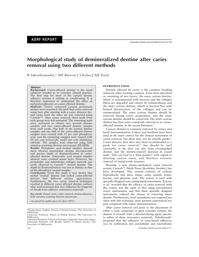
ADRF REPORT Australian Dental Journal 2002;47:(2):116-122 Morphological study of demineralized dentine after caries removal using two different methods R Sakoolnamarka,* MF Burrow,† S Kubo,‡ MJ Tyas§ Abstract Background: Caries-affected dentine is the usual substrate bonded to in everyday clinical practice. The first step for most of the current dentine adhesive systems is etching or conditioning. It is therefore important to understand the effect of etchant/conditioner on caries-affected dentine. Methods: Twelve extracted carious permanent molars were examined. Six teeth had caries removed using burs after staining with a caries detector dye, and caries from the other six was removed using CarisolvTM. After caries removal, three teeth from each group were left untreated. The remaining teeth were sectioned to obtain two normal dentine samples and two caries-affected dentine samples from each tooth. One half of the normal dentine samples and one half of the caries-affected dentine samples were treated with 35 per cent phosphoric acid, and the remaining samples were treated with 20 per cent polyacrylic acid/3 per cent aluminium chloride. The samples were observed using field emission scanning electron microscopy (FE-SEM). Results: Phosphoric acid treated samples showed more obvious intertubular dentine microporosity and greater depth of demineralization in cariesaffected dentine. Polyacrylic acid treated samples showed some residual smear layer. However, the peritubular and intertubular collagen network was easily observed in CarisolvTM treated dentine. The depth of demineralization was not as distinct as that of the phosphoric treated samples. Conclusions: From this study it was shown that etched ‘normal’ dentine and etched caries-affected dentine had different surface appearances. Furthermore, the two caries removal techniques resulted in different caries-affected dentine surfaces after acid treatment that may influence the longevity of bonds from adhesive restorative materials. Key words: Dental caries, caries removal, demineralization, field-emission scanning electron microscope. (Accepted for publication 26 April 2001.) *Postgraduate Student, School of Dental Science, The University of Melbourne. †Associate Professor, School of Dental Science, The University of Melbourne. ‡Assistant Professor, School of Dentistry, Nagasaki University, Japan. §Associate Professor and Reader, School of Dental Science, The University of Melbourne. 116 INTRODUCTION Dentine affected by caries is the common bonding substrate when treating a patient. It has been described as consisting of two layers;1 the outer carious dentine, which is contaminated with bacteria and the collagen fibres are degraded and cannot be remineralized, and the inner carious dentine, which is bacteria-free with limited denaturation of the collagen and can be remineralized. The outer carious dentine should be removed during cavity preparation, and the inner carious dentine should be conserved. The inner carious dentine has been more commonly referred to as ‘cariesaffected’ dentine in the recent literature.2,3 Carious dentine is routinely removed by rotary and hand instrumentation. Colour and hardness have been used as the main criteria for the clinical assessment of caries removal, but these may not be reliable guides.4 Caries detector dyes have also been recommended as a guide for caries removal,2,3 but should be used cautiously, as the dyes can also stain circumpulpal dentine and the dentino-enamel junction in sound teeth.5 This can lead to a ‘false positive’ with regard to detecting carious tissue, and therefore excessive removal of sound tooth structure. Recently, a new chemo-mechanical caries removal system, CarisolvTM (Medi-Team, Sävedalen, Sweden), has been developed. This system consists of sodium hypochlorite and three amino acids, namely lysine, leucine, and glutamic acid. The system is used with specially designed non-cutting hand instruments. Ericson et al.6 revealed that the efficacy of caries removal with this system was the same as that using conventional rotary instruments, and that these specially designed instruments reduced the chance of removing intact dentine. Furthermore, local anaesthesia was required less often with this technique, since patients experienced less pain compared with rotary instrumentation. After caries removal and prior to the placement of either a resin composite or glass ionomer cement, the dentine is treated with a demineralizing agent specific to either of these restorative materials. Nakabayashi7 proposed that the adhesion of resin-based adhesives was by infiltration into peritubular and intertubular Australian Dental Journal 2002;47:2. Fig 1. Scanning electron microscopy micrograph of dentine after caries removal using rotary instrumentation and caries detector dye: a) the smeared surface is characteristic of cut dentine; b) fractured surface showing the remaining dentine structure with mineralized collagen fibres. PT, peritubular dentine; IT, intertubular dentine. dentine of which the collagen network had been exposed by demineralization, to form the ‘hybrid layer’. Nakajima et al.8 revealed that moist bonding to normal dentine produced no significant difference in bond strength, compared with that to caries-affected dentine when Scotchbond Multi-Purpose Plus was used with 35 per cent phosphoric acid. The degree of demineralization by a demineralizing agent may affect the adhesion of ‘single-bottle’ dentine adhesives to caries-affected dentine, e.g., Nakajima et al.9 reported that bond strengths of those adhesives to such dentine were different when using different concentrations of phosphoric acid. However, there was no effect on bond strengths to normal dentine. The purpose of this study was to examine the surface morphology of caries-affected dentine after it had been exposed using either rotary instrumentation in conjunction with caries detector dye or a chemomechanical caries removal system (CarisolvTM), and then conditioned with one of two different demineralizing agents, namely phosphoric acid and polyacrylic acid. M AT E R I A L S A N D M E T H O D S Twelve extracted carious human permanent molars, which had been stored at 4ºC in normal saline containing thymol, were used within three months following extraction. Each tooth had occlusal caries to a depth of approximately 1-2mm below the central fissure, as assessed with a probe by one of the investigators (RS). The occlusal enamel was removed using a slow-speed diamond saw under copious water spray. Six teeth had the caries removed using slow-speed round steel burs (ISO #012; ELA, Engelskirchen, Germany) after staining with a caries detector dye (Caries Detector (Batch No 0250D); Kuraray Co, Osaka, Japan) until the dentine was no longer stained by the dye and was firm to probing with a blunt dental explorer. The caries from the other six teeth was removed using CarisolvTM gel (Batch No 10122; Medi-Team, Sävedalen, Sweden) according to the Australian Dental Journal 2002;47:2. manufacturer’s instructions. The gel was applied to the lesion for 30 seconds, and the instruments supplied were used to remove the softened carious dentine. More gel was applied and caries removal continued until the gel was no longer cloudy, and the surface felt hard to a blunt dental probe. After caries removal, three teeth from each group were investigated without acid treatment for comparison purposes. The remaining teeth were lapped down using 600-grit SiC paper until the normal dentine surrounded the cavity at almost the same level as the exposed cavity floor, in order to act as a control surface. The teeth were sectioned to obtain four samples from each tooth; two of normal dentine and two of caries-affected dentine. A shallow groove was placed in each sample, on the opposite side to the surface under study, using a diamond disc (ISO #335220-74; Dentsply, Milford, Delaware, USA) in a slowspeed handpiece. One half of the normal dentine samples and one half of the caries-affected dentine samples were treated with 35 per cent phosphoric acid (Ultraetch (Batch No 61624DV); Ultradent Products Inc., South Jordan, Utah, USA) for 15 seconds and washed for 20 seconds. The remaining samples were treated with 20 per cent polyacrylic acid/3 per cent aluminium chloride solution (Cavity Conditioner (Batch No 270571); GC International, Tokyo, Japan) for 10 seconds and washed for 20 seconds. Samples were fixed in 10 per cent phosphate buffered formalin for 24 hours, rinsed with distilled water three times for 15 minutes each time and dehydrated in increasing concentrations of ethanol and water up to 90 per cent ethanol, and placed in 100 per cent ethanol three times for 15 minutes each time. The specimens were placed in a critical point drier (Samdri PVT-3; Tousimis Research Corp., Rockville, Maryland, USA) until all residual moisture had been removed, and fractured along the prepared grooves using pliers. The conditioned surfaces and the fractured surfaces were gold sputter-coated and observed using a field-emission scanning electron microscope (XL30 FEG; Philips, Eindhoven, The Netherlands). 117 Fig 2. Scanning electron microscopy micrograph of dentine after caries removal using CarisolvTM: a) dentine with irregular, rough and porous surface is exposed after CarisolvTM treatment; b) fractured surface showing mineralized collagen fibres with a few porosities in intertubular dentine. PT, peritubular dentine; IT, intertubular dentine. R E S U LT S Representative micrographs are displayed in Fig 1-5. Differences were noticed between the dentine surfaces obtained using the two different caries removal systems. A smear layer was apparent on the teeth prepared with rotary instrumentation (Fig 1a), whereas the CarisolvTM treated surfaces (Fig 2a) were irregular, porous and did not exhibit a smear layer. The fractured surface of the specimens from the bur-treated group (Fig 1b) exhibited intact peritubular dentine, which was similar to the peritubular dentine of CarisolvTM treated samples (Fig 2b). However, more porosities were Fig 3. Scanning electron microscopy micrograph of normal dentine: a) dentine surface treated with 35 per cent phosphoric acid showing patent dentinal tubules with peritubular collagen fibre network and intertubular microporosity; b) fractured surface showing regular oriented exposed collagen fibres (arrows) with the depth of demineralization ranging from 1.5-2µm; c) dentine surface treated with 20 per cent polyacrylic acid/Al2Cl3 showing openings of the dentinal tubules with some residual smear layer and fibrous structure inside the tubules (arrows); d) fractured surface showing collagen fibres with some mineral remaining. 118 Australian Dental Journal 2002;47:2. Fig 4. Scanning electron microscopy micrograph of dentine after caries removal using round steel burs and caries detector dye: a) the dentine surface with patent dentinal tubules and intertubular microporosity after treatment with 35 per cent phosphoric acid, some fibrous structure remained in the dentinal tubules; b) lateral view of the randomly oriented exposed collagen fibres (arrows), a cuff of peritubular dentine in the dentinal tubules was also noted, the depth of demineralization was ranging from 3.5-4.5µm; c) the dentine surface showing partial opening of dentinal tubules with the residual smear layer after being treated with 20 per cent polyacrylic acid/Al2Cl3 and; d) fractured surface showing collagen fibres with calcific deposits remaining. evident in the intertubular dentine of the CarisolvTM treated teeth. The normal dentine surface and the fractured longitudinal surface, both after treatment with phosphoric acid are shown in Fig 3a and 3b respectively. Patent dentinal tubules and the peritubular collagen network were clearly visible. The fractured view clearly showed exposed collagen fibres with the depth of demineralization ranging from 1.5-2µm. Comparable views after polyacrylic acid/aluminium chloride treatment are shown in Fig 3c and 3d. Some residual smear layer and a fibrous structure inside the tubules was revealed. The fractured view showed collagen fibres with some calcific deposits remaining. The dentine after caries removal using rotary instrumentation/caries detector dye is shown in Fig 4. After treatment with phosphoric acid (Fig 4a), the dentine surface showed patent dentinal tubules and peritubular collagen network. Some fibrous structures were found in the dentinal tubules and the circular orientation of the peritubular collagen network was revealed. The intertubular microporosity appeared to be more obvious than in normal dentine (Fig 3a). The fractured surface (Fig 4b) showed an exposed collagen network as found in normal dentine (Fig 3b), but the depth of demineralization appeared to be greater in Australian Dental Journal 2002;47:2. caries-affected dentine (approximately 3.5-4.5µm). In addition, the orientation of the exposed collagen fibres appeared to be irregular, in contrast to the regular orientation of the fibres in normal dentine. A cuff of peritubular dentine was also observed. The dentine surface after being treated with polyacrylic acid/aluminium chloride (Fig 4c) displayed a partial opening of dentinal tubules with a residual smear layer remaining. The lateral view showed a collagen fibre network with a considerable amount of residual mineral deposit (Fig 4d). The images of dentine after caries removal using CarisolvTM are shown in Fig 5. The dentine surface after treatment with phosphoric acid (Fig 5a) showed patent dentinal tubules and an exposed peritubular collagen network, as in the normal dentine sample. The exposed superficial intertubular collagen network is obviously different from that of the normal dentine sample. The distinctly reticular collagen fibre network with a random orientation was noted in the CarisolvTM samples. The fractured view (Fig 5b) showed exposed collagen fibres. The depth of demineralization ranges from 7-8µm, which appeared to be greater than in the dentine sample where burs and a caries detector were used. After treatment with polyacrylic acid/aluminium 119 Fig 5. Scanning electron microscopy micrograph of dentine after caries removal using CarisolvTM: a) dentine surface treated with 35 per cent phosphoric acid showing patent dentinal tubules with a clearly exposed peritubular and intertubular collagen network; b) lateral view of the exposed collagen fibres (arrows) with the depth of demineralization approximately 7-8µm; c) dentine surface treated with 20 per cent polycrylic acid/Al2Cl3 showing openings of dentinal tubules with distinguishable peritubular and intertubular collagen network; d) fractured surface showing collagen fibres with some calcific deposits remaining. chloride, the dentine surface showed the opening of dentinal tubules with a peritubular and intertubular collagen network (Fig 5c). The collagen fibres appeared to be more distinguishable in the CarisolvTM treated samples than in both the normal dentine (Fig 3c) and in the caries detector dye treated samples (Fig 4c). The fractured view showed collagen fibres with some calcific deposits remaining (Fig 5d). The depth of demineralization of the samples treated with polyacrylic acid/aluminium chloride was not as evident as that of the samples treated with phosphoric acid. DISCUSSION The adhesion between dentine and resin-based adhesives is due to micro-mechanical interlocking of resin into collagen exposed by demineralizing the dentine surface with phosphoric acid.10 Erickson11 described the typical action of dentine demineralizing agent on ‘normal’ dentine surfaces, which resulted in an excellent opportunity for micro-mechanical retention of resin and in high bond strengths. In the present study, the normal dentine surface after demineralization with 35 per cent phosphoric acid showed patent dentinal tubules and a peritubular collagen network, and a depth of demineralization ranging from 1.5-2µm, which is in agreement with the previous study of Perdigão et al.12 120 The morphology of demineralized ‘normal’ dentine differed from demineralized caries-affected dentine, which may be due to the surface which is left after caries removal being different in ultrastructure and character from normal dentine. Fusayama1 revealed that the peritubular and intertubular apatite crystals of caries-affected dentine were smaller in size and less numerous than normal dentine. Ogawa et al.13 also reported that the hardness of the caries-affected dentine was less than that of normal dentine. The morphology of etched dentine obtained after rotary instrumentation and caries detector dye use differed from that of the CarisolvTM treated sample. This is probably the result of the caries removal technique and the high pH of 11 of CarisolvTM.14 It is believed that the non-cutting hand instruments used with this technique were more conservative of dentine or that the sodium hypochlorite in the gel caused some change to the dentine, especially the collagen, thus leading to the different appearances. Since preparing caries-affected cavities using CarisolvTM gel is less destructive of sound tooth structure, bonded restorations might be suitable for such cavities as mechanical retention is not required, hence preserving more tooth structure. The depth of demineralization by phosphoric acid appeared to be greater in caries-affected dentine. This is Australian Dental Journal 2002;47:2. presumably because the caries-affected dentine is already partially demineralized and more porous than normal dentine,15 which may facilitate demineralization. The greater depth of demineralization may produce a thicker hybrid layer, as Nakajima et al.16 demonstrated when bonding to caries-affected dentine, but they also reported a poor correlation between bond strength of resin-based adhesive and hybrid layer thickness. The increased thickness of demineralization may affect the success of resin adhesive systems, since the deepest part of the demineralized dentine may be left unencapsulated by the resin that forms the hybrid layer.17 A study of nanoleakage within the hybrid layer by Sano et al.18 revealed that leakage occurred around collagen fibres that were not fully resin-infiltrated. Furthermore, different leakage patterns were revealed by Li et al.19 when using different resin-adhesive systems. Thus, more consideration is needed with regard to selection and timing of demineralizing agents for caries-affected dentine, in order to ensure a reliable bond of resinbased adhesives to such dentine. In the present study, for the surface of normal dentine after demineralization using 20 per cent polyacrylic acid/3 per cent aluminium chloride (Cavity Conditioner), some residual smear layer was evident, which was in consistent with the study of Bloxham et al.20 In their study, 25 per cent polyacrylic acid was applied to the ‘normal’ dentine surface for 15 seconds, which resulted in smear layer remaining in the openings of the dentinal tubules. The extent of dentine demineralization with Cavity Conditioner, (pH 0.97; Tanumiharja et al.21), was less than that of 35 per cent phosphoric acid (pH 0.02; Perdigão et al.12). Peutzfeldt and Asmussen22 revealed that the ability to demineralize the dentine surface was dependent upon factors such as the concentration of the acid, duration of application and the nature of the surface. This explains why Cavity Conditioner, which has a higher pH and shorter treatment time, was associated with less demineralization of the dentine than phosphoric acid. Cavity Conditioner is used as the demineralizing agent for resin-modified glass ionomer cement. This restorative material bonds to dentine by micromechanical interlocking of the polymer to etched dentine23 and also bonds chemically by forming ionic bonds to the mineral content of tooth structure.24 The decrease in calcium content of cariesaffected dentine may affect the chemical bond of glass ionomer cement to such a surface. The use of a caries detector dye as a guide in caries removal may result in excessive removal of sound dentine, since the dye was specific to not only the damaged collagen fibres of the infected dentine, but also stained the demineralized organic material.5 From the results of this study, the chemo-mechanical caries removal system is believed to be a good alternative for caries removal since it is more conservative of sound tooth structure. Moreover, this system appeared to correspond to the current concepts in operative dentistry, which are now focusing on the use of adhesive materials to bond restorations to tooth Australian Dental Journal 2002;47:2. structure in order to be conservative. However, a greater depth of demineralized dentine was exhibited in CarisolvTM treated and acid treated dentine than in acid treated normal dentine. Therefore, etching procedures need to be reviewed to ensure optimum adhesion of resin-based materials to such dentine. CONCLUSION Results from this study showed that etched normal dentine and etched caries-affected dentine revealed different arrangements of the exposed collagen fibres. In addition, caries-affected dentine exposed by the two caries removal techniques demonstrated different fibril networks after acid treatment. Further study is needed, particularly in the area of the resin-dentine interface, when CarisolvTM is used in the caries removal process, to provide additional evidence for the clinical efficacy of this technique. AC K N OW L E D G E M E N T S The research was supported by Australian Dental Research Foundation Inc, St Leonards, NSW 2065, Australia. The assistance of Jocelyn L Carpenter, School of Botany, The University of Melbourne, with SEM imaging is greatly appreciated. The CarisolvTM gel and instruments were kindly provided by Medi-Team, Sävedalen, Sweden. REFERENCES 1. Fusayama T. New concepts in the pathology and treatment of dental caries: A simple pain-free adhesive restorative system by minimal reduction and total etching. Tokyo: Ishiyaku EuroAmerica Inc, 1993:1-21. 2. Harnirattisai C, Inokoshi S, Shimada Y, Hosoda H. Interfacial morphology of an adhesivecomposite resin and etched cariesaffected dentin. Oper Dent 1992;17:222-228. 3. Nakajima M, Ogata M, Okuda M, Tagami J, Sano H, Pashley DH. Bonding to caries-affected dentin using self-etching primers. Am J Dent 1999;12:309-314. 4. Fusayama T, Okuse K and Hosoda H. Relationship between hardness, discoloration, and microbial invasion in carious dentin. J Dent Res 1966;45:1033-1046. 5. Yip HK, Stevenson AG and Beeley J. The specificity of caries detector dyes in cavity preparation. Br Dent J 1994;176:417-421. 6. Ericson D, Zimmerman M, Raber H, Gotrick B, Bornstein R, Thorell J. Clinical evaluation of efficacy and safety of a new method for chemo-mechanical removal of caries. A multi-centre study. Caries Res 1999;33:171-177. 7. Nakabayashi N. Bonding of restorative materials to dentine: the present status in Japan. Int Dent J 1985;35:145-154. 8. Nakajima M, Sano H, Zheng L, Tagami J, Pashley DH. Effect of moist vs. dry bonding to normal vs. caries-affected dentin with Scotchbond Multi-Purpose Plus. J Dent Res 1999;78:1298-1303. 9. Nakajima M, Sano H, Urabe I, Tagami J, Pashley DH. Bond strengths of single-bottle dentin adhesives to caries-affected dentin. Oper Dent 2000;25:2-10. 10. Pashley DH, Ciucchi B, Sano H, Horner JA. Permeability of dentin to adhesive agents. Quintessence Int 1993;24:618-631. 11. Erickson RL. Mechanism and clinical implications of bond formation for two dentin bonding agents. Am J Dent 1989;2:117-123. 12. Perdigão J, Lambrechts P, Van Meerbeek B, Tome AR, Vanherle G, Lopes AB. Morphological field emission-SEM study of the effect of six phosphoric acid etching agents on human dentin. Dent Mater 1996;12:262-271. 121 13. Ogawa K, Yamashita Y, Ichijo T, Fusayama T. The ultrastructure and hardness of the transparent layer of human carious dentin. J Dent Res 1983;62:7-10. 21. Tanumuharja M, Burrow MF, Tyas MJ. Microtensile bond strengths of glass ionomer (polyalkenoate) cements to dentine using four conditioners. J Dent 2000;28:361-366. 14. Beeley JA, Yip HK and Stevenson AG. Chemochemical caries removal: a review of the techniques and latest developments. Br Dent J 2000;188:427-430. 22. Peutzfeldt A, Asmussen E. Effect of polyacrylic acid treatment of dentin on adhesion of glass ionomer cement. Acta Odontol Scand 1990;48:337-341. 15. Hamid A, Hume WR. Diffusion of resin monomers through human carious dentin in vitro. Endod Dent Traumatol 1997;13:1-5. 23. Lin A, McIntyre NS and Davidson RD. Studies on the adhesion of glass-ionomer cements to dentin. J Dent Res 1992;71:18361841. 16. Nakajima M, Sano H, Burrow MF, et al. Tensile bond strength and SEM evaluation of caries-affected dentin using dentin adhesives. J Dent Res 1995;74:1679-1688. 24. Mount GJ. Adhesion of glass-ionomer cement in the clinical environment. Oper Dent 1991;16:141-148. 17. Eick JD, Gwinnett AJ, Pashley DH, Robinson SJ. Current concepts on adhesion to dentin. Crit Rev Oral Biol Med 1997;8:306-335. 18. Sano H, Yoshiyama M, Ebisu S, et al. Comparative SEM and TEM observations of nanoleakage within the hybrid layer. Oper Dent 1995;20:160-167. 19. Li H, Burrow MF, Tyas MJ. Nanoleakage patterns of four dentin bonding systems. Dent Mater 2000;16:48-56. 20. Bloxham GP, Dennison JD and Charbeneau GT. A clinical scanning electron microscope study of tooth surface preparation and bonding. Aust Dent J 1990;35:345-351. 122 Address for correspondence/reprints: Dr Michael F Burrow School of Dental Science The University of Melbourne 711 Elizabeth Street Melbourne, Victoria 3000 Email: mfburrow@unimelb.edu.au Australian Dental Journal 2002;47:2.
