Outline A vein is not an artery
advertisement
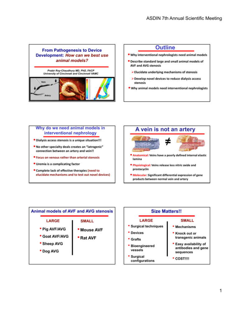
ASDIN 7th Annual Scientific Meeting From Pathogenesis to Device Development: How can we best use animal models? Outline • Why interventional nephrologists need animal models • Describe standard large and small animal models of AVF and AVG stenosis Prabir Roy-Chaudhury MD, PhD, FACP U i University it off Cincinnati Ci i ti and d Cincinnati Ci i ti VAMC ¾Elucidate underlying mechanisms of stenosis ¾Elucidate underlying mechanisms of stenosis ¾Develop novel devices to reduce dialysis access stenosis Vein • Why animal models need interventional nephrologists Artery Why do we need animal models in interventional nephrology A vein is not an artery ≠ • Dialysis access stenosis is a unique situation!!! • No other specialty deals creates an “iatrogenic” connection between an artery and vein!! • Focus on venous rather than arterial stenosis • Uremia is a complicating factor • Complete lack of effective therapies (need to elucidate mechanisms and to test out novel devices) • Anatomical: Veins have a poorly defined internal elastic A i l V i h l d fi d i l l i lamina • Physiological: Veins release less nitric oxide and prostacyclin • Molecular: Significant differential expression of gene products between normal vein and artery Size Matters!! Animal models of AVF and AVG stenosis LARGE • Pig AVF/AVG • Goat AVF/AVG • Sheep AVG • Dog AVG SMALL • Mouse AVF • Rat AVF LARGE SMALL • Surgical techniques • Devices • Grafts • Bioengineered • Mechanisms • Knock out or • Surgical • COST!!!! vessels configurations transgenic animals • Easy availability of antibodies and gene sequences 1 ASDIN 7th Annual Scientific Meeting Development of a pig model of venous neointimal hyperplasia in PTFE dialysis access grafts Specimen Preparation GRAFT VEIN A Suitable vascular tree Response of vessel wall to injury is similar to Man Pig Model (Day 2) Significant venous neointimal hyperplasia at 28 days V A C B Validation Time Points: Days 2, 4, 7, 14, 28 Pig Model (Day 14) Human/Pig Similarities D 28 Human GVA Pig GVA 2 ASDIN 7th Annual Scientific Meeting Placement of Perivascular Paclitaxel Polymers into a Pig Model of Venous Stenosis External radiation attenuates neointimal hyperplasia • • • Single dose of 16 Gy 5 x 6 cm area Within 24 hrs of surgery % Luminal S Stenosis % Luminall Stenosis Venous Neointimal Hyperplasia 60 50.5% 50 * 38.4% 40 30 20 10 0 Controls Radiated Vein: 50.5% vs 38.4% p = 0.021 24% reduction A V Kelly et al., Nephrol Dial Transplant 2006 The pig is ideally suited for devices (Sutureless anastomosis graft) Sutureless Anastomosis Hybrid Graft Converts an end to side anastomosis into an end to end venous anastomosis OptiflowTM is a sutureless anastomotic conduit for AVF creation Conduit Optiflow anastomotic conduit Vein Conduit Artery Flange Flange 3 ASDIN 7th Annual Scientific Meeting Optiflow Angiography AV Fistula Model Development (Dissection) Femoral Vein ??? Femoral Artery Wang et al. NDT 2008 AV Fistula Model Development (Arteriotomy) AV Fistula Model Development (Anastomosis) Wang et al. NDT 2008 AV Fistula Model Development Wang et al. NDT 2008 Neointimal budding in the 7d AVF 7d AVF, PV, SMA x 400 7d AVF, PV, SMA x 100 7d AVF, PV, BrdU x 400 Krishnamoorthy et al. P-PO1054, ASN 2006 Wang et al. NDT 2008 4 ASDIN 7th Annual Scientific Meeting Aggressive stenosis at 42d Alternative Origins for Neointimal Cells: Role of the Adventitia 42d AVF, PV, SMA x 400 BrDU x 400 A BrdU -ve M 2d SMA x 400 N Have we been targeting the wrong part of the vessel wall? Adventitia Media 2d Krishnamoorthy et al., F‐FC157, ASN 2007 Eccentric macrophage infiltration at 2d AV Fistula Model Development (Shear Stress Analysis) Neointima: NO Adventitia: YES Shi et al. Circ. 1996;7:1655 Eccentric neointimal hyperplasia at 42d Eccentric shear stress to macrophages to stenosis Proximal artery Proximal vein Inflammatory (macrophage) response AV anast. Different patterns of shear on opposite walls Chemokines and cytokines Intima‐media thickening Krishnamoorthy et al. Kidney International 2008 5 ASDIN 7th Annual Scientific Meeting The Pig is a good large animal model Can we make a uremic pig?? • Response to injury similar to humans • Animal model of choice for cardiovascular • Yes • Misra et al. use embolization of one research (comparisons) • Better availability of antibodies and gene sequences as compared to dogs and sheep • More aggressive response to injury is an advantage in intervention studies • Lack of thrombosis could be a disadvantage kidney followed by selective renal artery infarction • We have not made pigs uremic • Remember you don’t want them uremic enough to require HD (Dog) Mouse Model of AVF stenosis (14d) Mouse Model Creation b Arteryy Vein Uremic mouse model of AVF stenosis • Cauterize right kidney • Nephrectomy left kidney 2 wks Uremic mice have increased AV fistula stenosis Non-uremic Uremic X 2-3 later • Wait 6 weeks (BUN = 50-70) • Place AVF Neointimal Volume AV anastomosis Avoid morbidity and mortality due to uremia Non-uremic CKD Choi et al. JASN. 2008 6 ASDIN 7th Annual Scientific Meeting The Mouse is a good small animal model • Knock outs • Transgenics Do we really need animal models • Gene sequences • Molecular tools A Mouse is not a Man not a Man and neither is a Pig! Surgery is NOT easy A good human surgeon is not always a good mouse surgeon!! Do we need animal models? Do we need animal models? •Yes for safety •Yes for technique and •Proof of final human efficacy can only come from human clinical trials!! feasibility •Yes for mechanisms •Yes for an efficacy signal Animal models and interventional nephrology: a win-win situation Animal models and interventional nephrology: a win-win situation • Animal models need • Increases innovation • Develop and test out intervention skills • New devices will ??? mean new angiographic appearances • Animal model work gives you a heads up new devices ??? • Intellectual property • Starting a company! • Go from an idea to a product!! 7 ASDIN 7th Annual Scientific Meeting Paclitaxel Polymers Inhibit Neointimal Hyperplasia AXZ-100 Paclitaxel Control Kelly et al., Nephrol Dial Transplant 2006 Periadventitial injection Bullfrog Catheter Injections • Endovascular device such as the “Bullfrog” microinfusion catheter (Mercator-Med) Sheathed needle Tailor therapies to the biological course of vascular stenosis Extruded needle Drug A initially followed by Drug B at 6 monthly intervals Bullfrog Catheter Injections Bullfrog Catheter Injections 8 ASDIN 7th Annual Scientific Meeting PTFE grafts coated with Cu-II coatings generate a significant NO flux POST-IMPLANT Bullfrog Catheter Injections No Generating Control •2 weeks post implant Is there a difference in graft thrombosis? Surgery – 10/3/07 Sacrifice – 10/22/07 CONTROL @ 3 weeks Hemodynamics and Histology V A Surgery – 10/2/07 Sacrifice – 10/22/07 NITRIC OXIDE @ 3 weeks A V AV Fistula Model (CT Angiography) 3D Mesh Framework Proximal Artery AV Anastomosis Proximal Vein 9 ASDIN 7th Annual Scientific Meeting AV Fistula Model (Flow and Pressure Analysis) 3D Wall Shear Stress Profile Proximal Artery AV Anastomosis Proximal Vein Flow Pressure Wall Shear Stress Alterations in Systole and Diastole AV Fistula Model Development (Shear Stress Analysis) Proximal Artery Low Shear Stress AV anastomosis Proximal Vein Krishnamoorthy et al. In Revision Kidney International 2007 AV Fistula Model Development (Shear Stress Analysis) Implant Design Proximal artery Venous Flow Proximal vein Conduit (Vein) Flange (Artery) AV anast. Proximity of high and low shear stress “Heel Flange” “Toe Flange” “Side Flange” Confidential Krishnamoorthy et al. In Revision Kidney Int 2007 10 ASDIN 7th Annual Scientific Meeting Do we really need animal models A Mouse Is not A Man! Do we need animal models •Yes for safety •Yes for technique and feasibility •? for efficacy Optiflow Angiography AV Fistula Model Development (Curved) PA DA PV Wang et al. NDT 2008 OptiflowTM is a sutureless anastomotic conduit for AVF creation Conduit Vein Conduit Artery Flange Flange 11 ASDIN 7th Annual Scientific Meeting OptiFlow Sutureless Anastomotic Device 1. Mobilize Vessels 6. Insert flanges inside arteriotomy 2. Venotomy 5. Arteriotomy 4. Suture the vein and mark arteriotomy 3. Insert device into vein The adventitia is important! 2d AVF, PV, SMA x 200 2d AVF, PV, SMA x 200 2d AVF, PV, BrdU x 200 Normal Pig Vein, SMA x 200 Krishnamoorthy et al. F-PO1054, ASN 2006 Mouse model of AVF stenosis • End of jugular vein to side of carotid artery • 11/0 suture; finer than a human hair; barely visible • Significant neointimal hyperplasia by 2 wks • Some thrombus formation also 12
