Pop Bead Meiosis Lab
advertisement

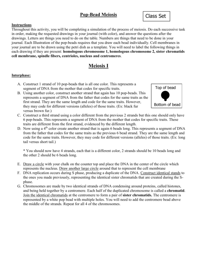
Pop-Bead Meiosis Class Set Instructions Throughout this activity, you will be completing a simulation of the process of meiosis. Do each successive task in order, making the requested drawings in your journal (with color), and answer the questions after the drawings. Letters are things you need to do on the table. Numbers are things that need to be done in your journal. Each Illustration of the pop-beads requires that you draw each bead individually. Cell membranes in your journal are to be drawn using the petri dish as a template. You will need to label the following things in each drawing if they are present: homologous chromosome 1, homologous chromosome 2, sister chromatids cell membrane, spindle fibers, centrioles, nucleus and centromeres. Meiosis I Interphase: A. Construct 1 strand of 10 pop-beads that is all one color. This represents a Top of bead segment of DNA from the mother that codes for specific traits. B. Using another color, construct another strand that again has 10 pop-beads. This represents a segment of DNA from the father that codes for the same traits as the first strand. They are the same length and code for the same traits. However, Bottom of bead they may code for different versions (alleles) of those traits. (Ex: black fur versus brown fur.) C. Construct a third strand using a color different from the previous 2 strands but this one should only have 6 pop-beads. This represents a segment of DNA from the mother that codes for specific traits. These traits are different from the first strand, evidenced by the different length. D. Now using a 4th color create another strand that is again 6 beads long. This represents a segment of DNA from the father that codes for the same traits as the previous 6 bead strand. They are the same length and code for the same traits. However, they may code for different versions (alleles) of those traits. (Ex: long tail versus short tail.) * You should now have 4 strands, each that is a different color, 2 strands should be 10 beads long and the other 2 should be 6 beads long. E. Draw a circle with your chalk on the counter top and place the DNA in the center of the circle which represents the nucleus. Draw another large circle around that to represent the cell membrane F. DNA replication occurs during S phase, producing a duplicate of the DNA. Construct identical stands to the ones you made previously, representing the identical sister chromatids that are created during the Sphase. G. Chromosomes are made by two identical strands of DNA condensing around proteins, called histones, and being held together by a centromere. Each half of the duplicated chromosome is called a chromatid. Join the identical chromatids at the centromere to form a pair of sister chromatids. The centromere is represented by a white pop bead with multiple holes. You will need to add the centromere bead above the middle of the strands. Repeat for all 4 of the chromosomes. 1. Draw a picture of the cell with chromosomes mixed together in the nucleus. Doing this represents the cell after S-phase of Interphase. In reality the strands would not be visible. The drawing should include each pop bead and color. Don’t forget to label. Interphase (Post S-phase) Prophase I A. The nucleus is dissolving. Remove sections of the nucleus from your cell on the table, making it dotted. The centrioles and small spindle fibers should appear off to the side. B. In prophase I, homologous chromosomes (chromosomes with the same lengths & genes) move close together and pair up along their entire length. A tetrad (group of 4 chromatids) is formed. Form tetrads and entwine the homologous pairs. C. Simulate crossing over by removing a few pop-beads from one area of one of the chromosomes and replacing it with the corresponding pop-beads of the other homologous chromosome. Do this for all pairs of homologous chromosomes. At the end of this step, all of the sister chromatids should look different. 2. Draw a picture of the cell in prophase I with the homologous pairs lined up next to each other after having undergone crossing over. In reality the homologous pairs would still be entangled. The drawing should include each pop bead and color. Don’t forget to label. Prophase I 3. Why doesn’t crossing over happen during mitosis? _________________________________________________________________________ Metaphase A. In this phase the chromosomes are pulled by the spindle fibers until they line up along the imaginary metaphase plate. Disentangle your chromosomes, and align the homologous chromosome pairs side by side at the metaphase plate. The long homologous pair should be together and the short pair should be together. Draw two centrioles at either end of the cell and spindle fibers should be connected to the chromosomes at the centromere. 4. Draw a picture of the chromosomes in metaphase. Drawing should include each pop bead and color. Don’t forget to label. Example Line Up Metaphase I Anaphase I A. The homologous chromosomes are separated and drawn towards the centrioles on opposite sides of the cell by the spindle fibers. One long and one short chromosome pair should be found on each side. B. Move the chromosomes by the centromeres; noting how the chromosome arms trail the centromere as movement occurs creating an arc with the edges curled towards the center of the cell. 5. Draw a picture of anaphase. Drawing should include each pop bead, color and labels. Anaphase I Telophase I / Cytokinesis I During meiosis I, cell division occurs resulting in two daughter cells still containing paired chromatids. A. During Telophase I the nucleus begins to regrow and the spindle fiber retract and the centrioles move out of the way. Cytokinesis begins during Telophase. Cytokinesis consists of the original cell breaking into two new cells. B. Create two cellular membranes by erasing the original circle and drawing two circles around the newly paired chromosomes. You will draw this on the lab table but DO NOT draw this in your journal as it will look very similar to Prophase II. 6. The sister chromatids of the daughter cell may be different from the sister chromatids in the parent cell. Why? ________________________________________________________________________ Meiosis II Prophase II The chromosomes move toward the center of the daughter cells. Centrioles move to the opposite ends of the cell. A. Place the chromosomes in the center of each daughter cell. Then make the nucleus a dotted line to show that it is disappearing. 7. Draw a picture of Prophase II. Drawing should include each pop bead, color and labels. Prophase II 8. What occurs in prophase I that doesn’t happen in prophase II of meiosis? ________________________________________________________________________ Metaphase II All of the chromosomes line up, single file, in the center of the cell. A. Center the chromosomes along an imaginary line across the center of the cell. Example Line Up 9. Draw a picture of metaphase II. Drawing should include each pop bead, color and labels. Daughter Cell 1: Metaphase II Daughter Cell 2: Metaphase II Anaphase II The chromosomes of each paired strand separate at the centromere and are drawn to opposite poles of the cell. A. The centromere is broken and the chromosomes are separated. The individual chromosomes are drawn towards opposite sides of the cell by the spindle fibers. Break the centromeres leaving the individual chromosomes next to each other in the center of the cell. B. Separate each chromosome, moving them towards the centrioles by pulling only the remaining piece of centromere. Move the chromosome by the centromeres; noting how the chromosome arms trail the centromere as movement occurs creating an arc with the edges curled towards the center of the cell. C. Repeat this procedure for both daughter cells. 10. Draw a picture of anaphase II. Drawing should include each pop bead and color. Daughter Cell 1: Anaphase II Daughter Cell 2: Anaphase II Telophase II / Cytokinesis II Cell division is completed and four daughter cells are formed. Each contains half of the chromosome number of the original parent cell. A nuclear membrane forms around each cell’s chromosomes and the daughter cells finish dividing. Here we are skipping the telophase step and showing you the final results of Telophase II and Cytokinesis II. A. Erase the original 2 circles and draw four circles to represent each of the new cells. Then draw a circle around each of the chromosome sets inside the new cells to represent the nucleus of the each cell. The centrioles and spindle fibers will not be in this illustration. There should be one long and one short chromosome in each of the new cells. 11. Draw a picture of the cells after telophase II & cytokinesis II. Drawing should include each pop bead, color, and labels. Daughter Cell 1 Daughter Cell 2 Daughter Cell 3 Daughter Cell 4 12. It would have been much easier to simulate mitosis. Give two ways the process of meiosis differs from mitosis. ________________________________________________________________________ 13. What is the general name of cells produced by mitosis? AND Produced by meiosis? ________________________________________________________________________ 14. In what step of meiosis is there a reduction in the chromosome number? ________________________________________________________________________ 15. Differentiate between homologous chromosomes and sister chromatids. ________________________________________________________________________ 16. Differentiate between haploid and diploid. ________________________________________________________________________


