Musculoskeletal Anatomy & Kinesiology LOWER APPENDICULAR
advertisement
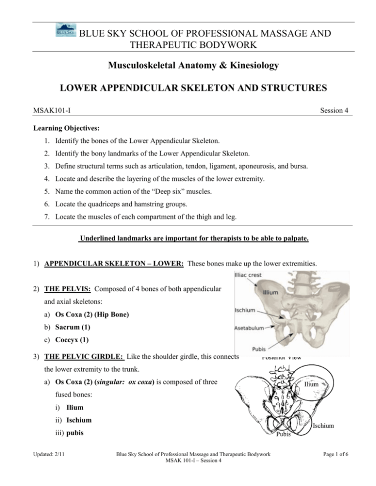
BLUE SKY SCHOOL OF PROFESSIONAL MASSAGE AND THERAPEUTIC BODYWORK Musculoskeletal Anatomy & Kinesiology LOWER APPENDICULAR SKELETON AND STRUCTURES MSAK101-I Session 4 Learning Objectives: 1. Identify the bones of the Lower Appendicular Skeleton. 2. Identify the bony landmarks of the Lower Appendicular Skeleton. 3. Define structural terms such as articulation, tendon, ligament, aponeurosis, and bursa. 4. Locate and describe the layering of the muscles of the lower extremity. 5. Name the common action of the “Deep six” muscles. 6. Locate the quadriceps and hamstring groups. 7. Locate the muscles of each compartment of the thigh and leg. Underlined landmarks are important for therapists to be able to palpate. 1) APPENDICULAR SKELETON – LOWER: These bones make up the lower extremities. 2) THE PELVIS: Composed of 4 bones of both appendicular and axial skeletons: a) Os Coxa (2) (Hip Bone) b) Sacrum (1) c) Coccyx (1) 3) THE PELVIC GIRDLE: Like the shoulder girdle, this connects the lower extremity to the trunk. a) Os Coxa (2) (singular: ox coxa) is composed of three fused bones: i) Ilium ii) Ischium iii) pubis Updated: 2/11 Blue Sky School of Professional Massage and Therapeutic Bodywork MSAK 101-I – Session 4 Page 1 of 6 b) Ilium i) Anterior Superior Iliac Spine (ASIS) ii) Anterior Inferior Iliac Spine (AIIS) iii) Iliac Crest iv) Iliac Fossa v) Posterior Superior Iliac Spine (PSIS) c) Ischium i) Ischial Tuberosity ii) Ischial Spine iii) Sciatic Notch d) Pubis i) Pubic Symphysis ii) Pubic Tubercles iii) Superior & Inferior Pubic rami (singular: ramus) e) Acetabulum (hip joint) i) Composed of all three bones. Updated: 2/11 Blue Sky School of Professional Massage and Therapeutic Bodywork MSAK 101-I – Session 4 Page 2 of 6 4) THIGH, LEG, ANKLE & FOOT (60 Bones) a) Femur (2): i) Head ii) Neck (Metaphysis) iii) Greater Trochanter iv) Lesser Trochanter v) Medial Epicondyle vi) Lateral Epicondyle vii) Linea Aspera viii) Medial Condyle ix) Lateral Condyle x) Adductor Tubercle b) Patella (2) c) Tibia (2): Medial leg bone i) Tibial Tuberosity ii) Lateral Tibial Condyle (or Plateau) iii) Medial Tibial Condyle (or Plateau) iv) Medial Malleolus d) Fibula (2): Lateral leg bone i) Head ii) Lateral Malleolus Updated: 2/11 Blue Sky School of Professional Massage and Therapeutic Bodywork MSAK 101-I – Session 4 Page 3 of 6 e) Tarsals (14): i) Talus ii) Calcaneus iii) Cuboid iv) Navicular v) Cuneiform #1-3 f) Metatarsals (10) and Phalanges (28): i) Base (proximal end) ii) Shaft iii) Head (distal end) iv) Proximal, middle and distal phalanges of toes #2-5 (Proximal and distal only for #1) g) Sesamoid Bones 5) COUNTING THE DIGITS: a) 1st digit = Big toe = Little toe b) 2nd – 4th c) 5th digit 6) STRUCTURES: a) Terminology: i) Articulation – A joint or point of contact between two or more bones. ii) Connective tissue: (T his topic will be covered in depth in CAPP.)The most abundant and widely distributed tissue in the body. It has various types with a variety of functions. For the purposes of MSAK, students are required to know the following forms, structures and functions: (1) Tendon –Connective tissue that attaches muscle to bone, non-contractile, each end of muscle has one or more tendons, can be short or long, broad or narrow. (2) Ligament – Connective tissue that attaches bone to bone to provide stability. Updated: 2/11 Blue Sky School of Professional Massage and Therapeutic Bodywork MSAK 101-I – Session 4 Page 4 of 6 (3) Aponeurosis – Sheet-like tendons that attach muscle to muscle or muscle to bone. (4) Fascia – a form of dense fibrous connective tissue that is found in abundance throughout the body. It not only wraps the entire body (superficial fascia) but also surrounds each organ and muscle (deep fascia). (5) Meniscus – Pad of cartilage that lies between articular surfaces of bones within some joints (e.g. knee). (6) Joint Capsule – A sleeve-like covering of ligamentous tissue surrounding the bony ends forming joints. (i) Characterized by articulating bones whose ends are capped with articular (hyaline) cartilage and are enclosed in a ligament-reinforced, sensitive, fibrous (joint) capsule. (ii) This joint capsule is lined internally with a vascular synovial membrane that secretes a lubricating fluid within the synovial cavity. (iii)The synovial membrane does not cover the articular cartilage. (iv) Bursas often exist between moving structures outside the joint. ii) Articular Cartilage – Cartilaginous surface at the ends of some bones; for articulation with another bone. iii) Bursae (purse) – Sac-like structures that alleviate pressure or reduce friction at some joints and cushion the movement of one body part over another. 7) MUSCLES OF THE PELVIS: a) Gluteal Group i) Gluteus Maximus ii) Gluteus Medius iii) Gluteus Minimus Updated: 2/11 Blue Sky School of Professional Massage and Therapeutic Bodywork MSAK 101-I – Session 4 Page 5 of 6 8) MUSCLES OF THE THIGH: a) Anterior Compartment: i) Quadriceps Group (1) Rectus Femoris (2) Vastus Lateralis (3) Vastus Medalis (4) Vastus Intermedius b) Medial Compartment: i) Adductor Group (Adduction is medial movement toward midline in frontal plane.) c) Posterior Compartment: i) Hamstring Group (1) Biceps femoris (2) Semitendinosis (3) Semimembransosis d) Lateral Compartment: i) Tensor Fascia Latae (TFL) 9) MUSCLES OF THE LEG: a) Posterior Compartment: i) Gastrocnemius ii) Soleus iii) Toe Flexor group b) Lateral Compartment: i) Fibularis group c) Anterior Compartment: i) Toe Extensor group Updated: 2/11 Blue Sky School of Professional Massage and Therapeutic Bodywork MSAK 101-I – Session 4 Page 6 of 6
