phylum nematoda
advertisement
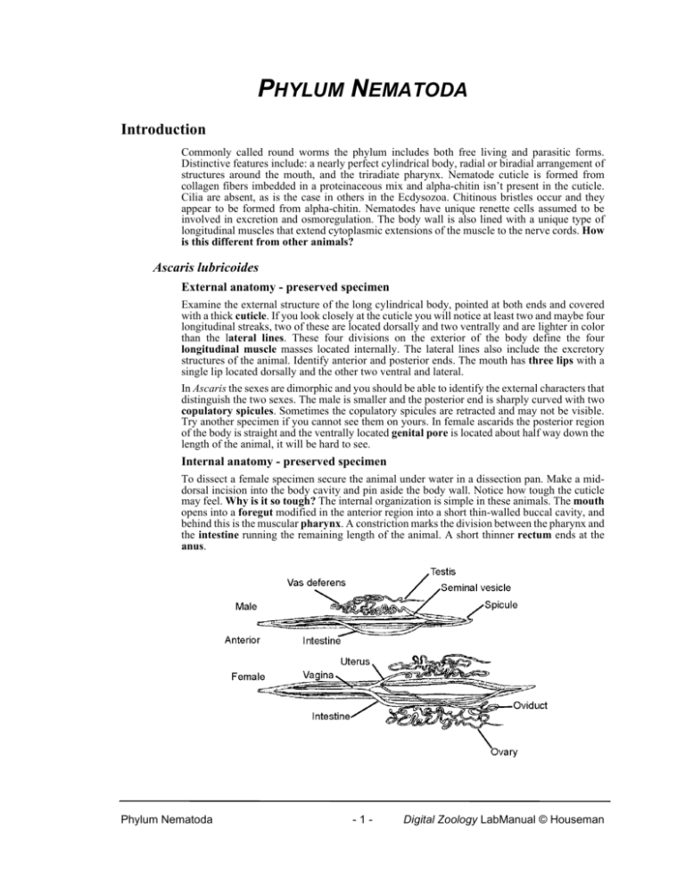
PHYLUM NEMATODA Introduction Commonly called round worms the phylum includes both free living and parasitic forms. Distinctive features include: a nearly perfect cylindrical body, radial or biradial arrangement of structures around the mouth, and the triradiate pharynx. Nematode cuticle is formed from collagen fibers imbedded in a proteinaceous mix and alpha-chitin isn’t present in the cuticle. Cilia are absent, as is the case in others in the Ecdysozoa. Chitinous bristles occur and they appear to be formed from alpha-chitin. Nematodes have unique renette cells assumed to be involved in excretion and osmoregulation. The body wall is also lined with a unique type of longitudinal muscles that extend cytoplasmic extensions of the muscle to the nerve cords. How is this different from other animals? Ascaris lubricoides External anatomy - preserved specimen Examine the external structure of the long cylindrical body, pointed at both ends and covered with a thick cuticle. If you look closely at the cuticle you will notice at least two and maybe four longitudinal streaks, two of these are located dorsally and two ventrally and are lighter in color than the lateral lines. These four divisions on the exterior of the body define the four longitudinal muscle masses located internally. The lateral lines also include the excretory structures of the animal. Identify anterior and posterior ends. The mouth has three lips with a single lip located dorsally and the other two ventral and lateral. In Ascaris the sexes are dimorphic and you should be able to identify the external characters that distinguish the two sexes. The male is smaller and the posterior end is sharply curved with two copulatory spicules. Sometimes the copulatory spicules are retracted and may not be visible. Try another specimen if you cannot see them on yours. In female ascarids the posterior region of the body is straight and the ventrally located genital pore is located about half way down the length of the animal, it will be hard to see. Internal anatomy - preserved specimen To dissect a female specimen secure the animal under water in a dissection pan. Make a middorsal incision into the body cavity and pin aside the body wall. Notice how tough the cuticle may feel. Why is it so tough? The internal organization is simple in these animals. The mouth opens into a foregut modified in the anterior region into a short thin-walled buccal cavity, and behind this is the muscular pharynx. A constriction marks the division between the pharynx and the intestine running the remaining length of the animal. A short thinner rectum ends at the anus. Phylum Nematoda -1- Digital Zoology LabManual © Houseman Fig. 1. Internal anatomy of the male and female Ascaris. © BIODIDAC Look at the body wall of your specimen and you will be able to see the two lateral lines that run the length of the animal and contain the excretory ducts. The reproductive system is wrapped around the intestine. From the female genital pore a single vagina leads back to join into the paired wider uteri which should be full of eggs. The uteri continue to run from the back and to the front of the animal as they narrow to oviducts that coil and twist and continue to narrow. They finally merge into the two filamentous ovaries. The ovaries are interwoven among the oviducal coils. Compare this to the reproductive system in a male that consists of a single testis. Open the animal the same way. Located at the posterior end is the ejaculatory duct that marks the end of the genital tract. As the ejaculatory duct passes forward into the straighter part of the body it enlarges into the seminal vesicle that winds back and forth in the pseudocoel until it changes to join the sperm duct, or vas deferens. The vas deferens, which also coils backwards and forwards, ultimately merges with the long blind-ended testis that in turn coils backwards and forwards. The simplicity of these animals means that other systems may not be present. Is a nervous system present? Can you locate it in your specimen? What about a circulatory system? Fig. 2. Cross section through a female Ascaris. © BIODIDAC Prepared slides If you have aligned the illumination of your microscope properly you should be able to see all of the structures mentioned. Examine the prepared slides of ascarid cross sections for the female and male. In both, the body wall consists of a thin outer cuticle protecting the worm from the digestive juices of the host. How do you distinguish the difference between the epithelium that secretes the cuticle and the cuticle itself? The underlying epidermis secretes the cuticle. Under this longitudinal muscles are arranged into four blocks separated by the lateral lines containing the excretory system and the dorsal and ventral nerve cords. In Ascaris the muscles themselves extend arms to the nerve cord rather than having the axons of the nervous system extending to the muscle cells, as would be the case in other organisms. Does Ascaris have circular muscles? Take a close look at the lateral line; you will be able to see the two components of the excretory system that form the excretory canal. You will also be able to see the dorsal and ventral nerves located in the body wall Phylum Nematoda -2- Digital Zoology Lab Manual © Houseman The body cavity is a pseudocoel and the alimentary tract is located within it. Are there muscles associated with the intestinal tract? How many different tissue layers can you see in the intestinal wall? The remaining organs contained inside the pseudocoel are parts of the convoluted reproductive tracts. Remember, from you dissection, that the ovary, oviduct and uterus of the female system are paired, and the two large sections filled with eggs surrounded by shells are the uteri. The small wheel-like sections with spoke-like divisions are the ovaries and the intermediate sections with eggs and without internal divisions are the oviducts. Fig. 3. Cross section through a male Ascaris © BIODIDAC The reproductive system in the male is less complex and unpaired. The largest section is the seminal vesicle containing sperm. Sections of both the testis and sperm duct are difficult to distinguish from each other. Ancylostoma caninum Although Ascaris is a convenient organism for dissection it is a rather unusual nematode because of its large size. Nematodes are usually only microscopic and we should take a look at some of the more traditional forms to familiarize ourselves with the general characters of the phylum. Ancylostoma caninum is the dog hookworm and adults live in the intestine feeding on the lining of the digestive tract. Eggs are released in the feces and hatch releasing a larval stage that feeds on bacteria and moults twice before becoming infective. Dogs are reinfected by either consuming contaminated food, or by hookworm larva that penetrate through the skin and migrate to the lungs where they are coughed up and into the digestive tract. Prepared slides Male and female hookworm slides are available. The body is long and cylindrical and covered by the ridged cuticle of the body wall. In both males and females the anterior end is curved forming the hook that gives the species its common name. The mouth is located at the end of the hook. Males are smaller than females who have a more pointed posterior end compared to the male. The posterior end of the male is large and forms the bursa that surrounds the female when they mate. The mouth opens at the anterior end and is located at the base of a buccal capsule lined with teeth used to not only hold onto the host, but to dig and cut away at the lining of the intestine ensuring a continuous blood supply for the feeding parasite. Are all the teeth the same? The mouth leads to the muscular pharynx and in the region of the nerve ring a constriction separates the pharynx and end bulb. The result is the two valves essential for swallowing the meal. Why are two valves necessary? Although you may not be able to see them, pharyngeal glands are embedded in the wall of the pharynx. You will only see glimpses of the rest of the Phylum Nematoda -3- Digital Zoology LabManual © Houseman digestive tract because it’s surrounded by the reproductive system. Behind the pharynx the intestine, of only a single layer of cells and without any musculature, extends to the muscular rectum and the anus. The “excretory system” will be hard to see and includes an excretory pore that opens near the cervical papilla in the same area as the nerve ring. The “excretory system” consists of renette cells forming glands joined in the region of the excretory pore. The male reproductive system has a single, coiled and tubular testis. After coiling forward and then back through the body, the testis drains into the seminal vesicle opening into the ejaculatory duct. Paired cement glands surround the ejaculatory duct. Two copulatory spicules inserted into the female working together with the secretions of the cement glands and the bursa help anchor the male and female during mating. The genital pore of the female reproductive system opens in the middle of the body and on the ventral surface. Paired filamentous ovaries coil throughout the body with one ovary anterior to the genital pore, the other posterior. Each ovary leads to a uterus that may contain eggs. In some slides the uterus may be so full of eggs it will make it hard to see any additional structures. The uteri join in a common vagina opening to the outside through the genital pore. Trichinella spirallia This particular nematode is the reason you’re often told to be sure and cook pork until it’s well done. That’s because pork (inspections keep our Canadian pork free of this problem and that may be why you’ve never heard this advisory) muscle can contain the cysts of Trichinella. Under unsanitary conditions pigs and rats often share the same food and both may be infected. Pigs will also eat rats and the nematode can cycle between the two. Consumption of contaminated pork results in trichinosis in humans. Fig. 4. Trichinella cysts in muscle. © BIODIDAC Trichinella encysts in the muscle tissue of infected hosts and if rats, pigs or humans consume that muscle the cyst is dissolved by the digestive system and the worms are released into the gut. Males and females mate and females burrow into the wall of the intestine where the ovoviviparous females produce juvenile worms that enter the circulatory system that transports them to their final site, the muscle. They encyst and the cycle repeats itself. Examine the slide of the muscle tissue and identify the encysted juveniles. The cyst itself is oriented parallel to the muscle fibers and consists of two components: an outer non-cellular hyaline sheath, and inner cellular fibrous sheath. Each worm is almost mature and the Phylum Nematoda -4- Digital Zoology Lab Manual © Houseman reproductive system hasn’t developed, making this is an excellent opportunity to see the alimentary tract. The mouth is located at the tip of the anterior end and is connected to a long esophagus followed by intestine and anus. Fig. 5. Anatomical features of male and female Trichinella spiralis. © BIODIDAC Adult males and females are also available and you should be able to identify the major anatomical features of both. Males are smaller than females. In both, the body is covered by a thin smooth cuticle. The mouth opens into a muscular pharynx followed by the long esophagus wrapped in glandular cells and referred to as the stichosome. The intestine connects the esophagus to the anus at the posterior end of the animal. In the males a large testis fills the posterior half of the animal and near the junction of the esophagus and intestine it bends back on itself forming the narrower sperm duct that opens through the cloaca at the posterior end. Why is this called a cloaca? Males hold onto the female using the lobes that surround the genital opening. The female reproductive system consists of a posterior ovary connected to the large uterus by the oviduct. The posterior end of the uterus serves as a seminal receptacle and as eggs pass down from the ovary, they are fertilized. Unlike any other nematode the eggs develop as they pass down the length of the uterus and by the time they reach its most anterior end they have already hatched into the small juveniles. They are released from the vulva on the ventral surface about half way down the length of the esophagus. Turbatrix Place a drop of Vinegar eel culture on a slide and watch the way the animal moves. The whip like motion is not efficient in water but when on a substrate they can push against it to move. Give it a try by preparing another preparation but this time add a few fine grains of sand. What happens? How is this movement related to the organization of muscles in the nematode? Phylum Nematoda -5- Digital Zoology LabManual © Houseman
