Nervous System: Organization, Neurons, and Physiology
advertisement
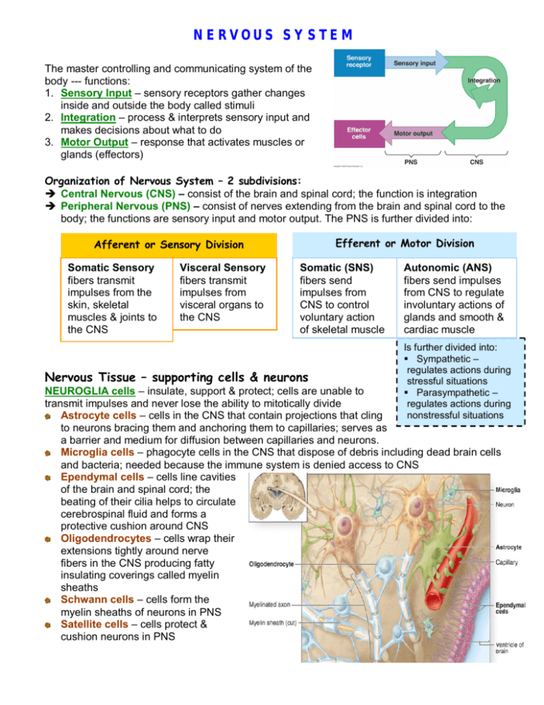
NERVOUS SYSTEM The master controlling and communicating system of the body --- functions: 1. Sensory Input – sensory receptors gather changes inside and outside the body called stimuli 2. Integration – process & interprets sensory input and makes decisions about what to do 3. Motor Output – response that activates muscles or glands (effectors) Organization of Nervous System – 2 subdivisions: Central Nervous (CNS) – consist of the brain and spinal cord; the function is integration Peripheral Nervous (PNS) – consist of nerves extending from the brain and spinal cord to the body; the functions are sensory input and motor output. The PNS is further divided into: Efferent or Motor Division Afferent or Sensory Division Somatic Sensory fibers transmit impulses from the skin, skeletal muscles & joints to the CNS Visceral Sensory fibers transmit impulses from visceral organs to the CNS Somatic (SNS) fibers send impulses from CNS to control voluntary action of skeletal muscle Nervous Tissue – supporting cells & neurons Autonomic (ANS) fibers send impulses from CNS to regulate involuntary actions of glands and smooth & cardiac muscle Is further divided into: Sympathetic – regulates actions during stressful situations Parasympathetic – regulates actions during nonstressful situations NEUROGLIA cells – insulate, support & protect; cells are unable to transmit impulses and never lose the ability to mitotically divide Astrocyte cells – cells in the CNS that contain projections that cling to neurons bracing them and anchoring them to capillaries; serves as a barrier and medium for diffusion between capillaries and neurons. Microglia cells – phagocyte cells in the CNS that dispose of debris including dead brain cells and bacteria; needed because the immune system is denied access to CNS Ependymal cells – cells line cavities of the brain and spinal cord; the beating of their cilia helps to circulate cerebrospinal fluid and forms a protective cushion around CNS Oligodendrocytes – cells wrap their extensions tightly around nerve fibers in the CNS producing fatty insulating coverings called myelin sheaths Schwann cells – cells form the myelin sheaths of neurons in PNS Satellite cells – cells protect & cushion neurons in PNS NEURONS (in CNS and PNS) – nerve cells that are able to transmit impulses and are amitotic. Neuron Anatomy Soma (Cell Body): contains nucleus & metabolic center Dendrite: slender fibers containing sensory receptors that conduct impulse toward soma Axon: conducts impulses away from soma Axon terminal: end branches of axons that contains neurotransmitter storage vesicles Synapse: junction of two neurons; space at synapse is called synaptic cleft Myelin: fatty material that protects and insulates fibers; speeds up impulse transmission Nodes of Ranvier: gaps between myelin sheaths Sensory receptors on dendrites of neuron receive impulse. Impulse is sent to cell body, then through the axon jumping from node to node. When impulse reaches terminals, a neurotransmitter is released to stimulate the receptors on dendrites of another neuron. Multiple Sclerosis (MS) – autoimmune disease resulting in gradually destruction of myelin sheaths around fibers; person loses ability to control muscles and becomes disabled. Types of Neurons 1. Sensory or Afferent neurons (unipolar) – carries impulse from sensory receptors on dendrite endings to CNS 2. Motor or Efferent neurons (multipolar) – carries impulse from CNS to effector organ; impulse brings about motor response 3. Interneurons or Mixed neurons (multipolar) – connect motor and sensory neurons in spinal cord; transmit impulse to and from brain Bipolar neurons – rare in adults, found only in some special sense organs … act as sensory receptors Neuron Physiology Irritability – ability to respond to stimulus and convert into nerve impulse Conductivity – ability to transmit impulse to other neurons, muscles or glands PHYSIOLOGY in action – 1. Plasma membrane of a resting (inactive) neuron is polarized, which means the outside of cell is more positive than the inside 2. Stimuli activate neurons to create an impulse; stimuli may be sensory or neurotransmitters released from other neurons 3. Stimulus changes permeability of membrane and Na+ diffuse into cell to change polarity – this is called depolarization (inside is more positive). 4. Action potential is created – impulse travels through neuron jumping from node to node 5. K+ diffuse out of cell to restore the electrical conditions – this is called repolarization. Repolarization must occur to conduct another impulse! 6. Na/K pump uses ATP to restore the initial concentrations of Na & K to resting conditions. 7. These events continue to spread across the membrane of the neuron until the impulse reaches the axon terminal. 8. At axon terminal, impulse causes Ca ions to enter cell triggering vesicles to release neurotransmitters 9. Neurotransmitters bind to receptors on dendrite of next neuron – process occurs again on next neuron. Factors that impair conduction of impulse: 1. alcohol, sedative & anesthetics block nerve impulse by reducing membrane permeability to Na+ 2. cold & pressure interrupt blood flow (“goes to sleep”) Reflexes Arcs – neural pathway for reflexes, rapid involuntary responses to stimuli Autonomic – reflex that stimulates smooth, cardiac and glands Somatic – reflex that stimulate skeletal muscle Reflex testing is used to evaluate the condition of the nervous system. Central Nervous System (CNS) BRAIN – 4 parts Brain weighs – 3 lbs Contains 100 billion neurons & trillions of glial cells Cerebrum Diencephalon Cerebellum Brain Stem 1. Cerebrum - 2 hemispheres & 4 lobes Cerebrum longitudinal fissure separates right & left hemispheres central fissure separates frontal & parietal lobes lateral fissure separates frontal & temporal lobes Corpus Collosum – large fiber tract (bundles of nerve fibers) that allows hemispheres to communicate with each other Frontal conscious intellect primary motor area – allows us to consciously move skeletal muscles Broca’s area – ability to speak language comprehension reasoning & memory Temporal auditory memory olfactory Parietal somatic sensory area – sensation of pain, temperature or touch understand & connect w/ speech Occipital visual area Recognizing patterns & speech areas Gray & White MATTER Cerebral cortex - outermost area consists of GRAY MATTER (unmyelinated fibers) –integration occurs here! Cerebral medulla – inner surface consist of WHITE MATTER (myelinated fiber tracts) – carry impulse to and from cortex Basal nuclei – islands of gray matter within white matter; regulates voluntary motor activities Huntington’s disease – degeneration of basal nuclei and later the cerebral cortex Parkinson’s disease – basal nuclei become overactive due to degeneration of dopamine-releasing neurons Alzheimer’s disease – degenerative changes in brain; gyri shrink and brain atrophies 2. Diencephalon (interbrain) – enclosed by the cerebral hemispheres Thalamus – relay station for sensory impulses passing upward to the sensory cortex Epithalamus – contains the pineal gland which regulates day and night cycles. It also contains the choroid plexus, knot of capillaries that form the cerebrospinal fluid. Hypothalamus – an important ANS center because it regulates body temperature, water balance and metabolism. It is considered part of the limbic system as it serves as the center for drives & emotions. It regulates the pituitary gland and produces two hormones. mammillary bodies – reflex centers for olfaction – found below hypothalamus Limbic system – involves cerebral and diencephalon structures that control emotions and memory o Amygdala – recognizes, assesses danger & elicits fear response; plays a role in memory processing o Hippocampus – plays a role in memory processing 3. Cerebellum – has two hemispheres, a convoluted surface and an outer cortex of gray matter and inner region of white matter. It controls balance and equilibrium so timing of skeletal muscles activity is smooth and coordinated. If damaged, movements become clumsy and disorganized – a condition called ataxia. 4. Brainstem – pathway for ascending and descending tracts Midbrain – contains tracts that convey ascending & Brainstem descending impulses and reflex centers for vision & hearing Pons – contains tracts involved in the control of breathing Medulla Oblongata – merges with the spinal cord; tracts contain centers to regulate vital visceral activities and centers to control heart rate, blood pressure, breathing, vomiting, etc. Reticular Formation – gray matter in brainstem involved in motor control of visceral organs and plays a role in consciousness & awake/sleep cycles; damage can result in coma SPINAL CORD – The spinal cord is a continuation of the brain stem with a two-way conduction pathway to & from the brain and serves as a reflex center. It extends from the foramen magnum of the skull to about the 2nd lumbar vertebrae. Gray matter – unmyelinated tracts contain synapse of sensory, motor and interneurons which carry impulses for integration to the brain or in the spinal cord (reflexes) White matter – contains sensory & motor tracts which ascend and descend to the brain o o o o Damage to ventral root results in inability to stimulate muscles (paralysis) Damage to dorsal root results in inability to send stimuli to CNS Transection between T1 & L1 – become paraplegic Injury in cervical – become quadriplegic Protection of CNS Bone - The skull and the vertebral column enclose the brain and spinal cord. Meninges – connective tissue membranes covering CNS. dura mater – double layer with periosteal layer (periosteum) attached to bone and meningeal layer covering the brain & spinal cord; limits excessive movement of brain and cord arachnoid mater – middle layer has web-like extensions spanning the subarachnoid space for attachment to underlying pia mater; space is filled with cerebrospinal fluid (CSF) & contains largest vessels serving the brain pia mater – clings to brain and spinal cord; composed of tiny capillaries Cerebrospinal Fluid (CSF) – forms a watery cushion that gives buoyancy to the CNS structures; protects the CNS from injury & nourishes the brain; CSF forms and drains at a constant rate so that its normal pressure and volume are maintained. Blood-brain barrier – refers to the least permeable capillaries that selectively diffuse materials to neurons. Only water, glucose and essential amino acids pass easily. Metabolic waste and most drugs are prevented from entering brain tissue. Fats, respiratory gases, alcohol, nicotine and anesthetics can easily diffuse. Meningitis – inflammation of the meninges caused by virus or bacteria. Brain inflammation is called encephalitis. Concussion – occurs when brain injury is slight. Contusion – brain injury causing tissue destruction. After head injuries, death may occur due to intracranial hemorrhage (bleeding of ruptured blood vessels) or cerebral edema (swelling of brain). Cerebrovascular Accident (CVA) or Stroke – occurs when blood circulation to brain is blocked either by blood clot or ruptured blood vessel. Transient Ischemic Attack (TIA) – is caused from temporary restriction of blood flow; warning for stroke Hydrocephalus – CSF drainage is obstructed and fluid accumulates exerting pressure on the brain. Anencephaly – failure of cerebrum to develop resulting in a child who cannot hear, see or process sensory inputs and development of Spina bifida where the vertebrae form incompletely PERIPHERAL NERVOUS SYSTEM (PNS) Nerve – a bundle of neuron fibers found outside the CNS. o Each fiber is surrounded by endoneurium, a loose connective tissue. o Groups of fibers are bound into bundles called fascicles by a connective tissue wrapping called perineurium. o All fascicles are enclosed by the epineurium to form the nerve. Nerve Classification: Mixed nerves – carry both sensory and motor fibers Sensory nerves – carry impulses toward the CNS Motor nerves – carry impulses away from CNS Cranial nerves – 12 pairs of nerves originate from the brain to innervate the head and neck. Most cranial nerves are mixed, but some are sensory. Only the vagus nerve extends to thoracic and abdominal cavities. (Cranial nerves are listed in table 7.1.) Spinal nerves – 31 pairs of mixed nerves are formed by the union of dorsal and ventral roots of spinal cord. The spinal nerves divide into dorsal and ventral rami which serve different areas of the body. The dorsal rami serve the skin and muscles of the posterior body trunk. The ventral rami rami serve the skin and muscles of the anterior and lateral trunk. Some ventral rami form networks of nerves called plexus. These plexus serve the needs of the limbs. (Spinal nerves are listed in table 7.2.) Developmental Aspects of Nervous System System is formed in 1st month of embryonic development. The hypothalamus is the last to mature. Few neurons are formed after birth, but growth and maturation continues all through childhood, mostly as a result of myelination. The brain reaches it max weight as an adult, as we age, neurons are damaged and die. However, neural pathways are always available and being developed. Nervous tissue has the highest metabolic rate in the body so lack of oxygen for a few minutes leads to death of neurons. Cerebral Palsy may be caused by a temporary lack of oxygen to the brain resulting in poor control of voluntary muscles.

