Do You Get the Point? - Division of Biology and Medicine
advertisement
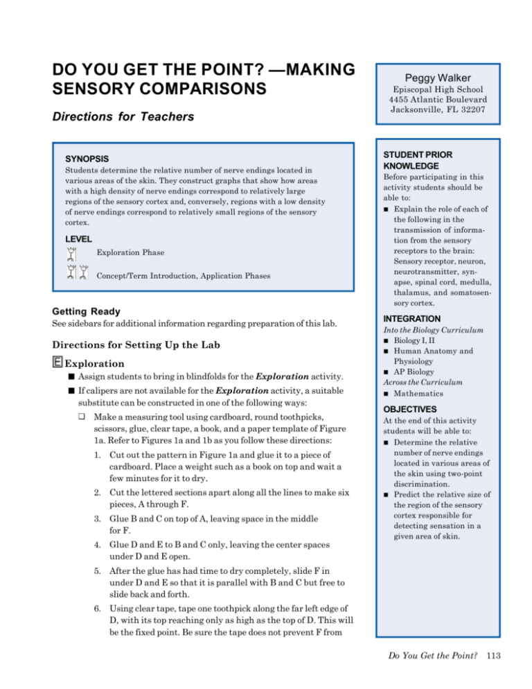
DO YOU GET THE POINT? —MAKING SENSORY COMPARISONS Directions for Teachers SYNOPSIS Students determine the relative number of nerve endings located in various areas of the skin. They construct graphs that show how areas with a high density of nerve endings correspond to relatively large regions of the sensory cortex and, conversely, regions with a low density of nerve endings correspond to relatively small regions of the sensory cortex. LEVEL Exploration Phase Concept/Term Introduction, Application Phases Getting Ready See sidebars for additional information regarding preparation of this lab. Directions for Setting Up the Lab Exploration ■ Assign students to bring in blindfolds for the Exploration activity. ■ If calipers are not available for the Exploration activity, a suitable substitute can be constructed in one of the following ways: ❏ Make a measuring tool using cardboard, round toothpicks, scissors, glue, clear tape, a book, and a paper template of Figure 1a. Refer to Figures 1a and 1b as you follow these directions: 1. Cut out the pattern in Figure 1a and glue it to a piece of cardboard. Place a weight such as a book on top and wait a few minutes for it to dry. 2. Cut the lettered sections apart along all the lines to make six pieces, A through F. 3. Glue B and C on top of A, leaving space in the middle for F. 4. Glue D and E to B and C only, leaving the center spaces under D and E open. Peggy Walker Episcopal High School 4455 Atlantic Boulevard Jacksonville, FL 32207 STUDENT PRIOR KNOWLEDGE Before participating in this activity students should be able to: ■ Explain the role of each of the following in the transmission of information from the sensory receptors to the brain: Sensory receptor, neuron, neurotransmitter, synapse, spinal cord, medulla, thalamus, and somatosensory cortex. INTEGRATION Into the Biology Curriculum ■ Biology I, II ■ Human Anatomy and Physiology ■ AP Biology Across the Curriculum ■ Mathematics OBJECTIVES At the end of this activity students will be able to: ■ Determine the relative number of nerve endings located in various areas of the skin using two-point discrimination. ■ Predict the relative size of the region of the sensory cortex responsible for detecting sensation in a given area of skin. 5. After the glue has had time to dry completely, slide F in under D and E so that it is parallel with B and C but free to slide back and forth. 6. Using clear tape, tape one toothpick along the far left edge of D, with its top reaching only as high as the top of D. This will be the fixed point. Be sure the tape does not prevent F from Do You Get the Point? 113 Figures 1a and 1b. Measuring device made from cardboard and toothpicks. sliding. LENGTH OF LAB 7. Tape a second toothpick to the inside edge of the perpendicular arm of F. Be certain that the points of the two toothpicks are aligned. A suggested time allotment follows: Day 1 5 minutes — Conduct the introductory demonstration. 40 minutes — Conduct the Exploration. 8. The completed tester should look like the diagram in Figure 1b. ❏ A fixed measuring device can be made by sliding two round toothpicks into the end of a cork, as shown in Figure 2. 1. Slide two round toothpicks gently into the end of a cork, as shown in Figure 2. Make sure that the ends of the toothpicks are a specific measured distance apart, such as 5 mm. Label the cork “5 mm” with a ball-point pen. Make as many corks of this size as groups of students who will be using them. Day 2 30 minutes — Draw and interpret graphs in small groups. 15 minutes — Discuss graphs as a class. 2. Repeat Step 1 to make other corks with toothpicks of fixed distances apart, such as 2 mm, 3 mm, 5 mm, 7 mm, 8 mm. Day 3 3. To use these measuring devices, each group should have a set 15 minutes — Make predictions for Application I. 30 minutes — Conduct library research to check predictions (optional). Day 4 15 minutes — Make predictions for Application II (optional). 30 minutes — Conduct library research to check predictions (optional). Figure 2. Measuring device made from cork and toothpicks. 114 Neuroscience Laboratory Activities Manual of corks of each size. Instead of moving the points closer together during the Exploration, as done with a caliper, the students will instead use a cork with toothpicks a different distance apart for each measurement. 4. The corks can be saved from year to year in plastic bags. Concept/Term Introduction ■ Make photocopies of Table 1. Make overhead transparencies of blank graph paper (optional), Graphs 1 and 2 in two different colors (optional), and Figures 4 and 5 (optional). Photocopy these figures and graphs for students (optional). MATERIALS NEEDED You will need the following for the teacher-led introduction: ■ 1 caliper ■ 1 metric ruler ■ 1 blindfold, such as a handkerchief tied around the eyes or safety goggles stuffed with cotton (optional) Application ■ Schedule a class visit to the school library (optional). Teacher Background ■ ■ You will need the following for each group of three students in a class of 24: 1 caliper 1 metric ruler 1 blindfold (optional) 1 alcohol swab Neurologists, physicians who specialize in the brain and central nervous system, use two-pointed objects and ask patients if they feel one point or two. They are checking the sensitivity of the skin, but what does the sensitivity of the skin have to do with the brain? Humans learn a great deal about their immediate environment from the sense of touch. The brain is able to determine where the body has been touched, and can often identify the object that touched it, because it contains a kind of map that reflects the relative number of touch receptors in various parts of the body. ■ Human skin contains several different sense receptors that respond to mechanical and thermal stimuli (e.g., touch, pressure, pain, cold, heat). These receptors are used to help us explore and determine the characteristics of our external environment. See Figure 3. PREPARATION TIME REQUIRED A sense receptor is a specialized cell that transduces (converts) a physical or chemical stimulus into action potentials. These action potentials produced by the receptors are conducted to the spinal cord and brain for processing and interpretation. The message that is sent to the central nervous system (CNS) is always a train of action potentials, regardless of the kind of stimulus that excites a particular receptor. Sensory receptors that respond to touch send action potentials through axons that enter the dorsal columns of the spinal cord and ascend to the medulla of the brainstem. These axons then make connections (synapses) with another pathway within the medulla. It is here that the pathway crosses over the brain midline and then continues to the thalamus. The final pathway begins in the thalamus and continues to the specific region of the sensory cortex, as shown in Figure 4. This pathway effectively gives one side of the brain responsibility for the opposite side of the body. This phenomenon is observed when a head injury or disease of the right side of the brain leads to physiological problems in a patient’s left side. ■ ■ ■ ■ You will need the following for each group of three students in a class of 24: 6 sheets graph paper 3 metric rulers 1 brain model (optional) 10 minutes — Locate and obtain calipers, if available. 2 hours — Make calipers, if these are not available. 20 minutes — Locate and obtain all other materials. 45 minutes — Make photocopies of Table 1, and overhead transparencies and photocopies of Graphs 1 and 2, and Figures 4 and 5 (optional). 15 minutes — Schedule a class visit to the school library (optional). Most of the information about touch is centered in a thin, convoluted surface layer of the cerebrum of the human brain called the somatosensory cortex. Each point on this band of sensory cortex contains densely packed cells that correspond to sensory receptors from different parts of the body, as shown in Do You Get the Point? 115 SAFETY NOTES ■ ■ ■ ■ Some students may not wish to be subjects. They can be measurers or data recorders. The students should apply the pointed objects to the skin gently, because even these everyday materials must be used carefully. The pointed objects should never be applied hard enough to puncture the skin. Points should be discarded or disinfected after each student is tested and new or disinfected points used for each student. Students who volunteer to be the subject should bring their own blindfolds or cotton for goggles. These should not be shared, as viruses and bacteria can be spread in this manner. Figure 3. Skin cross section indicating different sense receptors. Figure 5. The specific amount of space on the somatosensory cortex of the brain that is dedicated to sensing each body part is proportional to the density of the sensory receptors in that particular body region. For example, relatively few receptors are located in the upper arm; therefore, the upper arm area in the somatosensory cortex is small. In contrast, the density of receptors in the TEACHING TIPS ■ ■ Be prepared for comments about the mapping of genitalia and questions about their relative size on the graph. The Exploration phase of this lab can be stopped at any point. Figure 4. A schematic diagram showing how touch information from the finger is carried to the sensory cortex. 116 Neuroscience Laboratory Activities Manual SUGGESTED MODIFICATIONS FOR STUDENTS WHO ARE EXCEPTIONAL Below are possible ways to modify this specific activity for students who have special needs, if they have not already developed their own adaptations. General suggestions for modification of activities for students with impairments are found in the AAAS Barrier-Free in Brief publications. Refer to p. 19 of the introduction of this book for information on ordering FREE copies of these publications. Some of these booklets have addresses of agencies that can provide information about obtaining assistive technology, such as Assistive Listening Devices (ALDs); light probes; and talking thermometers, calculators, and clocks. Blind or Visually Impaired ■ Figure 5. Diagrams to illustrate the location of the somatosensory cortex on the surface of the brain (a) and the sensory homunculus of the somatosensory cortex (b). The line drawn through the somatosensory cortex in (a) shows where the cross section of the brain was drawn in (b). lips is very high, so the lip area of the cortex is large. The two-point discrimination method described in the Exploration phase of this activity can be used to approximate a map of the entire body as “sensed” by the cortex. The “picture” of the body on the somatosensory cortex is called the “homunculus,” which means “little person.” The two-point discrimination method determines the minimum distance that can be sensed between two points of touch. This technique can be used to determine the approximate size of the receptive field that senses light touch. In this procedure the two points of a caliper lightly touch the skin at the same time and the subject is asked to determine whether two points of ■ ■ A student who is blind due to diabetes would be unable to participate as a subject in this activity due to the lack of tactile ability. This student, however, could serve as the data recorder. Students who are blind due to causes other than diabetes, however, could participate as either subjects or data recorders. Provide text and Table 1 in braille format or on audiotape for the student who is blind. For students with low vision, provide text, figures, and Table 1 in photo-enlarged copies. A student with low vision —Continued Do You Get the Point? 117 SUGGESTED MODIFICATIONS —Continued could serve as the subject, data recorder, and perhaps the measurer, if he/she has sufficient functional vision. Gifted ■ Students who are gifted can research the concept of plasticity of the brain. They can design an experiment to find out if increased activation of receptors will change the representation on the cortex. Mobility Impaired ■ A student with limited use of his/her arms could participate as the subject or the data recorder. the stimulus are felt or one. Two points placed on the tips of the fingers can be distinguished as two separate points even when the points are as close together as two millimeters. In contrast, on the subject’s back the points must be about 40 millimeters apart before they can be identified as two separate stimuli (Martin & Jessell, 1991, p. 346). The reason the discrimination of the finger is better than the back is that the finger has a much larger number of specialized touch receptors than the back or other regions of the body. In this activity, students will locate touch receptors on the skin and estimate their relative numbers in different parts of the body. By calculating the reciprocals of the two-point discrimination numbers from the skin, students will be able to predict the relative size of the region of the sensory cortex responsible for detecting sensation in a given area of skin. They can then consider how the resulting pattern may affect human life and evolution. The size of the head and body representation in the somatosensory cortex differs among species of mammals. In primates and carnivores, for example, the somatosensory representation of the paws, hands, and feet is large compared to the rabbit (Adey & Kerr, 1954; Adrian, 1941; Crosby, Humphrey & Lauer, 1962; Kandel & Jessell, 1991; Mickle & Ades, 1952; Woolsey & Wang, 1945). Most likely these regions of the body contain a large number of touch receptors. These distributions in primates and carnivores probably reflect how they hunt, collect, and handle their prey and other food. The rabbit has a large distribution for the face and snout because it uses them as the primary means for exploring the environment. The hand is well represented in all primates, but the thumb region is the most important in the toolmaking humans. See p. 373 of Kandel & Jessell (1991) for an artist’s conception of how the brain serves various body parts in different animals. Touch sensors are found throughout a person’s body. Students may wonder why they are not normally aware of the touch caused by everyday stimuli such as their clothes and sitting in a chair. These touch sensors are designed to be excited for a short period of time. After a while they do not respond any more to the constant stimulation; in other words, they adapt to a stimulus. If the touch is changed slightly by movement or touch shifts, the sensors again become responsive for a short period of time. Regions in the brain corresponding to touch receptors on the skin can be reconfigured when the area of the body they are responsible for is missing, as in amputation. In doing this the brain may remodel the sensory neurons and they will be used in another area. Procedure Introduce the activity by demonstrating a method of two-point discrimination on the skin: 1. Gently touch two slightly pointed objects to the forearm of one of the students. Ask the student whether he/she can feel one point or two. The student should be wearing a blindfold during the demonstration, or close his/her eyes. 118 Neuroscience Laboratory Activities Manual 2. Pick up the points, move them closer together, and gently touch the skin of the same area again — again asking whether the student feels one point or two. Gradually move the points closer together until the test subject can no longer distinguish two separate points, and then measure the distance between the points. Write this measurement on the board. 3. Ask students in the class whether they think this number would be the same if this procedure were done on skin of other areas of the body. Questions you might ask the class include: ■ Would the number on the fingertip be the same as the number on the arm? ■ Would the number on the palm of the hand be the same as the number on the back of the leg? ■ Why do you think the numbers may or may not differ in each case? 4. Tell students that they will now take measurements of these numbers in groups of three. Exploration The Exploration activity has students take measurements as was done in the introduction. The student procedures are listed below. Details about how to construct measurers, if calipers are not available, appear under Directions for Setting Up the Lab. The Teacher Background explains the neuroscience involved in this activity. 1. Assign each student in a group a role as follows: ■ Subject ■ Measurer ■ Data recorder. 2. The measurer should gently place the two points of the caliper on the subject’s skin and ask the subject if he/she feels just one point or two. The measurer should make sure that the two points are applied simultaneously, and that the subject does not see the skin as it is being touched. The subject can either wear a blindfold during the test or close his/her eyes. 3. The measurer should close the gap between the two points slightly and repeat Step 1. He/she should continue in this same area of skin until the subject can no longer identify two separate points. The measurer should measure the distance between the two points and tell the data recorder this number. The data recorder should record the number in Table 1. 4. The measurer should pick either the left or the right side of the body to test, and repeat Steps 2 and 3 only on that side. Only those skin areas listed in Table 1 should be tested. The measurer should check the reliability of the subject’s responses by touching randomly with just one point. 5. The data recorder should use the reciprocal of each measurement to estimate the relative number of touch receptors in various areas of skin. He/she should calculate the reciprocals by dividing each measurement Do You Get the Point? 119 Two-Point Discrimination Example Reciprocal (1/measurement) 2 mm .5 Scalp Forehead Cheek Nose Chin Front of Neck Back of Neck Upper Back Shoulder Upper Arm Elbow Forearm Wrist Back of Hand Palm of Hand Tip of Thumb Tip of Index Finger Tip of Third Finger Tip of Fourth Finger Tip of Fifth Finger Front of Knee Back of Knee Lower Leg Back of Lower Leg Table 1. Student data table. into 1. For example, if the measurement is 2.0 mm, its reciprocal is 1 ÷ 2.0 = 0.5. The areas of the skin that have many touch receptors will have a small discrimination value. If the two-point discrimination on the knee is 2.0 mm, its reciprocal is 0.5; if the discrimination is 1.0 mm, the reciprocal is also 1.0. A higher reciprocal number means more touch receptors in an area, and a larger representation on the somatosensory cortex map. The data recorder should enter the reciprocals into Table 1. Concept/Term Introduction You may need to review terms such as sensory receptor, neuron, neurotransmitter, synapse, spinal cord, medulla, thalamus, and somatosensory cortex. 120 Neuroscience Laboratory Activities Manual Then, students should work in their groups and follow the Directions for Students for this section. As part of each group discussion, you may want to follow the suggested procedure below: 1. Refer to the sample graphs (Graph 1 and Graph 2) given for Question 1 under Answers to Questions in Directions for Students. Ideally, your students should construct their own graphs. 2. Check the student graphs. 3. Listen to the group discussions to be sure students do not have misconceptions. Address and try to correct any student misconceptions you hear. 4. For Question 4 in Directions for Students, the students can locate the somatosensory cortex by using a brain model, or an overhead transparency or photocopy of Figures 4 and 5. 5. After students have drawn and compared their graphs, have them present and discuss them with the class. You may find it helpful to work from overhead transparencies of blank graph paper as students discuss their graphs. You might make overhead transparencies of Graphs 1 and 2 in two different colors, and superimpose them on the overhead projector. Students will be able to compare skin measurements to relative brain areas by comparing the colored bars. 6. Show students an overhead transparency and/or a photocopy of correct graphs (Graphs 1 and 2). Be sure students correct any errors they made on the graphs they drew themselves. Be sure the skin measurements and reciprocals are each arranged in numerical order on the student graphs. Application Students can now build on their previous experiences and learn more about the sensory homunculus. Have them follow the instructions in Directions for Students for the Application section for one or both of the Application activities. Answers To Questions In “Directions For Students” Concept/Term Introduction Focus Questions 1. Graphs will vary, but sample graphs are found in Graph 1 and Graph 2. These graphs show the relative relationships between each region of the somatosensory cortex and the area of skin for which it is responsible. Graph 1 is arranged with the smallest measurements of two-point discrimination on the left, with measurements increasing as these bars move to the right. Graph 2 is arranged with the largest area of sensory perception in the brain on the left, with areas decreasing as these bars move to the right. 2a. Although answers will vary with the students’ individual results, many students probably will have arranged the measurements and reciprocals in numerical order so that they see a pattern, as shown in Graphs 1 and 2. Do You Get the Point? 121 2b. The larger the area of somatosensory cortex responsibility, the more dense the receptors in the given body area. 2c. Students should see that the area of the human face is small as a ratio of the total area of the human body; but that the area of the somatosensory cortex responsible for perception of touch for the face is large as a ratio of the total area of the somatosensory cortex. 2d. Answers will vary. Refer to the Teacher Background for more information. 2e. Answers will vary. Refer to the Teacher Background for more information. 3. Answers will vary, but refer to Graphs 1 and 2 for correct graphs. 4. Refer to the Teacher Background for an explanation of how touch information is relayed to the sensory cortex. 5. Refer to the answer to Question 1. References Adey, W.R. & Kerr, D.I.B. (1954). The cerebral representation of deep somatic sensibility in the marsupial phalanger and the rabbit: An evoked potential and histological study. Journal of Comparative Neurology, 100, 597–625. Adrian, E.D. (1941). Afferent discharges to the cerebral cortex from peripheral sense organs. Journal of Physiology (Lond), 100, 159–191. Crosby, E.C., Humphrey, T. & Lauer, E.W. (1962). The telencephalon. In: Correlative anatomy of the nervous system. pp. 442–446. New York: The Macmillan Company. Kandel, E.R. & Jessell, T.M. (1991). Touch. In E.R. Kandel, J.H. Schwartz & T.M. Jessell (Eds.), Principles of neural science. 3rd ed. pp. 367–384. New York: Elsevier Science Publishing Company. Martin, J.H. & Jessell, T.M. (1991). Modality coding in the somatic sensory system. In E.R. Kandel, J.H. Schwartz & T.M. Jessell (Eds.), Principles of neural science. 3rd ed. pp. 341-352. New York: Elsevier Science Publishing Company. Mickle, W.A. & Ades, H.W. (1952). A composite sensory projection area in the cerebral cortex of the cat. American Journal of Physiology, 170, 682–689. Walker, P. (1992). What does your “homunculus” look like? Natural history and ecology of homo sapiens. Princeton, NJ: Woodrow Wilson National Fellowship Foundation. Woolsey, C.N. & Wang, G. (1945). Somatic sensory areas I and II of the cerebral cortex of the rabbit. Federation Proceedings, 4, 79. Suggested Reading Marieb, E.N. (1995). Human anatomy and physiology. 3rd ed. Redwood City, CA: The Benjamin/Cummings Publishing Company, Inc. 122 Neuroscience Laboratory Activities Manual Melzack, R. (1992). Phantom limbs. Scientific American, 266, 120–126. Sacks, O. (1990). The man who mistook his wife for a hat and other clinical tales. New York: HarperPerennial, a division of HarperCollins Publishers. Tortora, G.J. & Anagnostakos, N.P. (1989). Principles of anatomy and physiology. 5th ed. New York: Harper and Row Publishers. Do You Get the Point? 123 Graph 1. Graph 2. Graphs showing relative relationships between each region of the somatosensory cortex and the area of the skin for which it is responsible. Graph 1: Two-point discrimination on skin. Graph 2: Sensory perception in brain. 124 Neuroscience Laboratory Activities Manual DO YOU GET THE POINT? — MAKING SENSORY COMPARISONS Directions for Students Introduction It’s a hot summer day and Rita and her sister Jean are walking downtown to shop. Both are dressed in shorts and sleeveless shirts. Rita lags behind Jean a few steps to look in a store window. When she catches up, she sees a large fly on the back of Jean’s leg. Jean doesn’t seem to notice. Rita yells, “Jean, get that fly off your leg!” Jean swats at the fly and then says, “That’s funny: A few minutes ago I felt something on my finger and looked down and saw a fly. I wonder why I couldn’t feel the fly on the back of my leg?” MATERIALS Materials will be provided by your teacher and consist of the following per group: ■ 1 caliper ■ 6 sheets graph paper ■ 3 metric rulers ■ 1 alcohol swab SAFETY NOTES ■ ■ What do humans learn about their immediate environment from the sense of touch? What role does the brain play in determining where the body has been touched? In this activity, you will gather some information about the sense of touch and try to answer some of these questions. Procedure Exploration ■ After your teacher conducts the initial demonstration, follow the directions your teacher gives you. ■ Concept/Term Introduction Work with your teacher and other students to better understand the measurements you just made. Let your teacher know if you do not wish to be a subject so that you can be assigned to another role. Apply the pointed objects to the skin gently. Even these everyday items should be used with care. The pointed objects should never be applied hard enough to puncture the skin. Discard or disinfect the points before measuring the next person, as directed by the teacher. If you volunteer to be the subject, bring your own blindfold or cotton for goggles. These should not be shared, as viruses and bacteria can be spread in this manner. Figure 1. Skin cross section indicating different sense receptors. Do You Get the Point? 125 FOCUS QUESTIONS 1. The reciprocals you obtained in the Exploration activity are an estimate of the relative number of touch receptors in the areas of skin that were measured. Your teacher may suggest that you work in your groups and draw graphs of the relative size of the sensory cortex and the area of the body it is responsible for to perceive sensation. Before you do, consider the following questions about your data from the Exploration activity: ■ Are the numbers measured on different areas of the subject’s skin the same? For example, is the number on the tip of the index finger the same as the number on the back of the lower leg? ■ For each measurement, how does the reciprocal compare to the number measured on the skin? Is the reciprocal larger, smaller, or the same? ■ Can the numbers measured be arranged in a particular order? ■ Can the reciprocals be arranged in a particular order? ■ How does the arrangement of numbers measured compare with the arrangement of corresponding reciprocals? 2. Answer the questions below on paper before going to Question 3: a. Describe the data represented in the graphs you have constructed, paying special attention to any patterns you may observe. b. How does the area of somatosensory cortex responsibility correspond to density of receptors in a given body area? c. Compare the area of human face/body to the area of the somatosensory cortex that perceives sensation for the face/body. d. From your lab observations, which part of the human’s body has the greatest number of touch receptors? e. Why do you feel it is important for the area you named in Question 2d to have the greatest number and another area to have fewer? Is any advantage conferred by this arrangement? 3. Compare your graphs with the graphs drawn by other students. Check your graphs against the ones presented by your teacher. How do they compare? What conclusions can you draw? 4. Locate the somatosensory cortex in Figure 5. Discuss how touch information is relayed to the somatosensory cortex. 5. Use the information you have learned about the functions of sensory receptors to explain the patterns of numbers on the graphs. 126 Neuroscience Laboratory Activities Manual Application Application I: Homunculus of Other Animals FOCUS QUESTIONS 1. Choose a mammal other than a human to explore information about its homunculus. Using the information you have learned about the human homunculus, predict the relative size of the somatosensory cortex in that animal that is responsible for given areas of skin. Base your predictions on the function of each region of the animal’s body corresponding to a given area of skin. You may want to draw a diagram illustrating your predicted homunculus for this animal. 2. Do library research to find the actual sizes of these areas of somatosensory cortex for the animal you chose. Compare your predictions with the most up-to-date understanding neuroscientists have for the homunculus of this animal. 3. Were there differences between your prediction and the homunculus understood by neuroscientists? Do you now understand the reasons for the neuroscientists’ explanation? Application II: Phantom Limb Pain FOCUS QUESTIONS 1. Based on what you now know about the role of the brain in the sense of touch, explain why you think phantom limb pain occurs after a limb has been amputated. 2. Do library research to learn about the most up-to-date explanation neuroscientists have of phantom limb pain. 3. Were there differences between your explanation of phantom limb pain and that of neuroscientists? Do you now understand the reasons for the neuroscientists’ explanation? Do You Get the Point? 127 Measuring tool template. Chapter 6, Fig. 1b at 100% Finished measuring tool. Figures 1a and 1b. Measuring device made from cardboard and toothpicks. 128 Neuroscience Laboratory Activities Manual Figure 2. Measuring device made from cork and toothpicks. Do You Get the Point? 129 Figure 3. Skin cross section indicating different sense receptors. 130 Neuroscience Laboratory Activities Manual Figure 4. A schematic diagram showing how touch information from the finger is carried to the sensory cortex. Do You Get the Point? 131 Figure 5. Diagrams to illustrate the location of the somatosensory cortex on the surface of the brain (a) and the sensory homunculus of the somatosensory cortex (b). The line drawn through the somatosensory cortex in (a) shows where the cross section of the brain was drawn in (b). 132 Neuroscience Laboratory Activities Manual Figure 5. Diagrams to illustrate the location of the somatosensory cortex on the surface of the brain (a) and the sensory homunculus of the somatosensory cortex (b). The line drawn through the somatosensory cortex in (a) shows where the cross section of the brain was drawn in (b). Do You Get the Point? 133 Two-Point Discrimination Example 2 mm Scalp Forehead Cheek Nose Chin Front of Neck Back of Neck Upper Back Shoulder Upper Arm Elbow Forearm Wrist Back of Hand Palm of Hand Tip of Thumb Tip of Index Finger Tip of Third Finger Tip of Fourth Finger Tip of Fifth Finger Front of Knee Back of Knee Lower Leg Back of Lower Leg Table 1. Student data table. 134 Neuroscience Laboratory Activities Manual Reciprocal (1/measurement) .5 Graph 1. Graphs showing relative relationships between each region of the somatosensory cortex and the area of the skin for which it is responsible. Graph 1: Two-point discrimination on skin. Graph 2: Sensory perception in brain. Do You Get the Point? 135 Graph 2. Graphs showing relative relationships between each region of the somatosensory cortex and the area of the skin for which it is responsible. Graph 1: Two-point discrimination on skin. Graph 2: Sensory perception in brain. 136 Neuroscience Laboratory Activities Manual
