The effect of endurance exercise on the morphology of muscle
advertisement
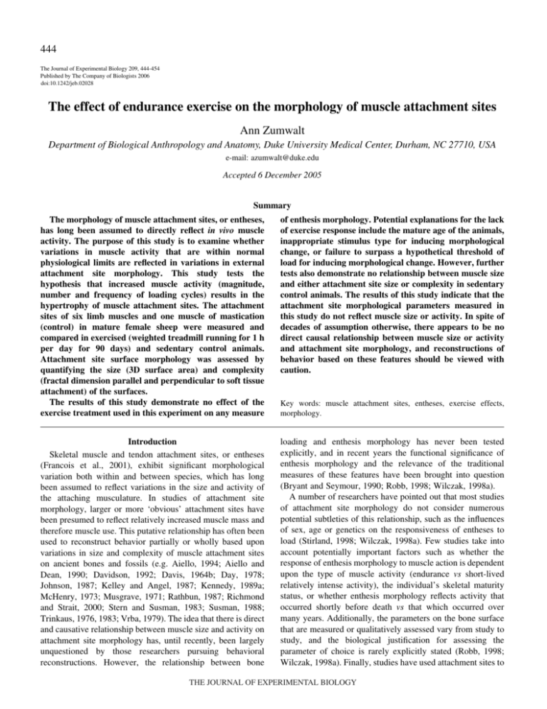
444 The Journal of Experimental Biology 209, 444-454 Published by The Company of Biologists 2006 doi:10.1242/jeb.02028 The effect of endurance exercise on the morphology of muscle attachment sites Ann Zumwalt Department of Biological Anthropology and Anatomy, Duke University Medical Center, Durham, NC 27710, USA e-mail: azumwalt@duke.edu Accepted 6 December 2005 Summary of enthesis morphology. Potential explanations for the lack of exercise response include the mature age of the animals, inappropriate stimulus type for inducing morphological change, or failure to surpass a hypothetical threshold of load for inducing morphological change. However, further tests also demonstrate no relationship between muscle size and either attachment site size or complexity in sedentary control animals. The results of this study indicate that the attachment site morphological parameters measured in this study do not reflect muscle size or activity. In spite of decades of assumption otherwise, there appears to be no direct causal relationship between muscle size or activity and attachment site morphology, and reconstructions of behavior based on these features should be viewed with caution. The morphology of muscle attachment sites, or entheses, has long been assumed to directly reflect in vivo muscle activity. The purpose of this study is to examine whether variations in muscle activity that are within normal physiological limits are reflected in variations in external attachment site morphology. This study tests the hypothesis that increased muscle activity (magnitude, number and frequency of loading cycles) results in the hypertrophy of muscle attachment sites. The attachment sites of six limb muscles and one muscle of mastication (control) in mature female sheep were measured and compared in exercised (weighted treadmill running for 1·h per day for 90 days) and sedentary control animals. Attachment site surface morphology was assessed by quantifying the size (3D surface area) and complexity (fractal dimension parallel and perpendicular to soft tissue attachment) of the surfaces. The results of this study demonstrate no effect of the exercise treatment used in this experiment on any measure Key words: muscle attachment sites, entheses, exercise effects, morphology. Introduction Skeletal muscle and tendon attachment sites, or entheses (Francois et al., 2001), exhibit significant morphological variation both within and between species, which has long been assumed to reflect variations in the size and activity of the attaching musculature. In studies of attachment site morphology, larger or more ‘obvious’ attachment sites have been presumed to reflect relatively increased muscle mass and therefore muscle use. This putative relationship has often been used to reconstruct behavior partially or wholly based upon variations in size and complexity of muscle attachment sites on ancient bones and fossils (e.g. Aiello, 1994; Aiello and Dean, 1990; Davidson, 1992; Davis, 1964b; Day, 1978; Johnson, 1987; Kelley and Angel, 1987; Kennedy, 1989a; McHenry, 1973; Musgrave, 1971; Rathbun, 1987; Richmond and Strait, 2000; Stern and Susman, 1983; Susman, 1988; Trinkaus, 1976, 1983; Vrba, 1979). The idea that there is direct and causative relationship between muscle size and activity on attachment site morphology has, until recently, been largely unquestioned by those researchers pursuing behavioral reconstructions. However, the relationship between bone loading and enthesis morphology has never been tested explicitly, and in recent years the functional significance of enthesis morphology and the relevance of the traditional measures of these features have been brought into question (Bryant and Seymour, 1990; Robb, 1998; Wilczak, 1998a). A number of researchers have pointed out that most studies of attachment site morphology do not consider numerous potential subtleties of this relationship, such as the influences of sex, age or genetics on the responsiveness of entheses to load (Stirland, 1998; Wilczak, 1998a). Few studies take into account potentially important factors such as whether the response of enthesis morphology to muscle action is dependent upon the type of muscle activity (endurance vs short-lived relatively intense activity), the individual’s skeletal maturity status, or whether enthesis morphology reflects activity that occurred shortly before death vs that which occurred over many years. Additionally, the parameters on the bone surface that are measured or qualitatively assessed vary from study to study, and the biological justification for assessing the parameter of choice is rarely explicitly stated (Robb, 1998; Wilczak, 1998a). Finally, studies have used attachment sites to THE JOURNAL OF EXPERIMENTAL BIOLOGY Exercise and attachment site morphology reconstruct a variety of parameters, including muscle size (Aiello, 1994; Churchill and Morris, 1998; Trinkaus, 1976), frequency of muscle use (Davidson, 1992; Hawkey and Merbs, 1995; Kelley and Angel, 1987; Stern and Susman, 1983; Wilczak, 1998b) and the magnitudes of forces produced during muscle contractions (Churchill and Morris, 1998; Kelley and Angel, 1987; Rathbun, 1987; Trinkaus, 1976). Again, the justifications for connecting enthesis morphology to these aspects of muscle morphology or activity are rarely stated (with one notable exception in Davidson, 1992). Clearly, the functional significance of attachment site morphology and the degree to which these features are responsive to load are still poorly understood and must be clarified before reliable behavioral reconstructions from these features are possible. The idea that more active or larger muscles should induce skeletal hypertrophy at the sites of their attachment appears intuitively reasonable. By definition, the mechanical stress experienced by a surface is proportional to the force experienced in each unit area of that surface (Biewener, 1992). It is therefore theoretically advantageous for bony attachment sites to hypertrophy in response to increased or unusual muscle forces as a mechanism of reducing stress at the interface between soft and hard tissues. There is indirect evidence that applications of external force to bone induce periosteal bone cell proliferation, indicating the existence of a mechanism by which bone can respond to muscle activity at the site of muscle attachment (Raab-Cullen et al., 1994). Additionally, abnormally strong or frequent muscle contractions may increase blood flow to periosteal bone, potentially hypertrophying and therefore strengthening the attachments of soft tissue fibers into the bone (Hawkey and Merbs, 1995; Herring, 1994). An example of such a relationship may be found in myostatin-null mice, a hypermuscular strain of genetic knock-out mice that have significantly larger deltoid crests and third trochanters than normal mice (Hamrick et al., 2000; Hamrick et al., 2002). Although it is currently unclear whether their hypertrophied attachment sites develop in response to stronger muscle pulls or simply because the large muscles require more area for attachment, myostatin-null mice provide an interesting example in which attachment site morphology reflects the attaching musculature. The functional significance of enthesis morphology is not, however, as straightforward as many believe. There are a number of studies that indicate that the visible features on bony surfaces do not fully or reliably reflect the actual extent of muscle attachment, and that the degree to which muscle scars reflect soft tissue attachment appears to vary between vertebrate lineages (Bryant and Seymour, 1990; Davis, 1964a; McGowan, 1979, 1986). Additionally, the asymmetry or relative robusticity of an individual’s skeleton may skew an observer’s assessment of the degree to which the sites are developed (Robb, 1998; Weiss, 2002). Therefore, the perception of an attachment site as being particularly faint or well-developed can be biased if the observer does not control for normal variations between lineages or the relative robusticity of the underlying bone. 445 Additionally, the degree to which attachment sites respond to external loads is poorly understood and certainly more complex than most interpretations of their morphology would suggest. Bone does not respond to all stimuli, and when it does, it responds differently in different conditions (Burr et al., 2002; Cullen et al., 2001; Currey, 2002; Judex and Zernicke, 2000b; Kontulainen, 2002; Lanyon et al., 1982; Matsuda et al., 1986; McLeod et al., 1998; Robling et al., 2000; Rubin and Lanyon, 1985; Turner, 1998; Turner et al., 1995a; Zernicke et al., 2001). Muscle attachments are sometimes, but not always, associated with osteogenesis at their points of attachment, and sometimes a muscle may attach to an area that is both depositional and resorptive in different locations at the same time (Hoyte and Enlow, 1966). A complicating issue is that there are a number of factors besides muscle size or activity that may contribute to the relative size or development of an attachment site or suite of attachment sites. An individual’s sex, age, hormone levels and genetics may all influence entheseal response to muscle activity (Stirland, 1998; Wilczak, 1998a), but the extents of these influences are currently entirely unknown. Additionally, an issue that is rarely considered in speculations about attachment sites’ response to load is that the interface between tendon or muscle and bone is likely designed to buffer the underlying bone from the strain created by muscle pulls. Most attachment sites are subjected to loads by muscle activity many times every day, and it is reasonable to suspect that mechanisms exist to protect the soft tissue–hard tissue interface from the effects of these loads. Indeed, at least two such mechanisms appear to exist. The first is a gradual change in tissue type at the sites of tendon attachments. From superficial to deep, a tendon’s fibers pass through four transitional zones to attach to bone: (I) tendon, (II) fibrocartilage, (III) calcified fibrocartilage and (IV) bone (Benjamin et al., 1986; Benjamin et al., 1991; Benjamin and Ralphs, 1998; Cooper and Misol, 1970; Dolgo-Saburoff, 1929; Evans et al., 1990; Thomas et al., 1999; Woo et al., 1988). This gradual transition between tissue types with distinctly different elastic moduli is thought to enhance the ability of tendons to dissipate force evenly during muscle contraction, thus resisting shear stresses at the bone surface (Cooper and Misol, 1970). Other muscles attach over very broad expanses of bone, reducing stress by dissipating force over a large area. The protective effects of these mechanisms imply that the bone at the site of a muscle or tendon attachment may not even be subjected to all variations in the activity of the attaching musculature, and that, by extension, its responsiveness is proportionately reduced. The extent and manner in which the bone at an attachment site responds to load is likely much less straightforward than many studies have implied in the past. The goal of the current study is to determine whether and how physiologically normal variations in muscle activity are reflected in the skeletal morphology of the sites to which the muscles are attaching. The morphological parameters traditionally used to reconstruct behavior are the size (Churchill and Morris, 1998; Davidson, 1992; Stirland, 1993; Wilczak, 1998a) and complexity (Hawkey, 1988; Hawkey THE JOURNAL OF EXPERIMENTAL BIOLOGY 446 A. Zumwalt and Merbs, 1995; Robb, 1998; Steen and Lane, 1998) of attachment sites. This study tests the hypothesis that moderate endurance exercise by a large mammal increases the size and complexities of the attachment sites of certain muscles that are active during the gait cycle. Materials and methods In this experiment, adult female sheep Ovis aries L., 4 years or older at the beginning of the study, were trained to an endurance exercise regimen that increased the magnitude, number and frequency of loading cycles on the attachment sites of interest. Adult animals were used for several reasons. First, most studies that reconstruct activity levels using attachment site morphology examine adult skeletons (Churchill and Morris, 1998; Hawkey and Merbs, 1995; Hawkey and Street, 1992; Steen and Lane, 1998), so this study examines the relationships between these factors in a population similar to those that are often studied. Second, the use of skeletally mature individuals removes potentially confounding effects of growth from this experiment (Biewener and Bertram, 1993, 1994; Loitz and Zernicke, 1992; Woo et al., 1981). In addition, the attachment sites of juvenile mammals are faint, variably developed and difficult to identify, because the muscles and tendons do not firmly attach to bone until skeletal maturity (Herring, 1994; Hurov, 1986; Lacroix, 1951; Matyas et al., 1990; J. Robb, unpublished; Woo et al., 1988). Finally, there is some evidence to indicate that attachment sites become more apparent with age (Lovejoy, 1973; J. Robb, unpublished; Wilczak, 1998b; Wilczak and Kennedy, 1997). However, it is unclear whether such age-related change is due to slowly accumulating microdamage from an ever-increasing number of loads throughout life, or to more global influences such as changes in metabolic or hormonal controls. Using only skeletally mature adults of the same sex in this study controls for these factors and examines a population that mimics those that are often studied in the paleontological literature. Exercise treatment In order to investigate the influence of muscle activity within normal (i.e. non-pathological) limits, the exercise regimen in this experiment was designed to provide a moderate increase in activity. The animals were divided into two weight-matched treatment groups (‘Exercised’ and ‘Controls’; N=10 per group). The exercised animals were trained to run on Marquette 1800 treadmills (Marquette Electronics Inc., Milwaukee, WI, USA) while carrying additional mass on their backs in backpacks designed for dogs. The animals were trained for 3 weeks, during which time their running speed and duration and the mass in the backpacks were slowly increased to their experiment levels. Care was taken not to train the animals too quickly to avoid the possibility of creating pathological effects due to injury at their muscle attachment sites. The training period culminated when the animals were able to run while wearing backpacks loaded with 20% of the animal’s body mass (Biewener and Bertram, 1994) for 60·min/day in 15·min intervals (2–4·min rests between intervals) at a constant Froude number of 0.65 (5.5–7·km·h–1), which is just below the trot–gallop transition. This exercise regimen was designed to increase the magnitude and frequency of muscle contractions on the attachment sites within normal (i.e. non-pathological) limits. It added approximately 7000 loading cycles to each limb per day, 5 days a week for 90 days. The ground reaction forces experienced by the limbs during this exercise was 65% greater in the forelimbs and 50% greater in the hind limbs than those experienced during unloaded walking (Zumwalt, 2005b). The experiment lasted for 90 days, which is longer than the bone formation period for sheep (74 days; Turner and Villanueva, 1994). The animals’ body masses were assessed three times during the duration of the experiment: on days 0, 45 and 90. The groups were housed in separate enclosures that permitted them to move and walk normally on natural sod and provided them with optional shelter from the elements. All animals were given access to identical diets and water ad libitum. At the end of the experiment, the animals were euthanized in accordance with the guidelines of the Institutional Animal Care and Use Committee of Harvard University. Muscle attachment sites A total of six muscles and their origins and/or insertions were analyzed in this study: the insertion of the infraspinatus muscle on the humeral head; the insertion of the biceps brachii muscle onto the radial tuberosity of the radius; the insertion of the patellar tendon of the quadriceps femoris into the tibial tuberosity on the proximal tibia; the lateral origin of the gastrocnemius muscle from the posterior distal femur; and the insertion of the gastrocnemius tendon onto the calcaneal tuberosity of the calcaneus. The attachment sites included here were chosen primarily because they all have distinct, obvious edges, thus minimizing the potential for measurement error during the quantification of various aspects of their forms. The insertion of the masseter muscle on the lateral surface of the mandible was also included as an internal control, as all of the animals in this study had identical diets and the level of exercise the animals underwent should not have affected the activity of this muscle. It should be noted that the tendons of two muscles (the flexor digitorum superficialis and the gastrocnemius) act on the site referred to here as the ‘gastrocnemius insertion’, and analyses of this site incorporated this information as relevant. The part of the flexor digitorum superficialis tendon that was analyzed in this study was the portion of the tendon between the muscle belly and the calcaneal tuberosity, but not the portion that continues distal to the tuberosity into the foot. Muscle, tendon, and bone preparation The soft tissue was dissected from the bones with great care so as not to damage the attachment sites of interest, and the masses of all muscles and tendons associated with the attachment sites examined here were immediately measured THE JOURNAL OF EXPERIMENTAL BIOLOGY Exercise and attachment site morphology (Anapol and Barry, 1996; Anton, 2000). To prepare them for detailed analyses of their attachment sites, the bones were placed in a dermestid beetle colony for 4–6 months, allowing the beetles to remove the majority of the soft tissue. At the end of this treatment, a small amount of tendon collagen still remained on some of the tendinous attachment sites. These bones were soaked in water until the collagen softened, and the collagen was carefully removed by hand. Great care was taken to not scratch or otherwise mar the bone at the site of attachment with the dissecting tools. Surface analysis Size and complexity assessments were made on digital three-dimensional (3D) reconstructions of the attachment sites, following the method described in detail in Zumwalt (2005a), which can be summarized as follows. The surfaces of the attachment sites on the dry bones were scanned using a Surveyor Model 810 laser scanner outfitted with an RPS-120 laser sensor (Laser Design, Inc., Minneapolis, MN, USA). Data collection was managed by Surveyor Scan Control (SSC) software (Laser Design, Inc; Minneapolis, MN, USA), which integrated all scan data for digital reconstruction of the surface. Geomagic Studio Reverse Engineering Software (Raindrop Geomagic, Research Triangle Park, NC, USA) handled data filtering and reverse engineering of the digital surface. The resolution of the scans used in this study was 0.023–0.027·mm, with a single point precision of 0.00635·mm. To remove redundant points, reduce file size and maintain a constant density in the point cloud, all scans were ultimately passed through a 3D Proximity Filter (Geomagic Studio Reverse Engineering Software) that removed points until the minimum distance between any two points in 3D space was 0.025·mm. The scans were then exported for analysis using ArcGIS 8.3 software (ESRI, Inc., Redlands, CA, USA), a popular Geographic Information Systems (GIS) program that is designed to analyze the shapes and distributions of landmasses. As ArcGIS cannot process the ASCII files exported by the Geomagic software, it was necessary to translate these files into a file format that was recognizable by ArcGIS. A Java program was written by the author to translate these files into text files and is freely available upon request. Analysis of 3D data The laser scan point cloud data were interpolated in ArcGIS using Inverse Distance Weighted nearest-neighbor resampling into 3D surface reconstructions. This resampling method dictates that the resolution of the reconstruction be as precise as the coarsest input, so each interpolated surface in this study had a slightly different resolution (mean=0.094±0.04·mm2). The attachment sites examined here have clear edges and delineation of these edges in two dimensions on a computer screen was straightforward; its accuracy was confirmed using an error study (Zumwalt, 2005b). Elevation ranges were assigned colors, so scans appeared as color-coded topographical maps in which variations in height appeared as variations in color. To isolate the attachment site surface from 447 the non-insertion bone, the edge of the attachment site was defined by the observer on the reconstruction while using the real bone as a reference to ensure accuracy. A new raster was then interpolated from the data within the digitized attachment site borders. All further analyses were performed on these reconstructions of the attachment sites themselves. The 3D surface area of each attachment site was calculated in ArcGIS. This calculation accounted for all 3D topography of the surface within the traced boundaries of the site. The surface complexities of the attachment sites were assessed by calculating the fractal dimensions of profiles extracted from the surfaces of the sites. The fractal dimension of a line indicates the degree to which the line appears complex at multiple levels of magnification. For example, examination of an extremely complex line at high levels of magnification reveals more complexity that was not necessarily apparent at lower levels of magnification (Kaye, 1994). A less complex line, however, appears simple at both high and low levels of magnification. Numerous algorithms exist that can quantify this phenomenon; the algorithm used in this study is described in detail elsewhere (Zumwalt, 2005a). By convention, a line can have a fractal dimension between 1 (simple) to 2 (infinitely complex). In this study, profiles were extracted at three equidistant locations along two perpendicular axes of the attachment site’s surface, one corresponding to the longitudinal axis of the soft tissue attachment (STA; Fig.·1A) (method described in detail in Zumwalt, 2005a). The locations of these profiles were determined relative to the maximum heights and widths of the attachment sites, except in the case of the masseter insertion. The majority of the masseter insertion’s surface is indistinguishable from non-insertion bone (though its edges are relatively obvious), except for a small portion along its anterior edge that projects distinctly from the bone’s surface. The complexity of this site was therefore assessed within this smaller, well-defined area. The fractal dimensions of these profiles were calculated using the R/S rescaled range algorithm in Benoit 1.3 (TruSoft International, Inc., St Petersburg, FL, USA) fractal analysis software to determine the complexity of the surface at that location. This method allows variations in surface complexity to be assessed both within attachment sites (Zumwalt, 2005a) and between the sites of different individuals. The fractal dimensions of the three profiles extracted along each major axis were also averaged to provide an assessment of average complexity in each axis. Non-parametric statistical tests were used in this study because many of the data sets that were examined had small sample sizes and were not normally distributed. All parameters describing the muscles, tendons and attachment site surface morphology of the exercised and sedentary control animals were compared between the treatment groups using Mann–Whitney U tests after controlling for body mass (Mb). To further examine the relationship between muscle size and attachment site morphology, correlations between muscle mass and both attachment site size and complexity were assessed individually for each attachment site in the sedentary controls using Spearman’s correlation tests. These correlations were run THE JOURNAL OF EXPERIMENTAL BIOLOGY 448 A. Zumwalt Profiles parallel to STA 1 A 2 3 Profiles perpendicular to STA Results Muscles and tendons For each limb attachment site examined in this study, either the attaching muscle or tendon was significantly larger in the exercised group than in the sedentary controls. Table·1 displays the median mass of each muscle and tendon assessed after controlling for each individual’s final body mass, and the results of comparisons of these values in the two groups. The infraspinatus, biceps brachii and quadriceps femoris muscles were significantly (15%, 9% and 12%, respectively) heavier in the exercised group than in the controls. The gastrocnemius tendon was significantly (about 16%) heavier in the exercised animals than in the control group. 4 5 6 Profile 5 B Z (elevation) Anterior–posterior scaling principles were used; i.e. lengths were divided by final Mb1/3, surface areas were divided by final Mb2/3, and masses and volumes were simply divided by final Mb. 1 cm Attachment site size After controlling for body mass, there were no significant differences between the groups in the size of any attachment site (Table·2, Fig.·2A), indicating that the exercise regime used in this study did not increase enthesis size, even in the cases in which the attaching muscle did hypertrophy. Correlations between muscle size and attachment site size were examined within the sedentary control animals to further examine whether there is a relationship between these parameters. There were no significant correlations between attachment site surface area and muscle size for any site at a significance level of P=0.05 (Table·3). The same patterns were observed both before and after controlling for body mass. The results of these tests Fig.·1. (A) The locations at which profiles were extracted from surface scans (shown: infraspinatus insertion). The white arrows indicate the direction of tendon attachment and solid black lines indicate the locations along which profiles were extracted. Three profiles (locations 1–3) were extracted at equidistant locations parallel to the axis of soft tissue attachment (STA) and three (locations 4–6) were extracted perpendicular to that attachment. The locations along each axis were defined at 25, 50 and 75% of the maximum width and height of the attachment, respectively. (B) Example of a profile extracted perpendicular to the STA (shown: Profile 5). both before and after controlling for Mb, in case muscle mass or attachment site size scale with Mb differently. Results were considered significant at P<0.05. To control for Mb, geometric Table·1. Soft tissue measurements in this study, summarized with the median (interquartile range) for each treatment group Tendon mass (⫻10–5) Muscle mass Exercised Sedentary Infraspinatus Biceps brachii 3.71 (0.747) 0.88 (0.11) 3.17 (0.41) 0.79 (0.08) Quadriceps Gastrocnemius Flexor digitorum superficialis Masseter 9.50 (1.32) 2.34 (0.67) 0.91 (0.59) 1.20 (0.35) 8.44 (0.67) 2.10 (0.33) 0.80 (0.51) 1.13 (0.33) † Exercised † 4.4 Proximal: 4.0† Distal: 0.7† 4.6† 10.3† 4.6† n/a Sedentary 3.6† 4.4† 0.5† 4.1† 9.3† 5.1† n/a indicates an interquartile range of approximately zero. All measures are controlled for final body mass. Muscle and tendon masses were divided by final body mass and the resulting numbers are dimensionless. For all significant differences, the exercised group is greater than the sedentary group (Mann–Whitney U tests). Exercised and sedentary pairs listed in bold are significantly different at P=0.05. n/a, not applicable. Values in parentheses are the interquartile range. THE JOURNAL OF EXPERIMENTAL BIOLOGY Exercise and attachment site morphology 449 Table·2. Surface morphology analysis Complexity 2 –2/3 3D Surface area (mm ·g Attachment site Infraspinatus Biceps brachii Quadriceps Gastrocnemius origin Gastrocnemius insertion Masseter ) Parallel to STA Perpendicular to STA Exercised Sedentary Exercised Sedentary Exercised Sedentary 12.60 (3.7) 4.20 (1.5) 10.65 (4.37) 37.1 (9.11) 40.7 (2.5) 96.6 (27.4) 10.50 (6.0) 4.54 (2.36) 9.15 (2.43) 37.6 (13.0) 25.0 (10.1) 103.0 (23.9) 1.62 (0.11) 1.65 (0.38) 1.56 (0.25) 1.36 (0.31) 1.49 (0.17) 1.45 (0.19) 1.65 (0.24) 1.68 (0.14) 1.52 (0.22) 1.56 (0.15) 1.52 (0.26) 1.50 (0.27) 1.61 (0.17) 1.78 (0.26) 1.62 (0.18) 1.45 (0.21) 1.39 (0.28) 1.28 (0.34) 1.64 (0.22) 1.77 (0.21) 1.55 (0.19) 1.50 (0.06) 1.34 (0.17) 1.34 (0.17) Three-dimensional surface areas and complexities are summarized with the median (interquartile range) of each attachment site measured in this study. Three-dimensional surface areas are controlled for final body mass. Complexity refers to the average fractal dimension of the three profiles extracted along and perpendicular to the soft tissue attachment (STA); fractal dimensions for a profile can be between 1 (smooth) and 2 (infinitely complex). There are no significant or near significant differences between treatment groups in 3D surface area or complexity in either axis for any attachment site measured in this study (Mann–Whitney U tests). further indicate no strong relationship between attachment site size and muscle size. Attachment site complexity The complexities (average fractal dimensions) of the attachment site surfaces parallel and perpendicular to the soft tissue insertion sites in the two treatment groups were compared using Mann–Whitney U tests. There were no significant differences between the treatment groups in overall surface complexity in either axis of any of the muscle attachment sites (Table·2; Fig.·2B,C). Following the same rationale described above regarding attachment site size, correlations between muscle size and attachment site complexity were examined in the control animals. These tests examined the average complexity measures parallel and perpendicular to the attaching soft tissue. Again, no significant correlations were found between muscle size and either of these measures of surface complexity in any of the attachment sites, either before or after controlling for body mass (Table·3). Discussion The results of this study indicate that endurance exercise has no effect on the morphology of the muscle attachment sites examined here. There were no reliable differences between the 2.0 1.9 1.8 125 A x Surface area (controlled for final Mb) 105 95 85 75 50 40 30 䊊 20 x 10 0 Fractal dimension Perpendicular to STA Parallel to STA 115 Fig.·2. Box and whisker plots of the (A) body mass (Mb) standardized surface areas and average fractal dimensions of profiles extracted along the axes (B) parallel and (C) perpendicular to the axis of soft tissue attachment (STA) of the attachment sites, as illustrated in Fig.·1. Values for the control group are indicated with black boxes and for the exercised group with white boxes. Boxes represent the interquartile range, or 50% of the values, and the line across the box indicates the median. The whiskers indicate the spread of observed values that fall within 1.5 times the interquartile range. Outliers are designated with (X) if they are between 1.5⫻ and 3⫻ the interquartile range, and (䊊) if they fall outside 3⫻ this range. Attachment sites: INF, Infraspinatus insertion; BB, Biceps brachii insertion; QF, Quadriceps femoris insertion; GO, Gastrocnemius origin; GI, Gastrocnemius insertion; MSTR, Masseter insertion. 1.7 1.6 1.5 1.4 1.3 1.2 1.1 1.9 1.8 THE JOURNAL OF EXPERIMENTAL BIOLOGY x C x 1.7 1.6 1.5 x 1.4 1.3 INF BB QF GO GI MSTR B 1.2 䊊 x INF BB QF GO GI MSTR 450 A. Zumwalt Table 3. Correlations of parameters documenting morphologies of attachment sites with muscle mass (Spearman non-parametric rank-order correlation test) in the control group only Complexity 3D surface area Attachment site Before controlling for body mass: Tendinous sites Infraspinatus Biceps brachii Quadriceps femoris Gastrocnemius insertion Fleshy sites Gastrocnemius lat. origin Masseter After controlling for body mass: Tendinous sites Infraspinatus Biceps brachii Quadriceps femoris Gastrocnemius insertion Fleshy sites Gastrocnemius lat. origin Masseter Parallel to STA Correlation coefficient P Correlation coefficient –0.164 –0.280 –0.200 0.667 0.651 0.432 0.580 0.071 0.055 –0.177 0.232 –0.183 0.285 0.317 0.425 0.406 0.358 0.370 0.433 –0.771 0.147 0 Perpendicular to STA Correlation coefficient P 0.880 0.625 0.519 0.637 0.231 0.037 –0.321 0.050 0.521 0.920 0.365 0.898 0.073 –0.452 0.841 0.260 –0.335 –0.548 0.343 0.160 0.310 0.293 0.244 0.072 –0.127 –0.273 –0.212 –0.300 0.726 0.446 0.556 0.624 –0.139 –0.273 –0.406 –0.086 0.701 0.446 0.244 0.872 0.535 1.000 –0.407 –0.071 0.243 0.879 0.103 –0.464 0.777 0.294 P The correlations between the various parameters are presented both before and after controlling muscle size and attachment site size for body mass. STA, soft tissue attachment. There are no significant correlations at P=0.05. One correlation approaching significance at P=0.10 is indicated in italics. treatment groups in the sizes of the attachment sites or in the complexity parallel or perpendicular to the soft tissue insertion, despite the fact that either the muscle or the tendon associated with every limb attachment site hypertrophied in response to the exercise (Table·1). These results challenge the long-held assumption that attachment site morphology simply and directly reflects in vivo muscle activity. Moreover, the lack of correlation between muscle size and either attachment site size or complexity in the sedentary control animals in this study further indicates that these skeletal features do not accurately reflect the size of the attaching musculature. These results are consistent with previous studies that have reported discrepancies between muscle size and attachment site morphology in some birds and reptiles (McGowan, 1979, 1986; Nicholls and Russell, 1985) and other mammals (Bryant and Seymour, 1990; Davis, 1964a). The results of this experiment therefore do not substantiate the long-held assumption that attachment site morphology reflects in vivo activity and muscle size. The functional significance of attachment site morphology is clearly more complex than many would like to believe, with many factors likely influencing this relationship in ways that are still poorly understood. Although variation in attachment site morphology exists, this variation appears not necessarily to reflect variations in in vivo loads. One reason for this may be the protective mechanisms at the soft tissue–hard tissue interface that have been described above. If these mechanisms do indeed buffer the influence of muscle activity on the underlying bone, variations in muscle activity will not necessarily be transferred into variations in enthesis morphology. This study implies that moderate endurance exercise is not a sufficient stimulus to induce skeletal response at attachment sites. A number of factors should be considered when contemplating the broader implications of this study. Age of subjects One possible explanation for the lack of bony response in this study is the fact that the sheep were skeletally mature at the beginning of the study. Most studies that have shown a response of the skeleton to load have examined growing animals (e.g. Biewener and Bertram, 1993; Biewener and Bertram, 1994; Hillam and Skerry, 1995; Loitz and Zernicke, 1992; Mosley and Lanyon, 2002; reviewed in Pearson and Lieberman, 2004; Woo et al., 1981). Those studies that have demonstrated a response to load in adult animals show that adult bone is less sensitive to exercise-induced changes in peak strain than younger, growing bone (Biewener and Bertram, 1993; Rubin et al., 1992; Turner et al., 1995b). Therefore, it is possible that the lack of exercise response by the attachment sites observed in this study may be ameliorated by examining THE JOURNAL OF EXPERIMENTAL BIOLOGY Exercise and attachment site morphology growing animals. However, as stated previously, most paleontological studies that have reconstructed behavior have done so on adults. The results of this study indicate that use of attachment sites is inappropriate for reconstructing recent behavior of skeletally mature individuals. In order to pursue the question of whether muscle activity is reflected in attachment site morphology fully, this relationship should be investigated in growing animals. Unfortunately, as stated above, the attachment sites of juvenile animals are difficult to observe and measure because muscles do not firmly attach to long bones before the bones cease growth, and their attachment sites are correspondingly faint (Herring, 1994; Hurov, 1986; Lacroix, 1951; Matyas et al., 1990; J. Robb, unpublished; Woo et al., 1988). Despite this methodological difficulty, the greater responsiveness of juvenile than adult bone to load suggests that the effect of exercise on the attachment sites of growing animals should be investigated as well. A separate but related question is whether the morphology of attachment sites varies with increasing age regardless of the activity level of the individual, as some evidence has suggested (Lovejoy, 1973; J. Robb, unpublished; Wilczak, 1998b; Wilczak and Kennedy, 1997). It is also currently unclear whether morphological variations that are induced by activity (if any prove to exist) are maintained in the absence of activity. Therefore, future work towards fully understanding attachment site functional morphology should investigate the relative influences that multiple age-related factors (e.g. activity levels before skeletal maturity, maintenance of morphological variations in the absence of activity, and age-related changes regardless of activity) have on adult attachment site morphology. Stimulus type The present study examined the response of the periosteal surface of cortical bone to increases in the magnitude and frequency of applied forces. While strain magnitude (Cullen et al., 2001; Mosley et al., 1997; Rubin and Lanyon, 1985) and frequency (Hsieh et al., 2001; Turner et al., 1995a; Warden and Turner, 2004) have been shown to influence osteogenesis, recent studies have demonstrated that cortical bone response to load is also influenced by other factors, such as the rate at which strains are applied (Burr et al., 2002; Judex and Zernicke, 2000b; Mosley and Lanyon, 1998; Turner et al., 1995a), the number of loading cycles (Forwood and Turner, 1994; Robling et al., 2002a; Turner and Villanueva, 1994), the distribution and gradient of applied strains (Judex et al., 1997) and periods of rest between loading bouts (Robling et al., 2000; Robling et al., 2002a,b). These studies suggest types of stimuli that may more dramatically alter the bone at the sites of muscle attachment than those induced in the present study. It is important to note, however, that most of the studies cited above examine the morphological response of bone to load by applying compressive, tensile and/or bending strains to the length of the entire bone. There are very few studies that have examined whether discrete, localized loads such as those 451 created by muscle contractions on attachment sites have local osteogenic effects. In one study, tensile forces induced the proliferation of osteoblast-like cells at the site of load application (Hirukawa et al., 2005), indicating that such loads may be able to induce local osteogenesis. However, the relative influences of the frequency, magnitude or rate of localized load applications on an osteogenic response are almost entirely unknown. For example, extremely forceful (high-magnitude) muscle pulls may more strongly influence attachment site morphology than the forces examined in the current study. Many examples of enthesis hypertrophy appear to be associated with the repetitive and powerful muscle pulls created during manual labor or competitive sports (Dutour, 1986; Hill et al., 1995; Kelley and Angel, 1987; Kennedy, 1989b; Priest et al., 1974; Wilczak and Kennedy, 1997). However, as entheses appear to be designed to protect the underlying bone from injury during muscle contraction (see above), forceful muscle pulls may not affect enthesis morphology until they reach pathological levels. Further study is therefore needed to determine the relative influences of different types of localized loads on bone. Threshold of bone response It has often been posited that a mechanical stimulus must differ substantially from a bone’s habitual milieu for an osteogenic response to occur in bone (Judex and Zernicke, 2000a). Many researchers have speculated that bone only responds to strains that are beyond a hypothetical threshold (Frost, 1987; Rubin and Lanyon, 1984), and there is evidence that the threshold of response varies with location in the bone (Currey, 2002; Hsieh et al., 2001). Given that muscle attachment sites inherently experience a higher proportion of localized forces than other locations on the bone, the response thresholds of these locations to such forces may be set very high. Perhaps the muscle forces produced during the endurance exercise experienced by the animals in this experiment were simply not high enough to surpass such a threshold. Future work that is focused on fully understanding the relationship between load and enthesis morphology should attempt to document whether such a threshold exists at these sites, and, if it does, determine the loads necessary to surpass it and induce osteogenesis. Conclusions This study demonstrates that there is not a simple direct relationship between attachment site morphology and the action or size of the attaching muscle. However, variation in attachment site morphology does exist and likely reflects some aspect or aspects of in vivo stimuli. As discussed above, the present study explored the influence of only one of many possible parameters on attachment site morphology, and many questions remain about the functional significance of these features. More work is needed before the influence of muscle size or activity and attachment site morphology may be fully understood. Specifically, future work should investigate whether attachment sites are more responsive to load prior to THE JOURNAL OF EXPERIMENTAL BIOLOGY 452 A. Zumwalt skeletal maturity (Biewener and Bertram, 1993; Rubin et al., 1992; Turner et al., 1995b), and whether attachment site morphology varies with age, regardless of activity level (Lovejoy, 1973; J. Robb, unpublished; Wilczak, 1998b; Wilczak and Kennedy, 1997). Additionally, future work should examine whether loads that more closely approach pathological levels influence enthesis morphology more dramatically than did the endurance-type exercise performed in this study (Kennedy, 1989a; Wilczak and Kennedy, 1997). Finally, the extent to which normal variations in morphology reflect an individual’s age or genetics rather than the size or activity of the attaching muscle should be explored (Stirland, 1998; Wilczak, 1998a). Muscle attachment sites provide an interesting challenge to scientists attempting to reconstruct activity levels using skeletal remains. Skeletal attachment sites are intimately connected with the attaching muscles, so have the potential to provide a window into the in vivo activity of those muscles. However, protective mechanisms appear to exist to shield the bone at these sites from strain caused by muscle pulls, and the evidence presented in this study implies that variations in load are not necessarily translated to the bone at these sites. While extensive literature exists documenting that the skeleton can and does respond to external loads, this relationship is almost entirely unexplored in the context of discrete, localized loads such as those at the sites of muscle attachments. At a minimum, it is clear that the relationship between muscle activity and attachment site morphology is neither as simple nor as obvious as morphologists have long believed it to be. Until the functional significance of attachment site morphology is more thoroughly understood, behavior reconstructions based on these features in ancient skeletons should be viewed with caution. The author would like to sincerely thank Drs Andrew Biewener and Daniel Lieberman for providing the use of the Concord Field Station at Harvard University for this study. Pedro Martinez, Craig McGowan, Russell Main and James Usherwood all provided extensive aid during the course of the experiment. Drs Suzanne Strait and Peter Ungar provided helpful support and advice relating to the laser scanning procedure. Finally, Drs Daniel Schmitt, Christopher Ruff and Mark Hamrick, and Jandy Hanna, all gave invaluable commentaries on earlier versions of this manuscript and their advice is sincerely appreciated. This study was supported by NSF DIG-0209411, Sigma Xi and a Journal of Experimental Biology Travelling Fellowship. References Aiello, L. (1994). Variable but singular. Nature 368, 399-400. Aiello, L. and Dean, C. (1990). An Introduction to Human Evolutionary Anatomy. New York: Academic Press. Anapol, F. and Barry, K. (1996). Fiber architecture of the extensors of the hindlimb in semiterrestrial and arboreal guenons. Am. J. Phys. Anthropol. 99, 429-447. Anton, S. C. (2000). Macaque pterygoid muscles: Internal architecture, fiber length and cross-sectional area. Int. J. Primatol. 21, 131-156. Benjamin, M. and Ralphs, J. (1998). Fibrocartilage in tendons and ligaments – an adaptation to compressive load. J. Anat. 193, 481-494. Benjamin, M., Evans, E. and Copp, L. (1986). The histology of tendon attachments to bone in man. J. Anat. 149, 89-100. Benjamin, M., Evans, E., Donthineni Rao, R., Findlay, J. and Pemberton, D. (1991). Quantitative differences in the histology of the attachment zones of the meniscal horns in the knee joint of man. J. Anat. 177, 127-134. Biewener, A. (1992). Overview of structural mechanics. In Biomechanics (Structures and Systems): A Practical Approach (ed. A. Biewener), pp. 120. New York: Oxford University Press. Biewener, A. A. and Bertram, J. E. A. (1993). Skeletal strain patterns in relation to exercise training during growth. J. Exp. Biol. 185, 51-69. Biewener, A. A. and Bertram, J. E. A. (1994). Structural response of growing bone to exercise and disuse. J. Appl. Physiol. 76, 946-955. Bryant, H. and Seymour, K. (1990). Observations and comments on the reliability of muscle reconstruction in fossil vertebrates. J. Morphol. 206, 109-117. Burr, D. B., Robling, A. G. and Turner, C. H. (2002). Effects of biomechanical stress on bones in animals. Bone 30, 781-786. Churchill, S. and Morris, A. (1998). Muscle marking morphology and labour intensity in prehistoric Khoisan foragers. Int. J. Osteoarchaeol. 8, 390-411. Cooper, R. and Misol, S. (1970). Tendon and ligament insertion. J. Bone Joint Surg. A 52, 1-20. Cullen, D. M., Smith, R. T. and Akhter, P. (2001). Bone-loading response varies with strain magnitude and cycle number. J. Appl. Physiol. 91, 19711976. Currey, J. (2002). Bones: Structure and Mechanics. Princeton: Princeton University Press. Davidson, K. (1992). Behavioral significance of variations in the morphology of the mastoid. In Department Anthropology, p. 69. Los Angeles: University of California. Davis, D. D. (1964a). The giant panda. A morphological study of evolutionary mechanisms. Fieldiana Zool. Mem. 3, 1-339. Davis, P. (1964b). Hominid fossils from Bed I, Olduvai Gorge, Tanganyika. Nature 201, 967-970. Day, M. (1978). Functional interpretations of the morphology of postcranial remains of early African hominids. In Early Hominids of Africa (ed. C. Jolly), pp. 311-346. New York: St Martin’s Press. Dolgo-Saburoff, B. (1929). Uber Ursprung und Insertion der Skelettmuskeln. Anat. Anz. 68, 30-87. Dutour, O. (1986). Enthesopathies (lesions of muscular insertions) as indicators of the activites of Neolithic Saharan populations. Am. J. Phys. Anthropol. 71, 221-224. Evans, E., Benjamin, M. and Pemberton, D. (1990). Fibrocartilage in the attachment zones of the quadriceps tendon and patellar ligament of man. J. Anat. 171, 155-162. Forwood, M. and Turner, C. H. (1994). The response of rat tibiae to incremental bouts of mechanical loading: A quantum concept of bone formation. Bone 15, 603-609. Francois, R., Braun, J. and Khan, M. (2001). Entheses and enthesitis: a histopathologic review and relevance to spondyloarthritis. Curr. Opin. Rheumatol. 13, 255-264. Frost, H. (1987). The mechanostat: A proposed pathogenic mechanism of osteoporoses and the bone mass effects of mechanical and nonmechanical agents. Bone Mineral 2, 73-85. Hamrick, M., McPherron, A., Lovejoy, C. and Hudson, J. (2000). Femoral morphology and cross-sectional geometry of adult myostatin-deficient mice. Bone 27, 343-349. Hamrick, M. W., McPherron, A. C. and Lovejoy, C. (2002). Bone mineral content and density in the humerus of adult myostatin-deficient mice. Calcified Tissue Int. 71, 63-68. Hawkey, D. (1988). Use of upper extremity enthesopathies to indicate habitual activity patterns. In Anthropology. Tempe: Arizona State University. Hawkey, D. and Merbs, C. (1995). Activity-induced musculoskeletal stress markers (MSM) and subsistence strategy changes among ancient Hudson Bay Eskimos. Int. J. Osteoarchaeol. 5, 324-338. Hawkey, D. E. and Street, S. R. (1992). Activity-induced stress markers in prehistoric human remains from the Eastern Aleutian Islands. In 61st Meeting of the American Association of Physical Anthropologists. Las Vegas: American Association of Physical Anthropologists. Herring, S. W. (1994). Development of functional interactions between skeletal and muscular systems. In Differentiation and Morphogenesis of Bone, Vol. 9 (ed. B. K. Hall). Boca Raton: CRC Press. THE JOURNAL OF EXPERIMENTAL BIOLOGY Exercise and attachment site morphology Hill, M. C., Blakey, M. L. and Mack, M. E. (1995). Women, endurance, enslavement: Exceeding the physiological limits. In 64th meeting of the American Association of Physical Anthropologists. Oakland: American Association of Physical Anthropologists. Hillam, R. A. and Skerry, T. M. (1995). Inhibition of bone resorption and stimulation of formation by mechanical loading of the modeling rat ulna in vivo. J. Bone Miner. Res 10, 683-689. Hirukawa, K., Miyazawaa, K., Maedab, H., Kameyamab, Y., Gotoa, S. and Togaric, A. (2005). Effect of tensile force on the expression of IGF-I and IGF-I receptor in the organ-cultured rat cranial suture. Arch. Oral Biol. 50, 367-372. Hoyte, D. A. N. and Enlow, D. H. (1966). Wolff’s law and the problem of muscle attachment on resorptive surfaces of bone. Am. J. Phys. Anthropol. 24, 205-214. Hsieh, Y.-F., Robling, A. G., Ambrosius, W. T., Burr, D. B. and Turner, C. H. (2001). Mechanical loading of diaphyseal bone in vivo: The strain threshold for osteogenic response varies with location. J. Bone Miner. Res. 16, 2291-2297. Hurov, J. R. (1986). Soft-tissue bone interface: How do attachments of muscles, tendons, and ligaments change during growth? A light microscopic study. J. Morphol. 189, 313-325. Johnson, R. (1987). A classification of Sharpey’s fibers within the alveolar bone of the mouse: A high-voltage electron microscope study. Anat. Rec. 217, 339-347. Judex, S. and Zernicke, R. (2000a). Does the mechanical milieu associated with high-speed running lead to adaptive changes in diaphyseal growing bone? Bone 26, 153-159. Judex, S. and Zernicke, R. (2000b). High-impact exercise and growing bone: relation between high strain rates and enhanced bone formation. J. Appl. Physiol. 88, 2183-2191. Judex, S., Gross, T. and Zernicke, R. F. (1997). Strain gradients correlate with sites of exercise-induced bone-forming surfaces in the adult skeleton. J. Bone Miner. Res. 12, 1737-1745. Kaye, B. H. (1994). A Random Walk Through Fractal Dimensions. New York: VCH Publishers. Kelley, J. and Angel, J. (1987). Life stresses of slavery. Am. J. Phys. Anthropol. 74, 199-211. Kennedy, K. (1989a). Skeletal markers of occupational stress. In Reconstruction of Life from the Skeleton (ed. M. Iscan and K. Kennedy), pp. 129-160. New York: Alan R. Liss. Kennedy, K. A. R. (1989b). Skeletal markers of occupational stress. In Reconstruction of Life from the Skeleton (ed. M. Iscan and K. Kennedy), pp. 129-160. New York: Alan R. Liss. Kontulainen, S. (2002). Training, detraining and bone – Effect of exercise on bone mass and structure with special reference to maintenance of the exercise-induced bone gain. In Studies in Sport, Physical Education and Health. Jyvaskyla, Finland: University of Jyvaskyla. Lacroix, P. (1951). The Organization of Bones. Philadelphia: The Blakiston Company. Lanyon, L. E., Goodship, A. E., Pye, C. J. and MacFie, J. H. (1982). Mechanically adaptive bone remodelling. J. Biomechanics 15, 141-154. Loitz, B. J. and Zernicke, R. F. (1992). Strenuous exercise-induced remodeling of mature bone: relationships between in vivo strains and bone mechanics. J. Exp. Biol. 170, 1-18. Lovejoy, C. (1973). Biomechanical perspectives on the lower limb of early hominids. In Primate Functional Morphology and Evolution (ed. R. Tuttle). Chicago: Mouton Publishers. Matsuda, J. J., Zernicke, R. F., Vailas, A. C., Pedrini, V. A., Pedrini-Mille, A. and Maynard, J. A. (1986). Structural and mechanical adaptation of immature bone to strenuous exercise. J. Appl. Physiol. 60, 2028-2034. Matyas, J., Bodie, D., Anderson, M. and Frank, C. (1990). The developmental morphology of a ‘periosteal’ ligament insertion: Growth and maturation of the tibial insertion of the rabbit medial collateral ligament. J. Orthop. Research 8, 412-424. McGowan, C. (1979). The hind limb musculature of the brown kiwi, Apteryx australis mantelli. J. Morphol. 160, 22-73. McGowan, C. (1986). The wing musculature of the Weka (Gallirallus australis), a flightless rail endemic to New Zealand. J. Zool. Lond. A 210, 305-346. McHenry, H. (1973). Early hominid humerus from East Rudolf, Kenya. Science 180, 739-741. McLeod, K., Rubin, C., Otter, M. and Qin, Y. (1998). Skeletal cell stresses and bone adaptation. Am. J. Med. Sci. 316, 176-183. Mosley, J. and Lanyon, L. (2002). Growth rate rather than gender determines 453 the size of the adaptive response of the growing skeleton to mechanical strain. Bone 30, 314-319. Mosley, J. R. and Lanyon, L. E. (1998). Strain rate as a controlling influence on adaptive modeling in response to dynamic loading of the ulna in growing male rats. Bone 23, 313-318. Mosley, J. R., March, B. M., Lynch, J. and Lanyon, L. E. (1997). Strain magnitude related changes in whole bone architecture in growing rats. Bone 20, 191-198. Musgrave, J. (1971). How dextrous was Neanderthal man? Nature 233, 538541. Nicholls, E. and Russell, A. (1985). Structure and function of the pectoral girdle and forelimb of Struthiomimus altus (Theropoda: Ornithomimidae). Paleontology 28, 643-677. Pearson, O. M. and Lieberman, D. E. (2004). The aging of Wolff’s ‘Law’: Ontogeny and responses to mechanical loading in cortical bone. Yearb. Phys. Anthropol. 47, 63-99. Priest, J., Jones, H. and Nagel, D. (1974). Elbow injuries in highly skilled tennis players. J. Sports Med. 2, 137-149. Raab-Cullen, D. M., Thiede, M., Petersen, D., Kimmel, D. and Recher, R. (1994). Mechanical loading stimulates rapid changes in periosteal gene expression. Calcified Tissue Int. 55, 473-478. Rathbun, T. (1987). Health and disease at a South Carolina plantation: 18401870. Am. J. Phys. Anthropol. 74, 239-253. Richmond, B. and Strait, D. (2000). Evidence that humans evolved from a knuckle-walking ancestor. Nature 404, 382-340. Robb, J. (1998). The interpretation of skeletal muscle sites: A statistical approach. Int. J. Osteoarchaeol. 8, 363-377. Robling, A. G., Burr, D. B. and Turner, A. S. (2000). Partitioning a daily mechanical stimulus into discrete loading bouts improves the osteogenic response to loading. J. Bone Miner. Res. 15, 1596-1602. Robling, A. G., Hinant, F. M., Burr, D. B. and Turner, C. H. (2002a). Improved bone structure and strength after long-term mechanical loading is greatest if loading is separated into short bouts. J. Bone Miner. Res. 17, 1545-1554. Robling, A. G., Hinant, F. M., Burr, D. B. and Turner, C. H. (2002b). Shorter, more frequent mechanical loading sessions enhance bone mass. Med. Sci. Sports Exercise 34, 196-202. Rubin, C. T. and Lanyon, L. E. (1984). Regulation of bone formation by applied dynamic loads. J. Bone Joint Surg. A 66, 397-402. Rubin, C. T. and Lanyon, L. E. (1985). Regulation of bone mass by mechanical strain magnitude. Calcified Tissue Int. 37, 411-417. Rubin, C. T., Bain, S. and McLeod, K. (1992). Suppression of the osteogenic response in the aging skeleton. Calcified Tissue Int. 50, 306-313. Steen, S. and Lane, R. (1998). Evaluation of habitual activities among two Alaskan Eskimo populations based on musculoskeletal stress markers. Int. J. Osteoarchaeol. 8, 341-353. Stern, J. and Susman, R. (1983). The locomotor anatomy of Australopithecus afarensis. Am. J. Phys. Anthropol. 60, 279-317. Stirland, A. (1993). Asymmetry and activity-related change in the male humerus. Int. J. Osteoarchaeol. 3, 105-113. Stirland, A. (1998). Musculoskeletal evidence for activity: Problems of evaluation. Int. J. Osteoarchaeol. 8, 354-362. Susman, R. (1988). Hand of Paranthropus robustus from Member 1, Swartkrans: Fossil evidence for tool behavior. Science 240, 781-784. Thomas, D., Inoue, N., Cosgarea, A. and Chao, E. (1999). A histomorphometric analysis of the human patellar tendon insertion. In Orthopaedic Research Society. Anaheim: Orthopaedic Research Society. Trinkaus, E. (1976). The evolution of the hominid femoral diaphysis during the Upper Pleistocene in Europe and the Near East. Zeitschr. Morphol. Anthropol. 67, 291-319. Trinkaus, E. (1983). Functional aspects of Neandertal pedal remains. Foot Ankle 3, 377-390. Turner, C. (1998). Three rules for bone adaptation to mechanical stimuli. Bone 23, 399-407. Turner, A. and Villanueva, A. (1994). Static and dynamic histomorphometric data in 9- to 11-year-old ewes. Vet. Comp. Orthop. Traumatol. 7, 101-109. Turner, C. H., Owan, I. and Takano, Y. (1995a). Mechanotransduction in bone: role of strain rate. Am. J. Physiol. 269, E438-E442. Turner, C. H., Takano, Y. and Owan, I. (1995b). Aging changes mechanical loading thresholds for bone formation in rats. J. Bone Miner. Res. 10, 15441549. Vrba, E. (1979). A new study of the scapula of Australopithecus africanus from Sterkfontein. Am. J. Phys. Anthropol. 51, 117-130. THE JOURNAL OF EXPERIMENTAL BIOLOGY 454 A. Zumwalt Warden, S. J. and Turner, C. H. (2004). Mechanotransduction in cortical bone is most efficient at loading frequencies of 5-10Hz. Bone 34, 261270. Weiss, E. (2002). The mystery of muscle markers: Aggregation and contruct validity. In American Association of Physical Anthropologists. Buffalo: American Association of Physical Anthropologists. Wilczak, C. (1998a). Consideration of sexual dimorphism, age, and asymmetry in quantitative measurements of muscle insertion sites. Int. J. Osteoarchaeol. 8, 311-325. Wilczak, C. (1998b). A new method for quantifying musculoskeletal stress markers (MSM): A test of the relationship between enthesis size and habitual activity in archaeological populations. In Physical Anthropology. Ithaca: Cornell University. Wilczak, C. and Kennedy, K. (1997). Mostly MOS: Technical aspects of identification of skeletal markers of occupational stress. In Forensic Osteology: Advances in the Identification of Human Remains (ed. K. J. Reichs), pp. 461-490. Springfield: Charles C. Thomas. Woo, S. L., Kuei, S. C., Amiel, D., Gomez, M. A., Hayes, W. C., White, F. C. and Akeson, W. H. (1981). The effect of prolonged physical training on the properties of long bone: a study of Wolff’s Law. J. Bone Joint Surg. Am. 63, 780-787. Woo, S., Maynard, J., Butler, D., Lyon, R., Torzilli, P., Akeson, W., Cooper, R. and Oakes, B. (1988). Ligament, tendon and joint capsule insertions to bone. In Injury and Repair of the Musculoskeletal Soft Tissues (ed. S. L.-Y. Woo and J. Buckwalter), pp. 133-166. Park Ridge: American Academy of Orthopaedic Surgeons. Zernicke, R., Wohl, G., Boyd, S. and Judex, S. (2001). Functional adaptation of bone. J. Med. Biol. Eng. 21, 75-78. Zumwalt, A. (2005a). A new method for quantifying the complexity of muscle attachment sites. Anat. Rec. B 286, 21-28. Zumwalt, A. C. (2005b). The effect of endurance exercise on the morphology of muscle attachment sites: An experimental study in sheep (Ovis aries). In Center for Functional Anatomy and Evolution, PhD Vol. pp. 272. Baltimore: Johns Hopkins University School of Medicine. THE JOURNAL OF EXPERIMENTAL BIOLOGY
