12.4 G Protein–Coupled Receptors and Second Messengers
advertisement
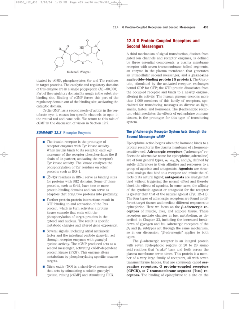
8885d_c12_435 2/20/04 1:19 PM Page 435 mac76 mac76:385_reb: 12.4 O N HN N S N N 435 12.4 G Protein–Coupled Receptors and Second Messengers O O G Protein–Coupled Receptors and Second Messengers O N Sildenafil (Viagra) tivated by cGMP, phosphorylates Ser and Thr residues in target proteins. The catalytic and regulatory domains of this enzyme are in a single polypeptide (Mr ∼80,000). Part of the regulatory domain fits snugly in the substratebinding site. Binding of cGMP forces this part of the regulatory domain out of the binding site, activating the catalytic domain. Cyclic GMP has a second mode of action in the vertebrate eye: it causes ion-specific channels to open in the retinal rod and cone cells. We return to this role of cGMP in the discussion of vision in Section 12.7. SUMMARY 12.3 Receptor Enzymes ■ The insulin receptor is the prototype of receptor enzymes with Tyr kinase activity. When insulin binds to its receptor, each monomer of the receptor phosphorylates the chain of its partner, activating the receptor’s Tyr kinase activity. The kinase catalyzes the phosphorylation of Tyr residues on other proteins such as IRS-1. ■ P –Tyr residues in IRS-1 serve as binding sites for proteins with SH2 domains. Some of these proteins, such as Grb2, have two or more protein-binding domains and can serve as adaptors that bring two proteins into proximity. ■ Further protein-protein interactions result in GTP binding to and activation of the Ras protein, which in turn activates a protein kinase cascade that ends with the phosphorylation of target proteins in the cytosol and nucleus. The result is specific metabolic changes and altered gene expression. ■ Several signals, including atrial natriuretic factor and the intestinal peptide guanylin, act through receptor enzymes with guanylyl cyclase activity. The cGMP produced acts as a second messenger, activating cGMP-dependent protein kinase (PKG). This enzyme alters metabolism by phosphorylating specific enzyme targets. ■ Nitric oxide (NO) is a short-lived messenger that acts by stimulating a soluble guanylyl cyclase, raising [cGMP] and stimulating PKG. A third mechanism of signal transduction, distinct from gated ion channels and receptor enzymes, is defined by three essential components: a plasma membrane receptor with seven transmembrane helical segments, an enzyme in the plasma membrane that generates an intracellular second messenger, and a guanosine nucleotide–binding protein (G protein). The G protein, stimulated by the activated receptor, exchanges bound GDP for GTP; the GTP-protein dissociates from the occupied receptor and binds to a nearby enzyme, altering its activity. The human genome encodes more than 1,000 members of this family of receptors, specialized for transducing messages as diverse as light, smells, tastes, and hormones. The -adrenergic receptor, which mediates the effects of epinephrine on many tissues, is the prototype for this type of transducing system. The -Adrenergic Receptor System Acts through the Second Messenger cAMP Epinephrine action begins when the hormone binds to a protein receptor in the plasma membrane of a hormonesensitive cell. Adrenergic receptors (“adrenergic” reflects the alternative name for epinephrine, adrenaline) are of four general types, 1, 2, 1, and 2, defined by subtle differences in their affinities and responses to a group of agonists and antagonists. Agonists are structural analogs that bind to a receptor and mimic the effects of its natural ligand; antagonists are analogs that bind without triggering the normal effect and thereby block the effects of agonists. In some cases, the affinity of the synthetic agonist or antagonist for the receptor is greater than that of the natural agonist (Fig. 12–11). The four types of adrenergic receptors are found in different target tissues and mediate different responses to epinephrine. Here we focus on the -adrenergic receptors of muscle, liver, and adipose tissue. These receptors mediate changes in fuel metabolism, as described in Chapter 23, including the increased breakdown of glycogen and fat. Adrenergic receptors of the 1 and 2 subtypes act through the same mechanism, so in our discussion, “-adrenergic” applies to both types. The -adrenergic receptor is an integral protein with seven hydrophobic regions of 20 to 28 amino acid residues that “snake” back and forth across the plasma membrane seven times. This protein is a member of a very large family of receptors, all with seven transmembrane helices, that are commonly called serpentine receptors, G protein–coupled receptors (GPCR), or 7 transmembrane segment (7tm) receptors. The binding of epinephrine to a site on the 8885d_c12_436 436 2/20/04 1:19 PM Chapter 12 Page 436 mac76 mac76:385_reb: Biosignaling CH HO HO CH2 NH CH3 5 Epinephrine OH CH HO CH3 CH2 NH Isoproterenol (agonist) HO CH2 CH CH 0.4 CH3 CH3 OH O receptor deep within the membrane (Fig. 12–12, step 1 ) promotes a conformational change in the receptor’s intracellular domain that affects its interaction with the second protein in the signal-transduction pathway, a heterotrimeric GTP-binding stimulatory G protein, or GS, on the cytosolic side of the plasma membrane. Alfred G. Gilman and Martin Rodbell discovered that when GTP is bound to Gs, Gs stimulates the production of cAMP by adenylyl cyclase (see below) in the plasma membrane. The function of Gs as a molecular switch resembles that of another class of G proteins typified by Ras, discussed in Section 12.3 in the context of the insulin receptor. Structurally, Gs and Ras are quite distinct; G proteins of the Ras type are monomers (Mr ∼20,000), whereas the G proteins that interact with serpentine kd (M) OH CH2 NH CH 0.0046 CH3 Propranolol (antagonist) FIGURE 12–11 Epinephrine and its synthetic analogs. Epinephrine, also called adrenaline, is released from the adrenal gland and regulates energy-yielding metabolism in muscle, liver, and adipose tissue. It also serves as a neurotransmitter in adrenergic neurons. Its affinity for its receptor is expressed as a dissociation constant for the receptor-ligand complex. Isoproterenol and propranolol are synthetic analogs, one an agonist with an affinity for the receptor that is higher than that of epinephrine, and the other an antagonist with extremely high affinity. Alfred G. Gilman Martin Rodbell, 1925–1998 1 Epinephrine binds to its specific receptor. NH3 E AC GDP Gs OOC Rec 2 The occupied receptor causes replacement of the GDP bound to Gs by GTP, activating Gs. GTP Gs GTP GDP 3 Gs ( subunit) moves to adenylyl cyclase and activates it. ATP 4 Adenylyl cyclase cAMP catalyzes the formation of cAMP. FIGURE 12–12 Transduction of the epinephrine signal: the adrenergic pathway. The seven steps of the mechanism that couples binding of epinephrine (E) to its receptor (Rec) with activation of adenylyl cyclase (AC) are discussed further in the text. The same adenylyl cyclase molecule in the plasma membrane may be regulated by a stimulatory G protein (Gs), as shown, or an inhibitory G protein (Gi, not shown). Gs and Gi are under the influence of different hormones. Hormones that induce GTP binding to Gi cause inhibition of adenylyl cyclase, resulting in lower cellular [cAMP]. 5 cAMP activates PKA. cyclic nucleotide phosphodiesterase 5-AMP 7 cAMP is degraded, reversing the activation of PKA. 6 Phosphorylation of cellular proteins by PKA causes the cellular response to epinephrine. 8885d_c12_437 2/20/04 1:19 PM Page 437 mac76 mac76:385_reb: 12.4 receptors are trimers of three different subunits, (Mr 43,000), (Mr 37,000), and (Mr 7,500 to 10,000). When the nucleotide-binding site of Gs (on the subunit) is occupied by GTP, Gs is active and can activate adenylyl cyclase (AC in Fig. 12–12); with GDP bound to the site, Gs is inactive. Binding of epinephrine enables the receptor to catalyze displacement of bound GDP by GTP, converting Gs to its active form (step 2 ). As this occurs, the and subunits of Gs dissociate from the subunit, and Gs, with its bound GTP, moves in the plane of the membrane from the receptor to a nearby molecule of adenylyl cyclase (step 3 ). The Gs is held to the membrane by a covalently attached palmitoyl group (see Fig. 11–14). Adenylyl cyclase (Fig. 12–13) is an integral protein of the plasma membrane, with its active site on the cytosolic face. It catalyzes the synthesis of cAMP from ATP: G Protein–Coupled Receptors and Second Messengers 437 AC GTP Gs NH2 N N O O P O O O P O O O N N CH2 P O O FIGURE 12–13 Interaction of Gs with adenylyl cyclase. (PDB ID O H H H H OH OH 1AZS) The soluble catalytic core of the adenylyl cyclase (AC, blue), severed from its membrane anchor, was cocrystallized with Gs (green) to give this crystal structure. The plant terpene forskolin (yellow) is a drug that strongly stimulates the enzyme, and GTP (red) bound to Gs triggers interaction of Gs with adenylyl cyclase. ATP PPi NH2 N N 1 Gs with GDP bound is turned off; it cannot activate adenylyl cyclase. 2 Contact of Gs with hormone-receptor complex causes displacement of bound GDP by GTP. GTP N N 5 CH2 O O H H GDP H H 3 O O P O 3 Gs with GTP bound dissociates into a and bg subunits. Gsa-GTP is turned on; it can activate adenylyl cyclase. OH Adenosine 3,5-cyclic monophosphate (cAMP) The association of active Gs with adenylyl cyclase stimulates the cyclase to catalyze cAMP synthesis (Fig. 12–12, step 4 ), raising the cytosolic [cAMP]. This stimulation by Gs is self-limiting; Gs is a GTPase that turns itself off by converting its bound GTP to GDP (Fig. 12–14). The now inactive Gs dissociates from adenylyl cyclase, rendering the cyclase inactive. After Gs reassociates with the and subunits (Gs), Gs is again available to interact with a hormone-bound receptor. Trimeric G Proteins: Molecular On/Off Switches GDP Gs GTP Gs GDP Pi Gs 4 GTP bound to Gsa is hydrolyzed by the protein’s intrinsic GTPase; Gsa thereby turns itself off. The inactive a subunit reassociates with the bg subunit. FIGURE 12–14 Self-inactivation of Gs. The steps are further described in the text. The protein’s intrinsic GTPase activity, in many cases stimulated by RGS proteins (regulators of G protein signaling), determines how quickly bound GTP is hydrolyzed to GDP and thus how long the G protein remains active. 8885d_c12_438 438 2/20/04 1:20 PM Chapter 12 Page 438 mac76 mac76:385_reb: Biosignaling Inactive PKA Regulatory subunits: empty cAMP sites R R Catalytic subunits: substrate-binding sites blocked by autoinhibitory domains of R subunits C C 4 cAMP Regulatory subunits: autoinhibitory domains buried 4 cAMP R R + Active PKA Catalytic subunits: open substratebinding sites C C (a) (b) FIGURE 12–15 Activation of cAMP-dependent protein kinase, PKA. (a) A schematic representation of the inactive R2C2 tetramer, in which the autoinhibitory domain of a regulatory (R) subunit occupies the substrate-binding site, inhibiting the activity of the catalytic (C) subunit. Cyclic AMP activates PKA by causing dissociation of the C subunits from the inhibitory R subunits. Activated PKA can phosphorylate a variety of protein substrates (Table 12–3) that contain the PKA consensus sequence (X–Arg–(Arg/Lys)–X–(Ser/Thr)–B, where X is any residue and B is any hydrophobic residue), including phosphorylase b kinase. (b) The substrate-binding region of a catalytic subunit revealed by x-ray crystallography (derived from PDB ID 1JBP). Enzyme side chains known to be critical in substrate binding and specificity are in blue. The peptide substrate (red) lies in a groove in the enzyme surface, with its Ser residue (yellow) positioned in the catalytic site. In the inactive R2C2 tetramer, the autoinhibitory domain of R lies in this groove, blocking access to the substrate. One downstream effect of epinephrine is to activate glycogen phosphorylase b. This conversion is promoted by the enzyme phosphorylase b kinase, which catalyzes the phosphorylation of two specific Ser residues in phosphorylase b, converting it to phosphorylase a (see Fig. 6–31). Cyclic AMP does not affect phosphorylase b kinase directly. Rather, cAMP-dependent protein kinase, also called protein kinase A or PKA, which is allosterically activated by cAMP (Fig. 12–12, step 5 ), catalyzes the phosphorylation of inactive phosphorylase b kinase to yield the active form. The inactive form of PKA contains two catalytic subunits (C) and two regulatory subunits (R) (Fig. 12–15a), which are similar in sequence to the catalytic and regulatory domains of PKG (cGMP-dependent protein kinase). The tetrameric R2C2 complex is catalytically inactive, because an autoinhibitory domain of each R subunit occupies the substrate-binding site of each C subunit. When cAMP binds to two sites on each R subunit, the R subunits undergo a conformational change and the R2C2 complex dissociates to yield two free, catalytically active C subunits. This same basic mechanism—displacement of an autoinhibitory domain— mediates the allosteric activation of many types of protein kinases by their second messengers (as in Figs 12–7 and 12–23, for example). As indicated in Figure 12–12 (step 6 ), PKA regulates a number of enzymes (Table 12–3). Although the proteins regulated by cAMP-dependent phosphorylation have diverse functions, they share a region of sequence similarity around the Ser or Thr residue that undergoes phosphorylation, a sequence that marks them for regulation by PKA. The catalytic site of PKA (Fig. 12–15b) interacts with several residues near the Thr or Ser residue in the target protein, and these interactions define the substrate specificity. Comparison of the sequences of a number of protein substrates for PKA has yielded the consensus sequence—the specific neighboring residues needed to mark a Ser or Thr residue for phosphorylation (see Table 12–3). Signal transduction by adenylyl cyclase entails several steps that amplify the original hormone signal (Fig. 8885d_c12_439 2/20/04 1:20 PM Page 439 mac76 mac76:385_reb: 12.4 Epinephrine x molecules Epinephrine-receptor complex x molecules GSa Hepatocyte ATP adenylyl cyclase Cyclic AMP 20x molecules G Protein–Coupled Receptors and Second Messengers 439 kinase activates glycogen phosphorylase b, which leads to the rapid mobilization of glucose from glycogen. The net effect of the cascade is amplification of the hormonal signal by several orders of magnitude, which accounts for the very low concentration of epinephrine (or any other hormone) required for hormone activity. Cyclic AMP, the intracellular second messenger in this system, is short-lived. It is quickly degraded by cyclic nucleotide phosphodiesterase to 5-AMP (Fig. 12–12, step 7 ), which is not active as a second messenger: NH2 Inactive PKA Active PKA N N 10x molecules N N Inactive phosphorylase b kinase Active phosphorylase b kinase 5 O CH2 H 100x molecules Inactive glycogen phosphorylase b O H O Active glycogen phosphorylase a P H H 3 OH O O Cyclic AMP 1,000x molecules H2O Glycogen Glucose 1-phosphate 10,000x molecules NH2 many steps Glucose O 10,000x molecules FIGURE 12–16 Epinephrine cascade. Epinephrine triggers a series of reactions in hepatocytes in which catalysts activate catalysts, resulting in great amplification of the signal. Binding of a small number of molecules of epinephrine to specific -adrenergic receptors on the cell surface activates adenylyl cyclase. To illustrate amplification, we show 20 molecules of cAMP produced by each molecule of adenylyl cyclase, the 20 cAMP molecules activating 10 molecules of PKA, each PKA molecule activating 10 molecules of the next enzyme (a total of 100), and so forth. These amplifications are probably gross underestimates. 12–16). First, the binding of one hormone molecule to one receptor catalytically activates several Gs molecules. Next, by activating a molecule of adenylyl cyclase, each active Gs molecule stimulates the catalytic synthesis of many molecules of cAMP. The second messenger cAMP now activates PKA, each molecule of which catalyzes the phosphorylation of many molecules of the target protein—phosphorylase b kinase in Figure 12–16. This P N N O Blood glucose N N O 5 CH2 O O H H 3 OH H H OH Adenosine 5-monophosphate (AMP) The intracellular signal therefore persists only as long as the hormone receptor remains occupied by epinephrine. Methyl xanthines such as caffeine and theophylline (a component of tea) inhibit the phosphodiesterase, increasing the half-life of cAMP and thereby potentiating agents that act by stimulating adenylyl cyclase. The -Adrenergic Receptor Is Desensitized by Phosphorylation As noted earlier, signal-transducing systems undergo desensitization when the signal persists. Desensitization of the -adrenergic receptor is mediated by a protein kinase that phosphorylates the receptor on the intracellular domain that normally interacts with Gs (Fig. 12–17). When the receptor is occupied by epinephrine, 8885d_c12_440 440 2/20/04 1:20 PM Chapter 12 TABLE 12–3 Page 440 mac76 mac76:385_reb: Biosignaling Some Enzymes and Other Proteins Regulated by cAMP-Dependent Phosphorylation (by PKA) Enzyme/protein Sequence phosphorylated * Pathway/process regulated Glycogen synthase Phosphorylase b kinase subunit subunit Pyruvate kinase (rat liver) Pyruvate dehydrogenase complex (type L) Hormone-sensitive lipase RASCTSSS Glycogen synthesis VEFRRLSI RTKRSGSV GVLRRASVAZL GYLRRASV PMRRSV Phosphofructokinase-2/fructose 2,6-bisphosphatase Tyrosine hydroxylase LQRRRGSSIPQ FIGRRQSL Histone H1 Histone H2B Cardiac phospholamban (cardiac pump regulator) Protein phosphatase-1 inhibitor-1 PKA consensus sequence† AKRKASGPPVS KKAKASRKESYSVYVYK AIRRAST IRRRRPTP XR(R/K)X(S/T)B } Glycogen breakdown Glycolysis Pyruvate to acetyl-CoA Triacylglycerol mobilization and fatty acid oxidation Glycolysis/gluconeogenesis Synthesis of L-DOPA, dopamine, norepinephrine, and epinephrine DNA condensation DNA condensation Intracellular [Ca2] Protein dephosphorylation Many *The phosphorylated S or T residue is shown in red. All residues are given as their one-letter abbreviations (see Table 3–1). † X is any amino acid; B is any hydrophobic amino acid. 1 Binding of epinephrine (E) to b-adrenergic receptor triggers dissociation of Gsbg from Gsa (not shown). 2 Gsbg recruits bARK to the membrane, where it phosphorylates Ser residues at the carboxyl terminus of the receptor. E E Gsbg P P bARK Gsbg E barr P 3 barr binds to the phosphorylated carboxyl-terminal domain of the receptor. P P P P 4 Receptor-arrestin complex enters the cell by endocytosis. P 5 In endocytic vesicle, arrestin dissociates; receptor is dephosphorylated and returned to cell surface. FIGURE 12–17 Desensitization of the -adrenergic receptor in the continued presence of epinephrine. This process is mediated by two proteins: -adrenergic protein kinase (ARK) and -arrestin (arr; arrestin 2). 8885d_c12_441 2/20/04 1:20 PM Page 441 mac76 mac76:385_reb: 12.4 -adrenergic receptor kinase (ARK) phosphorylates Ser residues near the carboxyl terminus of the receptor. Normally located in the cytosol, ARK is drawn to the plasma membrane by its association with th Gs subunits and is thus positioned to phosphorylate the receptor. The phosphorylation creates a binding site for the protein -arrestin (arr), also called arrestin 2, and binding of -arrestin effectively prevents interaction between the receptor and the G protein. The binding of -arrestin also facilitates receptor sequestration, the removal of receptors from the plasma membrane by endocytosis into small intracellular vesicles. Receptors in the endocytic vesicles are dephosphorylated, then returned to the plasma membrane, completing the circuit and resensitizing the system to epinephrine. -Adrenergic receptor kinase is a member of a family of G protein– coupled receptor kinases (GRKs), all of which phosphorylate serpentine receptors on their carboxyl-terminal cytosolic domains and play roles similar to that of ARK in desensitization and resensitization of their receptors. At least five different GRKs and four different arrestins are encoded in the human genome; each GRK is capable of desensitizing a subset of the serpentine receptors, and each arrestin can interact with many different types of phosphorylated receptors. While preventing the signal from a serpentine receptor from reaching its associated G protein, arrestins can also initiate a second signaling cascade, by acting as scaffold proteins that bring together several protein kinases that function in a cascade. For example, the -arrestin associated with the serpentine receptor for angiotensin, a potent regulator of blood pressure, binds the three protein kinases Raf-1, MEK1, and ERK (Fig. 12–18), serving as a scaffold that facilitates any signaling process, such as insulin signaling (Fig. 12–6), that requires these three protein kinases to interact. This is one of many known examples of cross-talk between systems triggered by different ligands (angiotensin and insulin, in this case). G Protein–Coupled Receptors and Second Messengers Cyclic AMP Acts as a Second Messenger for a Number of Regulatory Molecules Epinephrine is only one of many hormones, growth factors, and other regulatory molecules that act by changing the intracellular [cAMP] and thus the activity of PKA (Table 12–4). For example, glucagon binds to its receptors in the plasma membrane of adipocytes, activating (via a Gs protein) adenylyl cyclase. PKA, stimulated by the resulting rise in [cAMP], phosphorylates and activates two proteins critical to the conversion of stored fat to fatty acids (perilipin and hormone-sensitive triacylglycerol lipase; see Fig. 17–3), leading to the mobilization of fatty acids. Similarly, the peptide hormone ACTH (adrenocorticotropic hormone, also called corticotropin), produced by the anterior pituitary, binds to specific receptors in the adrenal cortex, activating adenylyl cyclase and raising the intracellular [cAMP]. PKA then phosphorylates and activates several of the enzymes required for the synthesis of cortisol and other steroid hormones. The catalytic subunit of PKA can also move into the nucleus, where it phosphorylates a protein that alters the expression of specific genes. Some hormones act by inhibiting adenylyl cyclase, lowering cAMP levels, and suppressing protein phosphorylation. For example, the binding of somatostatin to its receptor leads to activation of an inhibitory G protein, or Gi, structurally homologous to Gs, that inhibits adenylyl cyclase and lowers [cAMP]. Somatostatin therefore counterbalances the effects of glucagon. In adipose tissue, prostaglandin E1 (PGE1; see Fig. 10–18b) inhibits adenylyl cyclase, thus lowering [cAMP] and slowing the TABLE 12–4 Some Signals That Use cAMP as Second Messenger Corticotropin (ACTH) Corticotropin-releasing hormone (CRH) Dopamine [D1, D2]* Epinephrine (-adrenergic) Follicle-stimulating hormone (FSH) Glucagon Histamine [H2]* Luteinizing hormone (LH) Melanocyte-stimulating hormone (MSH) Odorants (many) Parathyroid hormone Prostaglandins E1, E2 (PGE1, PGE2) Serotonin [5-HT-1a, 5-HT-2]* Somatostatin Tastants (sweet, bitter) Thyroid-stimulating hormone (TSH) E P Raf-1 MAPKKK P barr ERK1/2 MAPK MEK1 MAPKK FIGURE 12–18 -Arrestin uncouples the serpentine receptor from its G protein and brings together the three enzymes of the MAPK cascade. The effect is that one stimulus triggers two distinct response pathways: the path activated by the G protein and the MAPK cascade. 441 * Receptor subtypes in square brackets. Subtypes may have different transduction mechanisms. For example, serotonin is detected in some tissues by receptor subtypes 5-HT-1a and 5-HT-1b, which act through adenylyl cyclase and cAMP, and in other tissues by receptor subtype 5-HT-1c, acting through the phospholipase C–IP3 mechanism (see Table 12–5). 8885d_c12_442 442 2/20/04 1:21 PM Chapter 12 Page 442 mac76 mac76:385_reb: Biosignaling mobilization of lipid reserves triggered by epinephrine and glucagon. In certain other tissues PGE1 stimulates cAMP synthesis, because its receptors are coupled to adenylyl cyclase through a stimulatory G protein, Gs. In tissues with 2-adrenergic receptors, epinephrine lowers [cAMP], because the 2 receptors are coupled to adenylyl cyclase through an inhibitory G protein, Gi. In short, an extracellular signal such as epinephrine or PGE1 can have quite different effects on different tissues or cell types, depending on three factors: the type of receptor in each tissue, the type of G protein (Gs or Gi ) with which the receptor is coupled, and the set of PKA target enzymes in the cells. A fourth factor that explains how so many signals can be mediated by a single second messenger (cAMP) is the confinement of the signaling process to a specific region of the cell by scaffold proteins. AKAPs (A kinase anchoring proteins) are bivalent; one part binds to the R subunit of PKA, and another to a specific structure within the cell, confining the PKA to the vicinity of that structure. For example, specific AKAPs bind PKA to microtubules, actin filaments, Ca2 channels, mitochondria, and the nucleus. Different types of cells have different AKAPs, so cAMP might stimulate phosphorylation of mitochondrial proteins in one cell and phosphorylation of actin filaments in another. In studies of the intracellular localization of biochemical changes, biochemistry meets cell biology, and techniques that cross this boundary become invaluable (Box 12–2). Two Second Messengers Are Derived from Phosphatidylinositols CH3 A second class of serpentine receptors are coupled through a G protein to a plasma membrane phospholipase C (PLC) that is specific for the plasma membrane lipid phosphatidylinositol 4,5-bisphosphate (see Fig. 10–15). This hormone-sensitive enzyme catalyzes the formation of two potent second messengers: diacylglycerol and inositol 1,4,5-trisphosphate, or IP3 (not to be confused with PIP3, p. 431). O O O P O O O P H O 6 5 OH H 1 H 2 H 3 H O C (CH2)12 CH3 O CH3 CH3 C O O CH3 HO CH3 O HO CH2OH Myristoylphorbol acetate (a phorbol ester) O H 4 OH HO (step 2 ), activating it exactly as the -adrenergic receptor activates Gs (Fig. 12–12). The activated Gq in turn activates a specific membrane-bound PLC (step 3 ), which catalyzes the production of the two second messengers diacylglycerol and IP3 by hydrolysis of phosphatidylinositol 4,5-bisphosphate in the plasma membrane (step 4 ). Inositol trisphosphate, a water-soluble compound, diffuses from the plasma membrane to the endoplasmic reticulum, where it binds to specific IP3 receptors and causes Ca2 channels within the ER to open. Sequestered Ca2 is thus released into the cytosol (step 5 ), and the cytosolic [Ca2] rises sharply to about 106 M. One effect of elevated [Ca2] is the activation of protein kinase C (PKC). Diacylglycerol cooperates with Ca2 in activating PKC, thus also acting as a second messenger (step 6 ). PKC phosphorylates Ser or Thr residues of specific target proteins, changing their catalytic activities (step 7 ). There are a number of isozymes of PKC, each with a characteristic tissue distribution, target protein specificity, and role. The action of a group of compounds known as tumor promoters is attributable to their effects on PKC. The best understood of these are the phorbol esters, synthetic compounds that are potent activators of PKC. They apparently mimic cellular diacylglycerol as second messengers, but unlike naturally occurring diacylglycerols they are not rapidly metabolized. By continuously activating PKC, these synthetic tumor promoters interfere with the normal regulation of cell growth and division (discussed in Section 12.10). ■ O Calcium Is a Second Messenger in Many Signal Transductions O P O O Inositol 1,4,5-trisphosphate (IP3) When a hormone of this class (Table 12–5) binds its specific receptor in the plasma membrane (Fig. 12–19, step 1 ), the receptor-hormone complex catalyzes GTP-GDP exchange on an associated G protein, Gq In many cells that respond to extracellular signals, Ca2 serves as a second messenger that triggers intracellular responses, such as exocytosis in neurons and endocrine cells, contraction in muscle, and cytoskeletal rearrangement during amoeboid movement. Normally, cytosolic [Ca2] is kept very low (107 M) by the action of Ca2 pumps in the ER, mitochondria, and plasma membrane. Hormonal, neural, or other stimuli cause either an influx of Ca2 into the cell through specific 8885d_c12_443 2/20/04 1:21 PM Page 443 mac76 mac76:385_reb: 12.4 G Protein–Coupled Receptors and Second Messengers FIGURE 12–19 Hormone-activated phospholipase C and IP3. Two intracellular second messengers are produced in the hormone-sensitive phosphatidylinositol system: inositol 1,4,5-trisphosphate (IP3) and diacylglycerol. Both contribute to the activation of protein kinase C. By raising cytosolic [Ca2], IP3 also activates other Ca2-dependent enzymes; thus Ca2 also acts as a second messenger. 1 Hormone (H) binds to a specific receptor. H Receptor Extracellular space GDP Gq 2 The occupied receptor causes GDP-GTP exchange on Gq. Phospholipase C (PLC) GTP 3 Gq, with bound GTP, moves to PLC and activates it. GDP Plasma membrane GTP Gq 4 Active PLC cleaves phosphatidylinositol 4,5-bisphosphate to inositol trisphosphate (IP3) and diacylglycerol. Endoplasmic reticulum 5 IP3 binds to a specific receptor on the endoplasmic reticulum, releasing sequestered Ca2. IP3 Diacylglycerol Cytosol Protein kinase C 6 Diacylglycerol and Ca2activate Ca2 Ca2 channel protein kinase C at the surface of the plasma membrane. 7 Phosphorylation of cellular proteins by protein kinase C produces some of the cellular responses to the hormone. TABLE 12–5 Some Signals That Act through Phospholipase C and IP3 Acetylcholine [muscarinic M1] 1-Adrenergic agonists Angiogenin Angiotensin II ATP [P2x and P2y]* Auxin * 443 Receptor subtypes are in square brackets; see footnote to Table 12–4. Gastrin-releasing peptide Glutamate Gonadotropin-releasing hormone (GRH) Histamine [H1]* Light (Drosophila) Oxytocin Platelet-derived growth factor (PDGF) Serotonin [5-HT-1c]* Thyrotropin-releasing hormone (TRH) Vasopressin 8885d_c12_444 2/20/04 1:21 PM Chapter 12 444 Page 444 mac76 mac76:385_reb: Biosignaling Ca2 channels in the plasma membrane or the release of sequestered Ca2 from the ER or mitochondria, in either case raising the cytosolic [Ca2] and triggering a cellular response. Very commonly, [Ca2] does not simply rise and then decrease, but rather oscillates with a period of a few seconds (Fig. 12–20), even when the extracellular concentration of hormone remains constant. The mechanism underlying [Ca2] oscillations presumably entails feedback regulation by Ca2 of either the phospholipase (a) 0 0.5 1.0 [Ca2] (mM) (b) Cytosolic [Ca 2 ] (nM) 600 500 that generates IP3 or the ion channel that regulates Ca2 release from the ER, or both. Whatever the mechanism, the effect is that one kind of signal (hormone concentration, for example) is converted into another (frequency and amplitude of intracellular [Ca2] “spikes”). Changes in intracellular [Ca2] are detected by 2+ Ca -binding proteins that regulate a variety of Ca2dependent enzymes. Calmodulin (CaM) (Mr 17,000) is an acidic protein with four high-affinity Ca2-binding sites. When intracellular [Ca2] rises to about 106 M (1 M), the binding of Ca2 to calmodulin drives a conformational change in the protein (Fig. 12–21). Calmodulin associates with a variety of proteins and, in its Ca2bound state, modulates their activities. Calmodulin is a member of a family of Ca2-binding proteins that also includes troponin (p. 185), which triggers skeletal muscle contraction in response to increased [Ca2]. This family shares a characteristic Ca2-binding structure, the EF hand (Fig. 12–21c). Calmodulin is also an integral subunit of a family of enzymes, the Ca2/calmodulin-dependent protein kinases (CaM kinases I–IV). When intracellular [Ca2] increases in response to some stimulus, calmodulin binds Ca2, undergoes a change in conformation, and activates the CaM kinase. The kinase then phosphorylates a number of target enzymes, regulating their activities. Calmodulin is also a regulatory subunit of phosphorylase b kinase of muscle, which is activated by Ca2. Thus Ca2 triggers ATP-requiring muscle contractions while also activating glycogen breakdown, providing fuel for ATP synthesis. Many other enzymes are also known to be modulated by Ca2 through calmodulin (Table 12–6). 400 TABLE 12–6 Some Proteins Regulated 2 by Ca and Calmodulin 300 200 100 0 100 200 300 400 FIGURE 12–20 Triggering of oscillations in intracellular [Ca2] by extracellular signals. (a) A dye (fura) that undergoes fluorescence changes when it binds Ca2 is allowed to diffuse into cells, and its instantaneous light output is measured by fluorescence microscopy. Fluorescence intensity is represented by color; the color scale relates intensity of color to [Ca2], allowing determination of the absolute [Ca2]. In this case, thymocytes (cells of the thymus) have been stimulated with extracellular ATP, which raises their internal [Ca2]. The cells are heterogeneous in their responses; some have high intracellular [Ca2] (red), others much lower (blue). (b) When such a probe is used to measure [Ca2] in a single hepatocyte, we observe that the agonist norepinephrine (added at the arrow) causes oscillations of [Ca2] from 200 to 500 nM. Similar oscillations are induced in other cell types by other extracellular signals. Adenylyl cyclase (brain) Ca2/calmodulin-dependent protein kinases (CaM kinases I to IV) Ca2-dependent Na channel (Paramecium) Ca2-release channel of sarcoplasmic reticulum Calcineurin (phosphoprotein phosphatase 2B) cAMP phosphodiesterase cAMP-gated olfactory channel cGMP-gated Na, Ca2 channels (rod and cone cells) Glutamate decarboxylase Myosin light chain kinases NAD kinase Nitric oxide synthase Phosphoinositide 3-kinase Plasma membrane Ca2 ATPase (Ca2 pump) RNA helicase (p68) 8885d_c12_445 2/20/04 3:16 PM Page 445 mac76 mac76:385_reb: 12.4 G Protein–Coupled Receptors and Second Messengers (a) 445 (b) FIGURE 12–21 Calmodulin. This is the protein mediator of many Ca2-stimulated enzymatic reactions. Calmodulin has four high-affinity Ca2-binding sites (Kd 0.1 to 1 M). (a) A ribbon model of the crystal structure of calmodulin (PDB ID 1CLL). The four Ca2-binding sites are occupied by Ca2 (purple). The amino-terminal domain is on the left; the carboxyl-terminal domain on the right. (b) Calmodulin associated with a helical domain (red) of one of the many enzymes it regulates, calmodulin-dependent protein kinase II (PDB ID 1CDL). Notice that the long central helix visible in (a) has bent back on itself in binding to the helical substrate domain. The central helix is clearly more flexible in solution than in the crystal. (c) Each of the four Ca2-binding sites occurs in a helix-loop-helix motif called the EF hand, also found in many other Ca2-binding proteins. E helix Ca2+ F helix EF hand (c) characteristic of most hormone-activated systems. SUMMARY 12.4 G Protein–Coupled Receptors and Second Messengers ■ ■ ■ A large family of plasma membrane receptors with seven transmembrane segments act through heterotrimeric G proteins. On ligand binding, these receptors catalyze the exchange of GTP for GDP bound to an associated G protein, forcing dissociation of the subunit of the G protein. This subunit stimulates or inhibits the activity of a nearby membrane-bound enzyme, changing the level of its second messenger product. The -adrenergic receptor binds epinephrine, then through a stimulatory G protein, Gs, activates adenylyl cyclase in the plasma membrane. The cAMP produced by adenylyl cyclase is an intracellular second messenger that stimulates cAMP-dependent protein kinase, which mediates the effects of epinephrine by phosphorylating key proteins, changing their enzymatic activities or structural features. The cascade of events in which a single molecule of hormone activates a catalyst that in turn activates another catalyst, and so on, results in large signal amplification; this is ■ Some receptors stimulate adenylyl cyclase through Gs; others inhibit it through Gi. Thus cellular [cAMP] reflects the integrated input of two (or more) signals. ■ Cyclic AMP is eventually eliminated by cAMP phosphodiesterase, and Gs turns itself off by hydrolysis of its bound GTP to GDP. When the epinephrine signal persists, -adrenergic receptor–specific protein kinase and arrestin 2 temporarily desensitize the receptor and cause it to move into intracellular vesicles. In some cases, arrestin also acts as a scaffold protein, bringing together protein components of a signaling pathway such as the MAPK cascade. ■ Some serpentine receptors are coupled to a plasma membrane phospholipase C that cleaves PIP2 to diacylglycerol and IP3. By opening Ca2 channels in the endoplasmic reticulum, IP3 raises cytosolic [Ca2]. Diacylglycerol and Ca2 act together to activate protein kinase C, which phosphorylates and changes the activity of specific cellular proteins. Cellular [Ca2] also regulates a number of other enzymes, often through calmodulin. 8885d_c12_446 446 2/20/04 1:21 PM Chapter 12 Page 446 mac76 mac76:385_reb: Biosignaling BOX 12–2 WORKING IN BIOCHEMISTRY FRET: Biochemistry Visualized in a Living Cell Fluorescent probes are commonly used to detect rapid biochemical changes in single living cells. They can be designed to give an essentially instantaneous report (within nanoseconds) on the changes in intracellular concentration of a second messenger or in the activity of a protein kinase. Furthermore, fluorescence microscopy has sufficient resolution to reveal where in the cell such changes are occurring. In one widely used procedure, the fluorescent probes are derived from a naturally occurring fluorescent protein, the green fluorescent protein (GFP) of the jellyfish Aequorea victoria (Fig. 1). When excited by absorption of a photon of light, GFP emits a photon (that is, it fluoresces) in the green region of the spectrum. GFP is an 11-stranded barrel, and the light-absorbing/emitting center of the protein (its chromophore) comprises the tripeptide Ser65–Tyr66–Gly67, located within the barrel (Fig. 2). Variants of this protein, with different fluorescence spectra, can be produced by genetic engineering of the GFP gene. For example, in the yellow fluorescent protein (YFP), Ala206 in GFP is replaced by a Lys residue, changing the wavelength of light absorption Chromophore (Ser65 Tyr66 Gly67) FIGURE 2 Green fluorescent protein (GFP), with the fluorescent chromophore shown in ball-and-stick form (derived from PDB ID 1GFL). and fluorescence. Other variants of GFP fluoresce blue (BFP) or cyan (CFP) light, and a related protein (mRFP1) fluoresces red light (Fig. 3). GFP and its variants are compact structures that retain their ability to fold into their native -barrel conformation even when fused with another protein. Investigators are using these fluorescent hybrid proteins as spectroscopic rulers to measure distances between interacting components within a cell. CFP GFP 100 BFP mRFP1 YFP Relative fluorescence 80 60 40 20 0 400 FIGURE 1 Aequorea victoria, a jellyfish abundant in Puget Sound, Washington State. 500 600 Wavelength (nm) FIGURE 3 Emission spectra of GFP variants. 700 8885d_c12_447 2/20/04 1:22 PM Page 447 mac76 mac76:385_reb: 12.4 An excited fluorescent molecule such as GFP or YFP can dispose of the energy from the absorbed photon in either of two ways: (1) by fluorescence, emitting a photon of slightly longer wavelength (lower energy) than the exciting light, or (2) by nonradiative fluorescence resonance energy transfer (FRET), in which the energy of the excited molecule (the donor) passes directly to a nearby molecule (the acceptor) without emission of a photon, exciting the acceptor (Fig. 4). The acceptor can now decay to its ground state by fluorescence; the emitted photon has a longer wavelength (lower energy) than both the original exciting light and the fluorescence emission of the donor. This second mode of decay (FRET) is possible only when donor and acceptor are close to each other (within 1 to 50 Å); the efficiency of FRET is inversely proportional to the sixth power of the distance between donor and acceptor. Thus very small changes in the distance between donor and acceptor register as very large changes in FRET, measured as the fluorescence of the acceptor molecule when the donor is excited. With sufficiently sensitive light detectors, this fluorescence signal can be located to specific regions of a single, living cell. FRET has been used to measure [cAMP] in living cells. The gene for GFP is fused with that for the regulatory subunit (R) of cAMP-dependent protein kinase, and the gene for BFP is fused with that for the G Protein–Coupled Receptors and Second Messengers catalytic subunit (C) (Fig. 5). When these two hybrid proteins are expressed in a cell, BFP (donor; excitation at 380 nm, emission at 460 nm) and GFP (acceptor; excitation at 475 nm, emission at 545 nm) in the inactive PKA (R2C2 tetramer) are close enough to undergo FRET. Wherever in the cell [cAMP] increases, the R2C2 complex dissociates into R2 and 2C and the FRET signal is lost, because donor and acceptor are now too far apart for efficient FRET. Viewed in the fluorescence microscope, the region of higher [cAMP] has a minimal GFP signal and higher BFP signal. Measuring the ratio of emission at 460 nm and 545 nm gives a sensitive measure of the change in [cAMP]. By determining this ratio for all regions of the cell, the investigator can generate a false color image of the (continued on next page) cAMP-dependent protein kinase (PKA) BFP R R C C CFP 476 nm 433 nm FRET 527 nm 380 nm FRET (inactive) GFP 433 nm 447 545 nm cAMP cAMP 380 nm YFP R protein– protein interaction R 460 nm + Genetically engineered hybrid proteins FIGURE 4 When the donor protein (CFP) is excited with monochromatic light of wavelength 433 nm, it emits fluorescent light at 476 nm (left). When the (red) protein fused with CFP interacts with the (purple) protein fused with YFP, that interaction brings CFP and YFP close enough to allow fluorescence resonance energy transfer (FRET) between them. Now, when CFP absorbs light of 433 nm, instead of fluorescing at 476 nm, it transfers energy directly to YFP, which then fluoresces at its characteristic emission wavelength, 527 nm. The ratio of light emission at 527 and 476 nm is therefore a measure of the interaction of the red and purple protein. 433 nm C C R no emission at 545 nm (active) FIGURE 5 Measuring [cAMP] with FRET. Gene fusion creates hybrid proteins that exhibit FRET when the PKA regulatory and catalytic subunits are associated (low [cAMP]). When [cAMP] rises, the subunits dissociate, and FRET ceases. The ratio of emission at 460 nm (dissociated) and 545 nm (complexed) thus offers a sensitive measure of [cAMP]. 8885d_c12_448 448 2/20/04 1:22 PM Chapter 12 Page 448 mac76 mac76:385_reb: Biosignaling BOX 12–2 WORKING IN BIOCHEMISTRY (continued from previous page) cell in which the ratio, or relative [cAMP], is represented by the intensity of the color. Images recorded at timed intervals reveal changes in [cAMP] over time. A variation of this technology has been used to measure the activity of PKA in a living cell (Fig. 6). Researchers create a phosphorylation target for PKA by producing a hybrid protein containing four elements: YFP (acceptor); a short peptide with a Ser residue surrounded by the consensus sequence for PKA; a P –Ser-binding domain (called 14-3-3); and CFP (donor). When the Ser residue is not phosphorylated, 14-3-3 has no affinity for the Ser residue and the hybrid protein exists in an extended form, with the donor and acceptor too far apart to generate a FRET signal. Wherever PKA is active in the cell, it phosphorylates the Ser residue of the hybrid protein, and 14-3-3 binds to the P –Ser. In doing so, it draws YFP and CFP together and a FRET signal is detected with the fluorescence microscope, revealing the presence of active PKA. 12.5 Multivalent Scaffold Proteins and Membrane Rafts About 10% of the 30,000 to 35,000 genes in the human genome encode signaling proteins—receptors, G proteins, enzymes that generate second messengers, protein kinases (500), proteins involved in desensitization, and ion channels. Not every signaling protein is expressed in a given cell type, but most cells doubtless contain many such proteins. How does one protein find another in a signaling pathway, and how are their interactions regulated? As is becoming clear, the reversible phosphorylation of Tyr, Ser, and Thr residues in signaling proteins creates docking sites for other proteins, and many signaling proteins are multivalent in that they can interact with several different proteins simultaneously to form multiprotein signaling complexes. In this section we present a few examples to illustrate the general principles of protein interactions in signaling. Protein Modules Bind Phosphorylated Tyr, Ser, or Thr Residues in Partner Proteins We have seen that the protein Grb2 in the insulin signaling pathway (Fig. 12–6) binds through its SH2 domain to other proteins that contain exposed P –Tyr residues. The human genome encodes at least 87 SH2containing proteins, many already known to participate in signaling. The P –Tyr residue is bound in a deep 433 nm 476 nm 433 nm FRET 527 nm PKA consensus sequence ATP CFP Ser ADP PKA 14-3-3 (Phosphoserinebinding domain) P YFP FIGURE 6 Measuring the activity of PKA with FRET. An engineered protein links YFP and CFP via a peptide that contains a Ser residue surrounded by the consensus sequence for phosphorylation by PKA, and the 14-3-3 phosphoserine binding domain. Active PKA phosphorylates the Ser residue, which docks with the 14-3-3 binding domain, bringing the fluorescence proteins close enough to allow FRET to occur, revealing the presence of active PKA. pocket in an SH2 domain, with each of its phosphate oxygens participating in hydrogen-bonding or electrostatic interactions; the positive charges on two Arg residues figure prominently in the binding. Subtle differences in the structure of SH2 domains in different proteins account for the specificities of their interactions with various P –Tyr-containing proteins. The three to five residues on the carboxyl-terminal side of the P –Tyr residue are critical in determining the specificity of interactions with SH2 domains (Fig. 12–22). PTB domains (phosphotyrosine-binding domains) also bind P –Tyr in partner proteins, but their critical sequences and three-dimensional structures distinguish them from SH2 domains. The human genome encodes 24 proteins that contain PTB domains, including IRS-1, which we have already met in its role as a scaffold protein in insulin-signal transduction (Fig. 12–6). Many of the signaling protein kinases, including PKA, PKC, PKG, and members of the MAPK cascade, phosphorylate Ser or Thr residues in their target proteins, which in some cases acquire the ability to interact with partner proteins through the phosphorylated residue, triggering a downstream process. An alphabet soup of domains that bind P –Ser or P –Thr residues has been identified, and more are sure to be found. Each domain favors a certain sequence around the phosphorylated residue, so the domains represent families of highly specific recognition sites, able to bind to a specific subset of phosphorylated proteins. 8885d_c12_449 2/20/04 1:22 PM Page 449 mac76 mac76:385_reb: 12.5 Multivalent Scaffold Proteins and Membrane Rafts 449 FIGURE 12–22 Structure of an SH2 domain and its interaction with a P –Tyr residue in a partner protein. (PDB ID 1SHC) The SH2 domain is shown as a gray surface contour representation. The phosphorus of the phosphate group in the interacting P –Tyr is visible as an orange sphere; most of the residue is obscured in this view. The next few residues toward the carboxyl end of the partner protein are shown in red. The SH2 domain interacts with P –Tyr (which, as the phosphorylated residue, is assigned the index position 0) and also with the next three residues toward the carboxyl terminus (designated 1, 2, 3). The residues important in the P –Tyr residue are conserved in all SH2 domains. Some SH2 domains (Src, Fyn, Hck, Nck) favor negatively charged residues in the 1 and 2 positions; others (PLC-1, SHP-2) have a long hydrophobic groove that selects for aliphatic residues in positions 1 to 5. These differences define subclasses of SH2 domains that have different partner specificities. In some cases, the domain-binding partner is internal. Phosphorylation of some protein kinases inhibits their activity by favoring the interaction of an SH2 domain with a P –Tyr in another domain of the same enzyme. For example, the soluble protein Tyr kinase Src, when phosphorylated on a critical Tyr residue, is rendered inactive as an SH2 domain needed to bind to the substrate protein instead binds to an internal P –Tyr (Fig. 12–23). Glycogen synthase kinase 3 (GSK3) is inactive when phosphorylated on a Ser residue in its autoinhibitory domain (Fig. 12–23b). Dephosphorylation of that domain frees the enzyme to bind and phosphorylate its target proteins. Similarly, the polar head group of the phospholipid PIP3, protruding from the inner leaflet of the plasma membrane, provides points of attachment for proteins that contain SH3 and other domains. (a) SH3 Pro Src kinase Autoinhibited (b) SH2 P P Ser Tyr GSK3 SH3 SH2 Pro P Tyr Ser P Ser Tyr Active site Tyr Tyr HO Tyr FIGURE 12–23 Mechanism of autoinhibition of Src Tyr kinase and GSK3. (a) In the active form of Src kinase, an SH2 domain binds a P –Tyr in the substrate, and an SH3 domain binds a proline-rich region of the substrate, lining up the active site of the kinase with several target Tyr residues in the substrate. When Src is phosphorylated on a specific Tyr residue, the SH2 domain binds to the internal P –Tyr instead of to the P –Tyr of the substrate, preventing productive binding of the kinase to its protein substrate; the enzyme Autoinhibited is thus autoinhibited. (b) In the autoinhibited glycogen synthase kinase 3 (GSK3), an internal P –Ser residue is bound to an internal P –Ser-binding domain (top). Dephosphorylation of this internal Ser residue leaves the P –Ser-binding site of GSK3 available to bind P –Ser in a protein substrate, and thus to position the kinase to phosphorylate neighboring Ser residues (bottom). Active; substrate positioned for phosphorylation Active site GSK3 HO Ser Ser Glycogen synthase Active; substrate positioned for phosphorylation 8885d_c12_450 450 2/20/04 1:22 PM Chapter 12 Adaptor Page 450 mac76 mac76:385_reb: Biosignaling SH3 SH2 SH3 Binding domains Grb2 proline-rich protein or membrane lipid PIP3 Tyr– P Scaffold PTB SH2 Shc Tyr– P PIP3 Kinase SH3 SH2 Tyr kinase phospholipids (Ca2+-dependent) Src DNA transcriptional activation Tyr phosphatase Phosphatase SH2 SH2 Ras signaling SH2 SH3 Transcription DNA SH2 TA Signal regulation SH2 SOCS SOCS Phospholipid secondmessenger signaling PH PLC PH SH2 PH Shp2 C2 carboxyl-terminal domain marking protein for attachment of ubiquitin GTPase-activating RasGAP STAT SH2 SH2 FIGURE 12–24 Some binding modules of signaling proteins. Each protein is represented by a line (with the amino terminus to the left); symbols indicate the location of conserved binding domains (with specificities as listed in the key; PH denotes plextrin homology; other abbreviations explained in the text); green boxes indicate catalytic ac- Most of the proteins involved in signaling at the plasma membrane have one or more protein- or phospholipid-binding domains; many have three or more, and thus are multivalent in their interactions with other signaling proteins. Figure 12–24 shows a few of the many multivalent proteins known to participate in signaling. A remarkable picture of signaling pathways has emerged from studies of many signaling proteins and the multiple binding domains they contain (Fig. 12–25). An initial signal results in phosphorylation of the receptor or a target protein, triggering the assembly of large multiprotein complexes, held together on scaffolds made from adaptor proteins with multivalent binding capacities. Some of these complexes have several protein kinases that activate each other in turn, producing a cascade of phosphorylation and a great amplification of the initial signal. Animal cells also have phosphotyrosine phosphatases (PTPases), which remove the phosphate from P –Tyr residues, reversing the effect of phosphorylation. Some of these phosphatases are receptorlike SH3 PH PLC C2 PLCg tivities. The name of each protein is given at its carboxyl-terminal end. These signaling proteins interact with phosphorylated proteins or phospholipids in many permutations and combinations to form integrated signaling complexes. membrane proteins, presumably controlled by extracellular factors not yet identified; other PTPases are soluble and contain SH2 domains. In addition, animal cells have protein phosphoserine and phosphothreonine phosphatases, which reverse the effects of Ser- and Thrspecific protein kinases. We can see, then, that signaling occurs in protein circuits, effectively hard-wired from signal receptor to response effector and able to be switched off instantly by the hydrolysis of a single phosphate ester bond. The multivalency of signaling proteins allows for the assembly of many different combinations of signaling modules, each combination presumably suited to particular signals, cell types, and metabolic circumstances. The large variety of protein kinases and of phosphoproteinbinding domains, each with its own specificity (the consensus sequence required in its substrate), provides for many permutations and combinations and many different signaling circuits of extraordinary complexity. And given the variety of specific phosphatases that reverse 8885d_c12_451 2/20/04 1:22 PM Page 451 mac76 mac76:385_reb: 12.5 Insulin receptor PIP3 PIP3 Ras Sos Grb2 Raf-1 P 14-3-3 MEK P P P PI-3K PKB IRS-1 P P PKC ERK MP1 FIGURE 12–25 Insulin-induced formation of supramolecular signaling complexes. The binding of insulin to its receptor sets off a series of events that lead eventually to the formation of membrane-associated complexes involving the 12 signaling proteins shown here, as well as others. Phosphorylation of Tyr residues in the insulin receptor initiates complex formation, and dephosphorylation of any of the phosphoproteins breaks the circuit. Four general types of interaction hold the complex together: the binding of a protein to a second phosphoprotein through SH2 or PTB domains in the first (red); the binding of SH3 domains in the first with proline-rich domains in the second (orange); the binding of PH domains in one protein to the phospholipid PIP3 in the plasma membrane (blue); or the association of a protein (RAS) with the plasma membrane through a lipid covalently bound to the protein (yellow). Two proteins shown here are not described in the text: 14-3-3, which binds a P –Ser in Raf and mediates its interaction with MEK; and MP1, a scaffold protein that cements the links between Raf, MEK, and ERK. the action of protein kinases, some under specific types of external control, a cell can quickly “disconnect” the entire protein circuitry of a signaling pathway. Together, these mechanisms confer a huge capacity for cellular regulation in response to signals of many types. Membrane Rafts and Caveolae May Segregate Signaling Proteins Membrane rafts are regions of the membrane bilayer enriched in sphingolipids, sterols, and certain proteins, including many attached to the bilayer by GPI anchors (Chapter 11). Some receptor Tyr kinases, such as the receptors for epidermal growth factor (EGF) and platelet-derived growth factor (PDGF), appear to be localized in rafts; other signaling proteins, such as the small G protein Ras (which is prenylated) and the heterotrimeric G protein Gs (also prenylated, on the and subunits), are not. Growing evidence suggests that this sequestration of signaling proteins is functionally significant. When cholesterol is removed from rafts by treatment with cyclodextrin (which binds cholesterol and removes it from membranes), the rafts are disrupted and a number of signaling pathways become defective. How might localization in rafts influence signaling through a receptor? There are several possibilities. If a receptor Tyr kinase in a raft is phosphorylated, and the Multivalent Scaffold Proteins and Membrane Rafts 451 phosphotyrosine phosphatase that reverses this phosphorylation is in another raft, then dephosphorylation of the Tyr kinase will be slowed or prevented. If two signaling proteins must interact during transduction of a signal, the probability of encounters between these proteins is greatly enhanced if both are in the same raft. Interactions between scaffold proteins might be strong enough to pull into a raft a signaling protein not normally located there, or strong enough to pull receptors out of a raft. For example, the EGF receptor in isolated fibroblasts is normally concentrated in the specialized rafts called caveolae (see Fig. 11–21), but treatment with EGF causes the receptor to leave the raft. This migration depends on the receptor’s protein kinase activity; mutant receptors lacking this activity remain in the rafts during treatment with EGF. Caveolin, an integral membrane protein localized in caveolae, is phosphorylated on Tyr residues in response to insulin, and phosphorylation may allow the now-activated EGF receptor to draw its binding partners into the raft. Finally, another example of the clustering of signaling proteins in rafts is the -adrenergic receptor. This receptor is segregated in membrane rafts that also contain the G proteins, adenylyl cyclase, PKA, and a specific protein phosphatase, PP2, providing a highly integrated signaling unit. Spatial segregation of signaling proteins in rafts adds yet another dimension to the already complex processes initiated by extracellular signals. SUMMARY 12.5 Multivalent Scaffold Proteins and Membrane Rafts ■ Many signaling proteins have domains that bind phosphorylated Tyr, Ser, or Thr residues in other proteins; the binding specificity for each domain is determined by sequences that adjoin the phosphorylated residue. ■ SH2 and PTB domains bind to proteins containing P –Tyr residues; other domains bind P –Ser and P –Thr residues in various contexts. ■ Plextrin homology domains bind the membrane phospholipid PIP3. ■ Many signaling proteins are multivalent, with several different binding modules. By combining the substrate specificities of various protein kinases with the specificities of domains that bind phosphorylated Ser, Thr, or Tyr residues, and with phosphatases that can rapidly inactivate a pathway, cells create a large number of multiprotein signaling complexes. ■ Membrane rafts and caveolae sequester groups of signaling proteins in small regions of the plasma membrane, enhancing their interactions and making signaling more efficient.
