Worksheets: Handout 3B-1, 3B-2, 3B-3, 3B-4, and 3B-5
advertisement
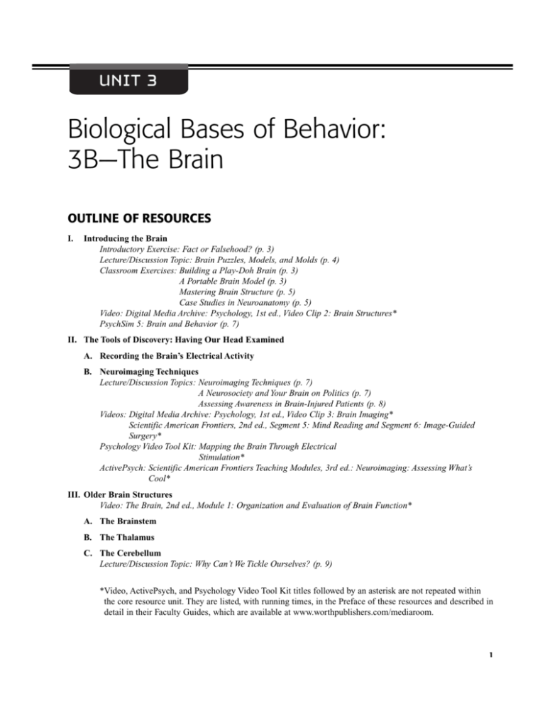
UNIT 3 Biological Bases of Behavior: 3B—The Brain OUTLINE OF RESOURCES I. Introducing the Brain Introductory Exercise: Fact or Falsehood? (p. 3) Lecture/Discussion Topic: Brain Puzzles, Models, and Molds (p. 4) Classroom Exercises: Building a Play-Doh Brain (p. 3) A Portable Brain Model (p. 3) Mastering Brain Structure (p. 5) Case Studies in Neuroanatomy (p. 5) Video: Digital Media Archive: Psychology, 1st ed., Video Clip 2: Brain Structures* PsychSim 5: Brain and Behavior (p. 7) II. The Tools of Discovery: Having Our Head Examined A. Recording the Brain’s Electrical Activity B. Neuroimaging Techniques Lecture/Discussion Topics: Neuroimaging Techniques (p. 7) A Neurosociety and Your Brain on Politics (p. 7) Assessing Awareness in Brain-Injured Patients (p. 8) Videos: Digital Media Archive: Psychology, 1st ed., Video Clip 3: Brain Imaging* Scientific American Frontiers, 2nd ed., Segment 5: Mind Reading and Segment 6: Image-Guided Surgery* Psychology Video Tool Kit: Mapping the Brain Through Electrical Stimulation* ActivePsych: Scientific American Frontiers Teaching Modules, 3rd ed.: Neuroimaging: Assessing What’s Cool* III. Older Brain Structures Video: The Brain, 2nd ed., Module 1: Organization and Evaluation of Brain Function* A. The Brainstem B. The Thalamus C. The Cerebellum Lecture/Discussion Topic: Why Can’t We Tickle Ourselves? (p. 9) *Video, ActivePsych, and Psychology Video Tool Kit titles followed by an asterisk are not repeated within the core resource unit. They are listed, with running times, in the Preface of these resources and described in detail in their Faculty Guides, which are available at www.worthpublishers.com/mediaroom. 1 2 Unit 3B The Brain D. The Limbic System Lecture/Discussion Topic and Video: The Case of Clive Wearing (p. 9) Videos: The Mind, 2nd ed., Module 6: Brain Mechanisms of Pleasure and Addiction* Digital Media Archive: Psychology, 1st ed., Video Clip 26: Self-Stimulation in Rats* Psychology Video Tool Kit: Compulsive Gambling and the Brain’s Pleasure Center* ActivePsych: Digital Media Archive, 2nd ed.: The Brain’s Reward Center* IV. The Cerebral Cortex Classroom Exercise: Individual Differences in Physiological Functioning and Behavior (p. 9) Lecture/Discussion Topics: Einstein’s Brain and Genius (p. 11) Kim Peek’s Brain (p. 12) Classroom Exercise: Neuroscience and Moral Judgments (p. 10) A. Structure of the Cortex Student Project: The Human Brain Coloring Book (p. 12) B. Functions of the Cortex Classroom Exercise: The Sensory Homunculus (p. 12) ActivePsych: Scientific American Frontiers Teaching Modules, 3rd ed.: Brain and Behavior: Phineas Gage Revisited* Scientific American Frontiers Teaching Modules, 3rd ed.: Brain Plasticity: Rewiring the Visual Cortex* Videos: Scientific American Frontiers, 2nd ed., Segment 8: Old Brain, New Tricks* The Mind, 2nd ed., Module 7: The Frontal Lobes: Cognition and Awareness* The Brain, 2nd ed., Module 25: The Frontal Lobes and Behavior: The Story of Phineas Gage* The Mind, 2nd ed., Module 18: Effects of Mental and Physical Activity on Brain/Mind* Psychology Video Tool Kit: Planning, Life Goals, and the Frontal Lobe* C. Language Lecture/Discussion Topic: The Smart-Talk Syndrome (p. 13) Videos: Psychology: The Human Experience, Module 16: Language Centers in the Brain* Digital Media Archive, 1st ed.: Psychology, Video Clip 22: Gleason’s Wug Test* The Brain, 2nd ed., Module 8: Language Processing in the Brain* The Brain, 2nd ed., Module 6: Language and Speech: Broca’s and Wernicke’s Areas Psychology Video Tool Kit: Planning, Life Goals, and the Frontal Lobe* Language and Brain Plasticity* D. The Brain’s Plasticity Lecture/Discussion Topics: Hemispherectomy (p. 14) Stem Cells Correct Fatal Skin Disease (p. 15) ActivePsych: Scientific American Frontiers Teaching Modules, 3rd ed.: Brain Plasticity: Rewiring the Visual Cortex* Videos: The Brain, 2nd ed., Module 7: Brain Anomaly and Plasticity: Hydrocephalus* The Brain, 2nd ed., Module 32: Neurorehabilitation* Psychology: The Human Experience, Module 4: A Case Study of Brain Damage* Psychology: The Human Experience, Module 5: Brain Plasticity* Psychology Video Tool Kit: Language and Brain Plasticity* Rewiring the Brain* V. Our Divided Brain PsychSim 5: Hemispheric Specialization (p. 15) A. Splitting the Brain Classroom Exercise: Behavioral Effects of the Split-Brain Operation (p. 15) Classroom Exercise/Student Project: The Wagner Preference Inventory (p. 16) Videos: Scientific American Frontiers, 2nd ed., Segment 7: Severed Corpus Callosum* The Brain, 2nd ed., Module 5: The Divided Brain* Unit 3B The Brain 3 Psychology Video Tool Kit: The Split Brain: Lessons on Language, Vision, and Free Will* The Split Brain: Lessons on Cognition and the Cerebral Hemispheres* ActivePsych: Scientific American Frontiers Teaching Modules, 3rd ed.: Achieving Hemispheric Balance: Improving Sports Performance* VI. Right-Left Differences in the Intact Brain Lecture/Discussion Topics: The Wada Sodium Amobarbital Test (p. 16) Left-Handedness (p. 18) The Right Brain Movement (p. 20) Student Project/Classroom Exercise: Hemispheric Specialization (p. 17) VII.The Brain and Consciousness ActivePsych: Digital Media Archive, 2nd ed., Video Clip 12: Consciousness and Artificial Intelligence* A. Cognitive Neuroscience Lecture/Discussion Topic: The Mind-Body Problem (p. 21) Classroom Exercise: The Dualism Scale (p. 22) B. Dual Processing Lecture/Discussion Topics: Automatic Processing (p. 22) Psychological Distance and Evaluative Judgments (p. 23) C. The Two-Track Mind Lecture/Discussion Topic: The Deliberation-Without-Attention Effect (p. 24) ActivePsych: Scientific American Frontiers Teaching Modules, 3rd ed.: Hidden Prejudice: The Implicit Association Test* UNIT OUTLINE I. Introducing the Brain (p. 66) Introductory Exercise: Fact or Falsehood? The correct answers to Handout 3B–1, as shown below, can be confirmed on the listed text pages. 1. 2. 3. 4. 5. T (p. 71) T (p. 72) F (p. 79) T (p. 82) F (p. 83) 6. 7. 8. 9. 10. T (p. 84) T (p. 87) T (p. 88) T (p. 89) T (p. 90) Classroom Exercise: Building a Play-Doh Brain Laura Valvatne of Shasta College passes along a delightful classroom exercise in which small groups build a human brain out of Play-Doh. Before the activity, tell students to read carefully the text unit’s section on the human brain, paying special attention to the illustrations. Ask each student to choose the five most important structures and to explain in writing why each was selected. In preparation for the working session, obtain enough Play-Doh for each group of four to build a brain. Then provide each group with five different colors as well as 10 toothpicks and 10 Post-it Notes. Each student should describe to his or her group the five structures he or she chose as most important and explain why. The group should then decide on a minimum of five structures to build into their Play-Doh brain, with at least one brain structure contributed by each student. In putting the brain together (they can decide whether it should be 3-D or a cross-section), they should write the name of each structure on a Postit Note and together decide what to say about its function on the opposite side. The groups will produce widely varied products. When they are done, have them set the brains on a piece of paper to display at the front of class. Have everyone vote for the “best” brain. Before they leave, encourage them to take the Play-Doh home to give to neighborhood kids, siblings, children, or relatives. Valvatne, L. (2000, October 14). Class demonstration suggestions (PSYCHTEACHER@list.kennesaw.edu). Classroom Exercise: A Portable Brain Model Susan J. Shapiro of Indiana University East suggests using students’ hands as a three-dimensional model of the human brain. The strategy can provide a very helpful starting point for students who are still attempting to grasp the basic vocabulary of brain structure. It is also a model they can take with them into a test! Shapiro uses a projected diagram of the brain in introducing the 4 Unit 3B The Brain model in class. What follows are a few highlights. See the reference at the end of the exercise for a more detailed model. Begin by having students hold their hands in front of them with palms outward. The skin on the hands represents the cortex, or “gray matter,” which controls much of our behavior and thought. Muscles in the hands represent the white matter and carry information from one part of the brain to another. Because the brain model could not fit inside the skull, we must curl the fingers down and bring the thumbs close in to our hands to make fists. Many brain structures are Cshaped because the brain tissue has been curled to fit inside the skull. Even when curled, however, there is still too much cortex and so we must pinch the skin on the back of the hands. This creates wrinkles—the cortex is both curled and wrinkled to fit inside the skull. Now have students cross their fists to reflect the two brain hemispheres. Have them place their hands next to each other, outer edges touching, thumbs on the outside. The right hand (on the left side) represents the left hemisphere and the left hand represents the right hemisphere. (This helps students to remember that the left and right hemispheres control movement and sensation on the opposite sides of the body). The wrists represent the brainstem. Continue by having them look at their right fist (the left hemisphere). The fingers of the right fist form the frontal lobe, which is responsible for such complex and abstract abilities as making plans and forming judgments. To move a finger, the student needs to move muscles at the base of the fingers. At about this location, just before the knuckles, is the motor cortex that controls most of the voluntary movement of the body. The area from the knuckles to halfway back on the hand represents the parietal lobe, which is responsible for combining sensory information. At about the knuckles, just behind the motor cortex and in the parietal lobe, is the sensory cortex. It registers and processes body sensations. The lower part of the back of the hand constitutes the occipital lobe, which is involved in vision. Obviously, the thumb can lift away from the rest of the brain model but is attached at the base. This is similar to the temporal lobe; although it is connected to the parietal and occipital lobes, its front section can be lifted away from the rest of the brain. The hand side of the thumb represents the area of the brain cortex that is responsible for hearing. Around this area in the parietal lobe are cells responsible for different aspects of language. Inside both temporal lobes (the thumbs) are the hippocampus and amygdala. The former is involved in memory; the latter is linked to emotional reactions such as fear and anger. Inside the brainstem (the wrists) is the medulla, which is responsible for heartbeat and breathing, and the reticular formation, which is responsible for general arousal. If you picked up a cluster of marbles and held them in your closed hand, they would be in the same location as the basal ganglia, thalamus, and hypothalamus. The basal ganglia are involved in initiating movement and in controlling fine movements. The thalamus is a relay station for messages between the lower brain centers and the cerebral cortex. The hypothalamus directs maintenance activities such as eating, drinking, and body temperature. It helps govern the endocrine system via the pituitary gland, and it is linked to emotion. Shapiro, S. (1999). The ultimate portable brain model. In L. T. Benjamin, B. F. Nodine, R. M. Ernst, & C. B. Broeker (Eds.), Handbook for the teaching of psychology (Vol. 4). Washington, DC: American Psychological Association. Lecture/Discussion Topic: Brain Puzzles, Models, and Molds A variety of items is available for use in discussing brain structure and function. Following are a few that have worked well. For great fun, either before or after discussing this unit, distribute a brain pop to each member of your class. These are 1.25 ounce lollipops, shaped in the form of a brain (called “sweet brain” or “sour brain”). They are available from the Maredy Candy Company, 12155 Kirkham Road, Poway, CA 92064-6870. Call 1800-462-7339 to verify price of a case of 300 at $109.13. Donald Leach uses a two-layer wooden puzzle to teach brain structure and function. The puzzle, which is 18” x 20”, contains approximately 16 colored pieces, each representing a separate structure. The two layers enable both external and internal views of the brain. This technique for reviewing the location, size, and function of the various regions of the brain works best with small groups. The puzzle is available for $60 plus shipping from the Puzzle Man, 21050 Placer Hills Road, Colfax, CA 95713, telephone 530-637-5575, www.thepuzzleman.com. Highly recommended! A variety of brain models useful for lecturing on brain structure is available from Ward’s Natural Science, P.O. Box 92912, Rochester, NY 14692-9012, telephone 1-800-962-2660. One of the simplest and least expensive is the Introductory Brain model for $106. It is bisected to show major structures both internally and externally. It is painted and numbered to distinguish the various components. Larger and more detailed models—for example, one that allows you to dissect the brain into eight parts and shows the intricate details of the limbic system—are available for $197 or more. 3B Scientific’s (www.3bscientific. com) numerous brain models, from the relatively simple and inexpen- Unit 3B The Brain 5 sive to the more elaborate, are available for purchase. You can view detailed descriptions and pictures of the models at the Web site. For something simple and inexpensive, yet sure to capture students’ attention, consider “The Brain Gelatin Mold” that enables you to make and bring a brain to class. Perhaps, as Gerald Peterson suggests, you might be able to implant some fruit to represent certain brain structures such as the limbic system. The plastic mold actually provides the left hemisphere of a jiggly brain about 71/2” x 61/2”. It is available for $7.95 from Archie McFee & Co., telephone 425-349-3009. Finally, introduce the cerebral cortex by noting that the deeply convoluted surface of the brain is most strongly linked to intelligence. Because of its wrinkled appearance in humans, only about a third of it is visible on the surface. Illustrate by taping together two 11.5 x 17 sheets of paper (large size copy paper) and saying to the class before crumpling it into a ball: “This sheet represents the approximate surface area of that thin sheet of neural tissue that we call the cerebral cortex. So how can we fit it inside a skull (a skull small enough to be vaginally delivered)? Nature’s answer: crumple it up.” Leach, D. C. (1996, January). The brain puzzle. Poster session presented at the 18th Annual National Institute on the Teaching of Psychology, St. Petersburg Beach, Florida. Classroom Exercise: Mastering Brain Structure You can use the following exercise, suggested by Tom Pusateri of Florida Atlantic University, to engage students as you introduce the various brain structures. To combat the myth that we use only 10 percent of our brain’s capacity, Pusateri suggests distributing Handout 3B–2 before you discuss brain function. As you introduce each structure, ask students to jot down a few notes regarding its activity while driving a car. Suggest that some brain structures may be more active under certain driving conditions, while others may be active regardless of conditions. After you have covered all the important brain structures, you might have students form small groups to compare their responses before reporting to the full class. The following are sample responses for each brain structure: Cerebellum: Coordinates left and right hand movements on the steering wheel. Medulla: Regulates breathing and heart rate while we concentrate on driving. Pons: Assists in the coordination of driving motions and in alertness. Reticular formation: Regulates our alertness or drowsiness as we drive. Ask students what actions they take to keep alert at the wheel (e.g., open windows, play music, drink caffeinated beverages). Thalamus: Relays visual and auditory cues to areas of the cerebrum. Hypothalamus: Makes us aware when we are too hot or too cold (to adjust the temperature controls), or too hungry, thirsty, or in need of a restroom stop. Amygdala: May be active during “road rage” (e.g., when another driver behaves recklessly). Hippocampus: Contributes to the formation of memories of road hazards for future trips. Corpus callosum: Shares sensory and motor driving information from both hemispheres. Frontal lobe (Helps us in planning our routes [e.g., if we notice a hazard or detour]). Motor cortex: Initiates driving actions (e.g., moves the right foot to the gas or brake pedals). Ask students to trace the pathway from the motor cortex to the right foot. Parietal lobe: Helps us determine if our car may fit into a parking space (right parietal lobe). Sensory cortex: Registers the pressure of the right foot on the gas pedal or brake. Ask students to trace the pathway from the right foot to the sensory cortex. Occipital lobe Visual cortex: Processes the visual road signals (e.g., stop lights, speed limit signs). Temporal lobe Auditory cortex: Processes the sounds of other vehicles (e.g., sirens, horns, passing vehicles). You might conclude by asking students which 90 percent of their brain would they like removed while driving. Source: “Use Your Brain: Critical Thinking in Action” by Thomas P. Pusateri, Florida Atlantic University, Boca Raton, FL from CTUP Symposium: It Can Be Taught! Hands-on Activities That Model the Process of Critical Thinking. Paper presented at the 72nd Annual Meeting of the Midwestern Psychological Association, May 5, 2000. Reprinted by permission of Thomas P. Pusateri. Classroom Exercise: Case Studies in Neuroanatomy Jane Sheldon uses an effective small-group exercise that challenges students to apply their knowledge of neuroanatomy mastered from the text and your classroom presentation. Divide the class into groups of four to six 6 Unit 3B The Brain students and ask them to analyze the case studies in Handout 3B–3. Using what they have learned about anatomy and the workings of the human brain, students should describe the brain areas activated in each situation and how such brain stimulation relates to the behavior in the scenario. Although many brain structures are obviously operating simply because the people in the cases are conscious and active, students should focus on the brain areas activated more than usual in these cases. They may use their notes and the textbook. Each group should record its answer to use for later class interaction (as well as for later studying). Reconvene the class and call on each group to give one interpretation. As necessary, ask the class to expand (or even correct) the answer given by a group. For example, a group may indicate that Anne’s motor cortex is operating as she moves her arm to paint. You might ask the class whether the left or right motor cortex is operating as the artist moves her right arm. You might also ask the class to indicate the lobe in which the motor cortex is located. Rarely does one group produce all possible answers so the exercise enables them to teach each other. Neuroanatomy structures and related functions for each of the cases are given below. Neuroanatomy structure Related function Anne Left motor cortex Left frontal lobe Visual cortex Both occipital lobes Auditory cortexes Both temporal lobes Right hemisphere Thalamus Frontal lobes Left sensory cortex Left parietal lobe Cerebellum Controls right hand Contains motor cortex Used for vision Contain visual cortexes Used for hearing music Contain auditory cortexes Spatial ability for painting Relays sensory information Deciding what to paint Feeling the paintbrush Contains sensory cortex Coordinates moving arm Eddie Both motor cortexes Frontal lobes Both sensory cortexes Parietal lobes Visual cortexes Both occipital lobes Right hemisphere Thalamus Frontal lobes Medulla Amygdala Reticular formation Cerebellum Hypothalamus Hippocampus Move muscles Contain motor cortexes Needed for sense of touch Contain sensory cortexes Used for vision Contain visual cortexes Spatial ability for wrestling Sensory relay Decision making and attention Regulates heart and breathing Aggression and fear Controls arousal Balance and coordination Regulates temperature Memory for moves Jill Hippocampus Amygdala Frontal lobes Hypothalamus Angular gyrus Remembering and learning Anger and fear about cases Decision making and attention Regulates hunger and thirst Needed for reading Source: TEACHING OF PSYCHOLOGY by Sheldon. Copyright 2000 by Taylor & Francis Informa UK Ltd. - Journals. Reproduced by permission of Taylor & Francis Informa UK Ltd. Journals in the format Other Book via Copyright Clearance Center. Unit 3B The Brain 7 PsychSim 5: Brain and Behavior This activity reviews the major divisions of the brain, the structures within them, and their functions. The student takes a tour of the brain, discovering the functions of each region or area. II. The Tools of Discovery: Having Our Head Examined (pp. 67–68) A. Recording the Brain’s Electrical Activity (p. 67) B. Neuroimaging Techniques (p. 68) Lecture/Discussion Topic: Neuroimaging Techniques The text indicates that the brain-imaging techniques are doing for psychological science what the microscope did for biology and the telescope for astronomy. Every year scientists announce new discoveries and also generate new interpretations of old discoveries. Scientists have long been able to map basic functions such as memory, language, and musical ability. Now some brain mappers are wondering if the neuroimaging techniques can also unravel the more complex mysteries of consciousness, morality, and empathy. For example, in one study researchers scanned the brains of participants as they reflected on a variety of moral dilemmas. Using earlier data about where emotions are processed, the researchers found that even when people think they are making strictly rational judgments, they also seem to be employing emotion, because both emotion and reasoning areas are highlighted. (See Classroom Exercise: Neuroscience and Moral Judgments on p. 10.) Marcus Raichle, a professor of radiology and neurology at Washington University in St. Louis, is trying to learn something of the sense of “self.” How might the brain generate the sense that “you’re you and I’m me and we know that”? He hypothesizes that some of the brain’s frenetic “resting” activity—it consumes about 20 percent of the body’s entire energy budget even when not engaged in any particular task—might be supporting self-awareness. In scans of people undertaking challenges that seem to lie outside the self, such as math problems, baseline resting activity dropped off in a portion of the brain’s frontal cortex, a couple of inches behind the center of the forehead. Raichle’s team then compared brain activity in situations that were identical, except in the self-involvement they required. In one case, the participants had to say whether pictures of mundane objects, say picnic scenes or kittens, belong indoors or outdoors. This task, which demanded that participants step outside themselves, caused activity in the prefrontal areas to decrease. In the other case, they were asked to consider whether the same pictures had pleasant or unpleasant associations. As the viewers con- sidered their own responses to the pictures, activity in the possible “self ” networks surged. The research team concluded that at least part of our sense of self depends on knots of neurons elaborately interconnected in the frontal cortex. Other researchers are looking for the basis of “other,” that is, our ability to put ourselves in other people’s shoes and imagine their beliefs and desires. Charles Frith and his colleagues at University College, London, asked participants to think about the following event: A burglar robs a shop, walks down the street, and unknowingly drops his glove. A police officer coming from behind stops him to tell him about the glove. The burglar turns around and surrenders. Why does he do this? The answer requires thinking about the “other”— the burglar thinks the police officer is about to arrest him. The neural circuits in the participants’ brains that light up at this moment of empathy paralleled the ones that typically light up when thinking about one’s self. “Thinking about yourself in a situation may be the way you think about other people,” suggests Frith. Researchers are quick to acknowledge the limits of their methodology. For example, Dartmouth’s Michael Gazzaniga notes that the fMRI traces brain activity by tracking blood flow, which rises whenever there is a surge in metabolism. Elements of some tasks, he suggests, “may be so automatic that they require no increase in metabolism,” thus allowing active brain regions to slip past the technique undetected. Eric Kandel of Columbia University College of Physicians and Surgeons adds, “If a number of areas show activation, we don’t know whether they are causally involved or going along for the ride.” Certainly, no one claims that research will identify a single brain area as the site of morality or consciousness. “Everything that happens in the brain is based on the work of systems, like music in an orchestra performed from a score,” says Antonio Damasio of University of Iowa. “It all sounds like one thing, but it’s coming from 100 or more individual parts. What we’re doing is finding out those little parts.” Sobel, R. K. (2001, November 12). Mind in a mirror. U.S. News and World Report, 64–65. Lecture/Discussion Topic: A Neurosociety and Your Brain on Politics Neurologist Richard Restak predicts the emergence of a neurosociety in which we will have to come to terms with advancements in brain imaging that include • tests that reveal certain thoughts and inclinations that we might prefer to keep private. • brain scans that assess our suitability for certain jobs. 8 Unit 3B The Brain • assessments that explain why we are romantically attracted to some people but not to others. • brain-imaging studies that predict the products we are likely to purchase. • brain response patterns that reveal the emotions elicited in us by television programs and movies. • brain-imaging profiles that assess which political candidates we are most likely to vote for. Relatively recently, neuroscientist Marco Iacoboni and his colleagues at the Brain Research Institute at University of California, Los Angeles, used functional magnetic resonance imaging to “read” the brains of swing voters as they responded to the 2008 presidential candidates. The 20 research participants had indicated they were open to choosing a candidate from either party in the 2008 election. While their brains were scanned, they viewed still photos of each candidate as well as video excerpts from their campaign speeches. The participants rated the favorability of the candidates on “before” and “after” questionnaires. The researchers’ intriguing conclusions included the following: • Activity in the amygdala when participants’ viewed the words “Democrat,” “Republican,” and “independent” suggested high levels of anxiety. Men who viewed “Republican” showed high activity in both the amygdala and the insular cortex indicating both anxiety and disgust. • Of all the candidates’ speech excerpts, Mitt Romney’s prompted the greatest activity in the amygdala (indicating high voter anxiety). However, as the participants continued to listen, their anxiety subsided. The researchers suggested that perhaps voters would become more comfortable with Mr. Romney as they saw more of him. • In swing voters who gave John Edwards lower ratings, photos of him elicited activity in the insular cortex, a brain area associated with disgust and other negative feelings. Swing voters who had given him higher ratings showed significant activation of the mirror neurons, brain cells activated when people feel empathy. Conclusion? Mr. Edwards elicits a strong effect on swing voters— both those who like him and those who don’t. • Voters who rated Hillary Clinton unfavorably showed significant activity in the anterior cingulate cortex, an emotional center of the brain that is aroused when a person feels compelled to act in two different ways but must choose one. The researchers suggested this finding might indicate unacknowledged impulses to like Mrs. Clinton. In contrast, those swing voters who rated her favorably showed very little activity in this brain area when viewing pictures of her. After the researchers published their Op-Ed piece in the New York Times, another team of cognitive neuroscientists voiced concern. They challenged the idea that it is possible to definitively read the minds of potential voters by looking at their brain activity while viewing presidential candidates. Brain regions, the critics claimed, are typically engaged by many mental states and thus a one-to-one mapping is difficult. For example, the amygdala is activated by positive emotions as well as by anxiety. Careful experimental design and peer review is crucial, they argued, to drawing sound conclusions. Nonetheless, the critics voiced excitement about the potential of using brain-imaging techniques to better understand the psychology of political decisions. Iacoboni, M., et al. (2007, November 11). This is Your Brain on Politics. New York Times. Retrieved December 18, 2007, from www.nytimes.com/2007/ 11/11/opinion/11freedman.html. Aron, A. et al. (2007, November 14). Letter: Politics and the Brain. New York Times. Retrieved December 18, 2007, from http://query.nytimes.com/ gst/fullpage. html?res=9907E1D91E3CF937A25752C1A9619C8B63. Restak, R. (2006). The naked brain: How the emerging neurosociety is changing how we live, work, and love. New York: Harmony Books. Lecture/Discussion Topic: Assessing Awareness in Brain-Injured Patients Nicholas Schiff and his colleagues’ work with “minimally conscious” patients provides an excellent example of how neuroimaging technology provides important insights into the living brain. The results of their work suggest that brain-damaged people who are unable to respond and thus have often been treated as though they are unaware may actually be hearing and registering what is happening in their environment. Using fMRI scans, the researchers compared the brain activity of men determined to be minimally conscious with that of healthy men and women. In terms of overall brain activity, the two groups were very different, with the brains of the minimally conscious men showing less than half the activity of the brains of healthy adults. However, when the researchers played an audiotape in which a relative or loved one reminisced, telling familiar stories and recalling shared experiences, the sound of the voice prompted a pattern of brain activity in the minimally conscious that was similar to that of the healthy participants. In fact, the brain-injured persons showed near-normal patterns in the language processing areas of their brains. This suggests, argues Schiff, that some neural networks “could be perfectly preserved under some conditions.” Unit 3B The Brain 9 In one case, a minimally conscious male heard his sister recount his toast at her wedding as well as times they played together as children. Although his eyes were closed, the researchers found that the visual areas of his brain were active, suggesting that he might be producing images. Mental states change over time. Some patients have recovered function almost completely after being thought vegetative. Neuroimaging provides one way to assess these changes and even link them to treatment efforts. “The most consequential thing about this is that we have opened a door, we have found an objective voice for these patients, which tells us that they have some cognitive ability in a way they cannot tell us themselves,” concludes Joy Hirsch, a member of the research team. The patients, she added, “are more human than we imagined in the past, and it is unconscionable not to aggressively pursue research efforts to evaluate them and develop therapeutic techniques.” As many as 6 million Americans live with the consequences of serious brain injuries. An estimated 100,000 to 300,000 are in a minimally conscious state, that is, bedridden and unable to communicate or feed and care for themselves, but typically breathing on their own. Schiff, N., et al. (2005). fMRI reveals large-scale network activation in minimally conscious patients. Neurology, 64, 514–523. III. Older Brain Structures (pp. 69–73) A. The Brainstem (pp. 69–70) B. The Thalamus (p. 70) C. The Cerebellum (pp. 70–71) Lecture/Discussion Topic: Why Can’t We Tickle Ourselves? The text reports that one function of the cerebellum, the “little brain” attached to the rear of the brainstem, is to help coordinate movement output and balance. Research indicates that part of the cerebellum’s function is to tell the brain what to expect from the body’s own movements. In this way, the brain can ignore expected pressure on the soles of the feet while walking and attend to more important sensations such as stubbing a toe. Sarah-Jayne Blakemore and her colleagues at University College London have addressed the interesting question, “Why can’t we tickle ourselves?” For their study, the researchers had six volunteers lie in a brainscanning machine with their eyes closed. A plastic rod with a piece of soft foam attached to it moved up and down, tickling the participants’ left palms. The experimenter and the volunteers took turns moving the rod, so the volunteers were either tickling themselves or were being tickled. In a third condition, the foam was secretly removed, so the volunteers moved the rod but felt nothing. Throughout this process, the researchers used functional MRI (fMRI) scans to compare activity in different parts of the brain. On the basis of the results, they concluded that during self-tickling one part of the brain tells another: “It’s just you. Don’t get excited.” The cerebellum is involved in predicting the specific sensory consequences of movement. It provides the signal that is used to cancel the sensory response to selfgenerated stimulation. In short, it tells the sensory cortex what sensation to expect and this dampens the tickling sensation. Blakemore, S., Wolpert, D., & Frith, C. (1998). Central cancellation of self-produced tickle sensation. Nature Neuroscience, 1, 635–640. D. The Limbic System (pp. 71–73) Lecture/Discussion Topic and Video: The Case of Clive Wearing The text notes that the hippocampus processes memory. When animals or people lose their hippocampus to surgery, illness, or injury, they become unable to lay down new memories of facts and experiences. Unit 7A of these resources describes the remarkable case of Clive Wearing, who, afflicted by encephalitis, experienced total destruction of the hippocampus as well as damage to other brain structures. Both the lecture/discussion topic and Modules 10 and 11 of The Mind, 2nd edition, series portray the devastating effects of his memory loss. You may choose to use it now. IV. The Cerebral Cortex (pp. 74–83) Classroom Exercise: Individual Differences in Physiological Functioning and Behavior Research on individual differences in physiological functioning provides another opportunity to drive home the importance of the biological perspective. Eysenck’s work on introversion–extraversion (see Handout 11–3, the last six items of which assess extraversion) and Zuckerman’s research on sensation-seeking (see Handout 8B–5) are excellent examples. Eysenck has suggested that differences in introversion–extraversion are closely linked to the cortical arousal of the brain’s ascending reticular activating system (reticular formation). Extraverts seem to have higher sensory thresholds and less-arousable cortexes. They must constantly seek stimulation to maintain their brain activity levels and avoid boredom. In contrast, introverts typically operate at an above-optimal cortical 10 Unit 3B The Brain arousal level. They are so easily aroused that they tend to avoid external stimulation, seeking solitude and nonstimulating environments in an attempt to keep their arousal level from becoming too aversive. They avoid the noisy parties that extraverts seek. Eysenck’s famous “lemon-juice” demonstration illustrates this arousal difference. Ask your students how strongly they salivate to lemon juice. Their personality type is likely to be a good predictor of their reaction, or vice versa. “Under conditions of equal stimulation,” predicted Eysenck, “effector output will be greater for introverts.” Research findings confirm that introverts do salivate more when pure lemon juice is placed on their tongues. Other studies have indicated that when exposed to the same level of various stimuli, introverts become more physiologically aroused than extraverts. Given their choice of an optimal level of stimulation, extraverts also choose higher levels. And, they are less likely to be inhibited by punishment and actually seem to experience less pain than introverts. Zuckerman’s concept of sensation-seeking is discussed in Unit 8B of these resources. Levels of sensation-seeking have been related not only to a wide range of cognitive and behavioral variables but also to differences in physiological functioning. For example, in response to new stimuli as well as to changes in such stimulation, high sensation seekers demonstrate greater electrical activity in the brain than low sensation seekers. They also show a rise in sex hormones and lower levels of monoamine oxidase (MAO), an enzyme in the brain and most other tissues that controls neurotransmitters. Zuckerman observes that as testosterone levels peak in males during their late teens and early 20s (and then tapers down), so does sensation seeking. MAO seems to serve some dampening or regulatory role, since drugs inhibiting its action can produce euphoria, excitement, and even hallucinations. Some researchers believe that the lower MAO level of high sensation seekers explains their greater activity level and sociability. More recent work by neuroscientist Brian W. Haas and his colleagues on how personality traits may be linked with specific brain responses suggests that extraverts exhibit different brain responses to positive and negative words. That is, while processing a positive word such as lucky, the extravert’s brain shows increased activation in the anterior cingulate. In fact, Haas claims he can assess a person’s tendency toward extraversion by merely observing (through fMRI) his or her anterior cingulate’s response to positive and negative words. It seems that the extravert’s brain is attracted to positive words and lingers on them a few milliseconds longer. The relationships between physiological functioning and personality types or traits are intriguing. However, much of the research is correlational and thus open to alternative interpretation. Questions of cause and effect and of possible third mediating factors must still be answered. Haas, B. W., et al. (2006). Functional connectivity with the anterior cingulate is associated with extraversion during the emotional Stroop task. Social Neuroscience, 1, 16–24. Liebert, R. M., & Spiegler, M. D. (1998). Personality: Strategies and issues (8th ed.). Belmont, CA: Wadsworth. Schultz, D., & Schultz, S. (2009). Theories of personality (9th ed.). Belmont, CA: Wadsworth. Classroom Exercise: Neuroscience and Moral Judgments Joshua Greene and his research team at Princeton University study the neural correlates of moral judgments. Their fascinating findings provide important insight into the interplay between emotion and reason in resolving moral dilemmas. Pose the following dilemma that the Princeton research team uses to your students: It’s wartime and you are hiding in the basement with a group of townspeople. Enemy soldiers are outside. Your baby starts to cry loudly; if nothing is done, the soldiers will find you and kill everyone including the baby. The only way to prevent this loss of life is to cover the baby’s mouth; if you do, the baby will smother. What should you do? Participants are about equally divided on the right course of action. Greene suggests that the dilemma is challenging because it creates a conflict between a strong emotional response (don’t ever kill a baby) and a strong cognitive response (if you don’t kill the baby, everyone dies). Two findings from neuroimaging studies support this interpretation. First, the anterior cingulate cortex, a brain region associated with response conflict, exhibits increased activity. This indicates that the moral dilemma is not difficult merely because it requires more time to process. Second, the dorsolateral prefrontal cortex and inferior parietal cortex, regions associated with cognitive control, show greater activity when people favor the promotion of the best overall consequences. That is, when people say it is okay to smother the baby, they exhibit increased activity in the brain area associated with high-level cognitive function. The Princeton team concludes that when a tough moral dilemma is posed, the reasoning processes of the brain conflict with the more automatic emotional response, and the decision takes longer. Our gut moral instinct, suggests Greene—that is, “what people know deep down is right”—was not designed for the modern world. Abstract reasoning goes on in the more recently evolved parts of the brain. “My hope,” writes Greene, Unit 3B The Brain 11 “is that by understanding how we think, we can teach ourselves to think better, that is, in ways that better serve the needs of humanity as a whole.” Continuing research on how the brain tackles difficult moral dilemmas highlights the potential tension between the cool cognition associated with the dorsolateral prefrontal cortex and the moral emotions such as compassion, empathy, and guilt that are associated with the ventromedial prefrontal cortex (VMPFC). For example, Michael Koenigs (a postdoctoral fellow at the National Institute of Neurological Disorders) and his colleagues compared moral judgments made by neurologically normal people with those who had damage to the VMPFC. They used variations on the Trolley dilemma (which you might also present to your class): A runaway trolley is hurtling down the tracks toward five people who will be killed if it proceeds on its present course. You can save these five people by diverting the trolley onto a different set of tracks, one that has only one person on it, but if you do this that person will be killed. Is it morally permissible to turn the trolley and thus prevent five deaths at the cost of one? Most respondents say yes. They make a utilitarian judgment (associated with the dorsolateral prefrontal cortex) that will lead to the greater aggregate welfare. Now consider a slightly different dilemma. Once again, the trolley is headed for five people. You are on a footbridge over the tracks next to a large man. The only way to save the five people is to push this man off the bridge and into the path of the trolley. Is that morally permissible? In this case, the majority of respondents say no. The much stronger emotion associated with the VMPFC conflicts with a purely utilitarian judgment. Interestingly, however, Koenigs and his research team reported that those with damage to the VMPFC were much more likely to say that it is okay to push a man in front of a moving trolley. Presumably, with less emotion aroused, they gave a cooler, cognitive response. Greene, J. D., Engell, A. D., Darley, J. M., & Cohen, J. D. (2004). The neural basis of cognitive conflict and control in moral judgment. Neuron, 44, 389–400. Hobson, K. (2005, February 28). Making those choices about right and wrong. U.S. News & World Report, 60–61. Koenigs, M., et al. (2007, April 19). Damage to the prefrontal cortex increases utilitarian moral judgments. Nature, pp. 908–911. Lecture/Discussion Topic: Einstein’s Brain and Genius When Albert Einstein died of a ruptured abdominal aneurysm in 1955, pathologist Dr. Thomas Harvey removed his brain and, with the family’s consent, kept the organ for scientific study. At the time, Harvey reported that from all appearances Einstein’s brain was well within the normal range. For example, it was no larger or heavier than anyone else’s. From time to time, Harvey has provided samples for other researchers to study. In 1996, Dr. Sandra F. Witelson and her colleagues at McMaster University obtained photos of Einstein’s brain before it had been sectioned as well as a significant portion of brain tissue itself. For comparative purposes, Witelson maintains a Brain Bank of normal, undiseased brains that have been donated by people whose intelligence has been carefully assessed before death (see also text p. 67). The Brain Bank enabled researchers to compare Einstein’s brain with those of men close to his age. Witelson, Kigar, and Harvey have reported that while the overall size of Einstein’s brain was average, the region called the inferior parietal lobe was 15 percent wider than normal. “Visual-spatial cognition, mathematical thought, and imagery of movement,” reported the researchers, “are strongly dependent on this region.” Einstein’s brilliant insights were often the result of visual images that he translated into the language of mathematics. For example, his special theory of relativity was based on his reflections of what it would be like to ride through space on a beam of light. The researchers also reported that a feature known as the sylvian fissure (a groove that normally runs through the brain tissue) was shorter than average. This meant that the brain cells were packed more closely together, permitting more interconnections and thus more cross-referencing of information and ideas. Although other researchers studying Einstein’s brain have reported differences such as more glial cells and more densely populated neurons, Witelson suggests that the most recent findings are compelling because “the differences occur in the region that supports psychological functions of which Einstein was master.” Critics have observed that while Einstein’s brain may well be different, the cause-effect relationship is unanswered. As Michael Lemonick concludes, the differences may be the result of strenuous mental exercise, not the cause of genius. “Bottom line,” writes Lemonick, “we still don’t know whether Einstein was born with an extraordinary mind or whether he earned it, one brilliant idea at a time.” Lemonick, M. D. (1999, June 28). Was Einstein’s brain built for brilliance? Time, p. 54. Witelson, S. F., Kigar, D. L., & Harvey, T. (1999). The exceptional brain of Albert Einstein. The Lancet, 353, 2149–2153. 12 Unit 3B The Brain Lecture/Discussion Topic: Kim Peek’s Brain Perhaps even more intriguing than the study of Einstein’s brain are brain scans of Kim Peek, who was the inspiration for Rain Man, the 1988 film about an autistic savant with astounding mathematical skills. Born November 11, 1951, Kim Peek is not autistic, but he does have a lot in common with Rain Man. He has the ability to memorize telephone directories at lightning speed and has total recall of 9000 books. Researchers have discovered that each of his eyes can read a separate page simultaneously, taking in every word. In 10 seconds, he reads a page that takes most people 3 minutes. Moreover, he never forgets what he reads. Peek is a megasavant. While most savants have expertise in one or two subjects, Kim is an expert in at least 15 different subjects, including history, sports, space, music, and geography. In fact, no one in the world is thought to possess a brain as extraordinary as Peek’s. Known as Kimputer to many, his knowledge-library includes world and American history, geography (roads and highways in the United States and Canada), professional sports (baseball, basketball, football, Kentucky Derby winners), the space program, movies, the Bible, and calendar calculations (he can tell the day of the week of any person’s birth, day of the week of his or her present year’s birthday, and year and day of the week the person will turn 65). When asked, he can also name the highways that lead into a person’s small town, the county, the area code and zip code, the television stations available in the town, and to whom telephone bills must be paid, and he can describe any historical events that may have occurred in the area. Kim’s musical abilities are similarly phenomenal. Having heard a symphonic work once as a boy, he remembers it today. If there is a mistake in the playing, he will observe, for example, “The second trombone player came in a few moments late.” At the same time, Kim is developmentally disabled. He did not walk until he was 4 and continues to have severe motor deficiencies. Kim needs help bathing and brushing his teeth. Often, his father, Fran, needs to redress him in the morning because he puts his shirt on backward. Fran takes care of his son full time and rarely has a moment to himself. In fact, he checks on him two or three times in the course of the night. Fran explains that from birth his son was different: “Physically, he was unusual. His head was a third larger than normal.” Across the back of his head, he had an encephalocele—a blister into which part of the brain protrudes. The blister retracted when Kim was 3, pulling a nodule into his cerebellum and destroying half of it. In 1983, Kim had his first brain scan, which indicated that his brain was highly unusual. It is not divided into separate hemispheres and it has no corpus callosum, the connecting tissue that normally connects the left and right hemispheres. There is no anterior commissure, and there is damage to the cerebellum. In fact, an MRI indicated that the right half of Kim’s cerebellum had exploded into eight or nine small pieces, likely caused by pressure when the blister retracted into his brain. Only a thin layer of skull covers the area of the previous encephalocele. Although Boyle cannot fully explain Kim’s capabilities at this point, he suggests that “Because he has no corpus callosum, there are theories about the right brain being freed from the dominance of the left. So instead of having the two sides of the brain competing, you have one megacomputer. But this is just a theory. Usually when someone has that condition, there are other conditions that are more detrimental to the individual.” The original Rain Man. (2005, March 4). The Week, pp. 40–41. A. Structure of the Cortex (p. 74) Student Project: The Human Brain Coloring Book In A Colorful Introduction to the Anatomy of the Human Brain: A Brain and Psychology Coloring Book psychologists John P. J. Pinel and Maggie E. Edwards introduce brain structures and their important psychological functions. The book promotes active learning by encouraging close attention through coloring. Students assess their progress through the use of a cover flap that conceals labels while they review. Each chapter ends with an extensive series of review exercises. Intended for those who have no background in neuroscience, the book is a particularly useful supplement for the introductory psychology course. Pinel, J. P. J., & Edwards, M. E. (2008). A colorful introduction to the anatomy of the human brain: A brain and psychology coloring book (2nd ed.). Boston: Allyn & Bacon. B. Functions of the Cortex (pp. 74–79) Classroom Exercise: The Sensory Homunculus Each nerve fiber carries a message about the location and intensity of touch to the sensory cortex, so that together the nerve fibers form a spatial “map” of the body skin surface in the cortex that is called the “sensory homunculus.” James Motiff provides a classroom demonstration that shows students how the amount of sensory cortex devoted to specific areas is closely related to the area’s touch sensitivity. The largest portions are devoted to areas having the greatest sensitivity, for example, the lips, tongue, and hands (see Figure 3B.13 in the text). Unit 3B The Brain 13 The children’s game of identifying the number of fingers on a person’s back provides the basis for this exercise. Ask for a volunteer to come to the front of the room. Have the volunteer close his or her eyes and report the number of fingers you press on the skin. Randomly press from one to four digits lightly on the back or on the hand and inform the class of the correct number after the volunteer’s guess. Greater accuracy for touch to the hand will be obvious. Far more cortex is devoted to the hand than to the back and this explains the difference in sensitivity. (You could do this same exercise by having students work in pairs.) Douglas Chute and Philip Schatz suggest a related classroom exercise or student project. It involves touching toes. (If you use this demonstration in class, you will want to obtain informed consent from your student volunteers.) Ask the volunteer to remove his or her shoe and sock. After a blindfold is in place, touch the second, third, or fourth toes gently with a pen or stylus. It is most effective to touch slightly to the left or right midline of each toe. Chute and Schatz report that student accuracy in identifying the toe being touched is only 80 or 90 percent. Less cortex is devoted to the toes than to, say, the fingers. At the same time, with feedback on accuracy, participants are 98 percent correct after as few as 10 trials. Performance, however, returns to baseline in a few days. Chute and Schatz suggest that “the toetouching phenomenon shows that the nervous system does not come with sensory relationships prewired but that these relationships can be learned and forgotten.” Chute, D., & Schatz, P. (1999) Observing neural networking in vivo. In L. T. Benjamin, B. F. Nodine, R. M. Ernst, & C. B. Broeker (Eds.), Handbook for the teaching of psychology (Vol. 4). Washington, DC: American Psychological Association. Motiff, J. P. (1987). Physiological psychology: The sensory homunculus. In V. P. Makosky, L. G. Whittemore, & A. M. Rogers (Eds.), Activities handbook for the teaching of psychology: Vol. 2 (pp. 51–52). Washington, DC: American Psychological Association. C. Language (pp. 80–82) Lecture/Discussion Topic: The Smart-Talk Syndrome Williams syndrome can be discussed in connection with the brain and language or as part of the relationship between thought and language. Educational psychologist Eleanor Semel captured the unique challenge that faces those teaching children with Williams syndrome: Educators are confused because the Williams syndrome child tests like the retarded child, talks like a gifted child, behaves like a disturbed child, and functions like a learning-disabled child. Each of these terms has a specific meaning in the world of special education, yet none seems to fit the characteristic peaks and valleys in Williams syndrome. The result is that children with Williams syndrome are generally not well served by schools. This intriguing disorder is a form of intellectual disability marked by the preservation of linguistic functioning in the face of severe cognitive deficits. In a sense, it could be labeled language without thought. Williams syndrome, a rare genetic disorder occurring in as many as 1 in 7500 births, seems to contradict the common assumption that certain cognitive abilities are prerequisites to language development. First identified in 1961, Williams syndrome results when a group of genes on one copy of chromosome 7 is deleted during embryonic development. Neurophysiologist Ursula Bellugi is presently studying victims of Williams syndrome at the Salk Institute for Biological Studies in La Jolla, California. Having low IQs (in the 50s), these people are unable to read, write, dress themselves, or cross the street without help. They typically have trouble remembering the routines of daily life. They can’t copy a figure as simple as a triangle, nor have they mastered Piaget’s principle of conservation. At the same time, they can string complex vocabulary words into complex, elegant sentences. Bellugi has observed appropriate use of such infrequently used words as surrender, sauté, nontoxic, commentator, and brochure. When asked to name all the animals he could think of, Ben, a 16-year-old with an IQ of 54, named such animals as the buffalo, sabertooth tiger, condor, and vulture. When treated in an emergency room for a broken toe during the Special Olympics, he spontaneously asked the doctor, “Are you going to use novocaine, xylocaine, or procaine?” Crystal, another victim of Williams syndrome, described her career plans this way: “You are looking at a professional book writer. I am going to write books, page after page, stack after stack. I’m going to start on Monday.” Putting the finishing touches on her drawing of an elephant she pointed to the mouth, trunk, and ears: “Fan ears,” she said, “ears that can blow in the wind.” However, the drawing itself is unrecognizable, having crude lines and blobs strewn over the page. The eloquent statements do not seem to reflect deep, insightful thinking. The children may give a detailed account of how to cross a street safely and then blindly walk into busy traffic. They may correctly identify all the contexts in which the word “contagious” is used; yet, when asked to choose the most common definition—easily spread— they may score as poorly as children with Down syndrome. Says Bellugi, “They can tell you an enormous amount in very complex grammar without getting to the point.” Bellugi’s studies suggest that the brain deals with elements of language, including grammar and syntax, separately from the rest of thinking. Some researchers believe that those with Williams syndrome may be able 14 Unit 3B The Brain to pull themselves up by their linguistic bootstraps. For example, to boost their concentration, Orlee Udwin teaches those with Williams syndrome to rehearse verbally: “I must sit still. I must pay attention.” The technique seems to help focus the attention of the typically hyperactive children. Bellugi initially hypothesized that Williams might damage the right hemisphere of the brain where spatial tasks are processed while leaving language in the left hemisphere intact. However, subsequent research indicated that people with Williams excel at recognizing faces, a task that utilizes the visual-spatial skills of the right hemisphere. By using functional brain imaging Bellugi also found that in those with Williams, both hemispheres carry the task of language. Research has focused on the neocerebellum, a part of the brain that is enlarged in people with Williams. Among the brain’s newest parts, the neocerebellum appeared in human ancestors about the same time as did the enlargement of the frontal cortex. Interestingly, this same brain structure is significantly smaller in people with autism, who are generally antisocial and poor at language, just the reverse characteristics of those suffering Williams syndrome. On the other hand, researchers have found that the posterior forebrain areas (posterior parietal lobe and occipital lobe) are quite small in those with Williams syndrome, which may explain their visuospatial deficits. They have also found cellular anomalies, such as exaggerated horizontal organization of neurons, particularly in striate cortex; an increased density of cells throughout brain areas; and abnormally clustered neurons. Brownlee, S. (1998, June 15). Rare disorder reveals split between language and thought. U.S. News and World Report, 52–53. Klein, S. B., & Thorne, B. M. (2007). Biological psychology. New York: Worth. Rubin, J. (1990, May/June). The smart-talk syndrome. In Health, 38–39. D. The Brain’s Plasticity (pp. 82–83) Lecture/Discussion Topic: Hemispherectomy The hemispherectomy provides a vivid example of brain plasticity. Dating back to 1928, the hemispherectomy was devised as a treatment for malignant brain tumors. However, not only did it fail to cure the patients, but it was also associated with high mortality and morbidity. The surgery was used again in the 1940s and 1960s as a treatment for seizure disorders, but each time it fell into disfavor because of postoperative complications. A number of medical advancements have contributed to its more recent success. In 1997, a Johns Hopkins medical team followed up on the 58 child hemispherectomies they had performed and were “awed” by how well the children retained their memory, personality, and humor after removal of either hemisphere. Jason Brandt, a Johns Hopkins neurologist, concluded, “That a child with half a brain can indeed be a whole person speaks to the malleability of both the human brain and human spirit. It’s amazing, it’s wonderful. I’m at a loss to describe it.” In a follow-up study, Hopkins researchers contacted many of the families of the 58 children who participated in the 1997 study as well as those of 53 other children who had a hemispherectomy more recently. They found that 65 percent of the patients were seizure-free; 21 percent had occasional, nonhandicapping seizures; and 14 percent had troublesome seizures. Most notably, 80 percent of the patients no longer use drugs or are taking only one anticonvulsant medication. The researchers concluded that those with Rasmussen’s syndrome, a nervous system disorder characterized by chronic inflammation of the brain, and those with congenital vascular injuries benefit most from the surgery. Surely some drawbacks will always remain. For example, some neurological functions do not transfer from one hemisphere to the other. All the “hemis” remain blind in one-half of each eye. They also continue to have some degree of paralysis on one side of their bodies. Fine motor movement is lost in one hand. In general, the effect of removing one hemisphere is inversely related to the age of the child at the time of surgery. If performed early enough, the surgery does not seem to cause deficits in higher mental functions in adulthood. Two different theoretical conclusions have been drawn from this finding. One is that no shift from one hemisphere to the other has occurred because lateralization of function is not present in early infancy. The other is that hemispheric differences are present very early in life, but the young brain has the ability to reorganize itself in the face of damage to specific areas. Studies comparing the abilities of persons with left and right hemispherectomies suggest that the latter plasticity explanation is more likely to be correct. For example, research on those who had hemispherectomies (some in the first few months of life) indicate that those who have had the left hemisphere removed have some continuing difficulty with both syntax and the processing of speech sounds. When asked to judge the acceptability of the three sentences “I paid the money by the man,” “I was paid the money to the lady,” and “I was paid the money by the boy,” those who had had the left hemisphere removed failed to recognize the first two as grammatically incorrect. The researchers concluded that the right hemisphere does not accurately comprehend the meaning of passive sentences. There are limits to the plasticity of the infant brain, and some Unit 3B The Brain 15 hemispheric differences seem to be present very early in life. Choi, Charles (2007, May 24). Strange but true: When half a brain is better than a whole one. Scientific American Newsletter. Retrieved December 11, 2007, from www.sciam.com/article.cfm?chanID=sa003&article ID=BE96F947-E7F2-99DF3EA94A4C4EE87581&ref =rss. Johns Hopkins Medical Institutions (2003, October 16). Study confirms benefits of hemispherectomy surgery. ScienceDaily. Retrieved December 11, 2007, from www.sciencedaily.com/releases/2003/10/031015030730. htm. Springer, S., & Deutsch, G. (1998). Left brain, right brain (5th ed.). New York: W. H. Freeman. Lecture/Discussion Topic: Stem Cells Correct Fatal Skin Disease The genetic disease recessive dystrophic epidermolysis bullosa (RDEB) is associated with painful wounds and mutilating scarring. The body is continuously bandaged to minimize the risk of tears and blistering. In June 2008, researchers announced that stem cell therapy was successfully used in curing 25-month-old Nate Liao from Clarksburg, New Jersey, who underwent treatment in October 2007. Until then, there had been no successful treatments of RDEB. It had always been fatal due to malnutrition, infection, or an aggressive skin cancer. The body is missing a critical protein called collagen type VII, which anchors the skin and lining of the gastrointestinal system to the body. Because of delicate skin, Nate and his older brother, Jake, had to wear special clothes and had never eaten solid food. Their fragile esophagus could handle only liquids. Research and clinical collaboration between researchers at Columbia University Medical Center in New York as well as Thomas Jefferson University in Philadelphia and physicians at the University of Minnesota and University of Minnesota Children’s Hospital, Fairview, led to the use of stem cells in an effort to replace the missing protein throughout the body. Research was first done with RDEB mice. Delivering marrow-derived stem cells in the bloodstream greatly lengthened the life expectancy of the mice and also healed existing blisters. Treatment of Nate involved a bone marrow transplant using stem cells harvested from umbilical cord blood. It proved successful and his skin is now strong enough for him to wear normal clothes and eat solid foods. His parents have decided to pursue the treatment for Jake as well. Dr. John Wagner, the lead University of Minnesota Medical School physician who developed the clinical trial stated, “We have developed a new standard of care for RDEB patients, beginning with Nate. . . . Nate’s quality of life is forever changed. If future treatments are as successful as Nate’s, maybe we can take one more disorder off the incurable list.” Columbia University Medical Center (2008, June 23). Stem Cells Correct Defect In Child’s Fatal Skin Disease. ScienceDaily. Retrieved June 23, 2008, from www.sciencedaily.com/releases/2008/06/080620213845.htm. V. Our Divided Brain (pp. 83–86) PsychSim 5: Hemispheric Specialization Hemispheric Specialization is a graphic demonstration of how messages reach the two sides of the brain and of the special functions of each side—for example, speech is controlled in the left hemisphere. The processing of a visual stimulus through the brain is the example used. Roger Sperry’s work with split-brain patients is also illustrated, and the responses of normal people are compared with those of split-brain patients. A. Splitting the Brain (pp. 83–86) Classroom Exercise: Behavioral Effects of the SplitBrain Operation Edward Morris suggests a classroom exercise that is most useful in providing a dynamic visual image of the consequences of the split-brain procedure. Choose two volunteers, both of whom are right-handed, and sit them next to each other at the same desk (if possible, have them sit in the same chair). The one on the left represents the brain’s left hemisphere, the one on the right represents the brain’s right hemisphere. Instruct the volunteers to place their outer hands behind their back, their inner hands on the desk, one crossing over the other. The two hands represent the split-brain patient’s left and right hands. The student sitting on the right (representing the patient’s left hand) should be instructed not to speak. Begin by simulating apparent visual deficits. Tell the volunteer on the right to look only to the left, and the volunteer on the left to look only to the right. Use a piece of poster board to separate the students and their lines of sight. Introduce a flashcard or actual object to the right visual field and ask the volunteer representing the right hand to name it or choose the correct object from a selection of objects. He or she should have no difficulty, demonstrating the link between the right visual field and the left hemisphere. Repeat this with another object in the right visual field, this time asking the volunteer representing the left hand to choose. Choosing incorrectly (or not at all) will demonstrate the right hemisphere’s lack of awareness of the right visual field. Follow the same procedure for the left visual 16 Unit 3B The Brain field. Although mute, this time the right hemisphere will pick the correct object. When asked, the left hemisphere will most likely guess and pick incorrectly. You might conclude that in more normal settings the splitbrain patient will be able to scan both visual fields, allowing both hemispheres to receive the information presented to only one hemisphere in the laboratory. Blindfold the students and place a familiar object, such as a quarter, in the left hand. Ask the volunteer representing the left hand to indicate only in a nonverbal manner whether he or she recognizes the object. Ask the speaker (the volunteer representing the right hand) to name the object. Often, the students representing the left hand will guess but be incorrect. Elicit audience reaction to the possible relationship between hemispheric language dominance and the experience of the split-brain patient. Encourage students to generate ideas concerning how the left hand may communicate knowledge in other ways. For example, the volunteer representing the left hand might select the object from an array of objects placed in front of the pair upon removal of the blindfolds. Morris describes additional activities in the article cited here. Morris, E. (1991). Classroom demonstration of behavioral effects of the split-brain operation. Teaching of Psychology, 18, 226–228. Classroom Exercise/Student Project: The Wagner Preference Inventory Handout 3B–4, the Wagner Preference Inventory, provides an opportunity to stimulate critical thinking about some of the controversial applications of research on the divided brain. The inventory is designed to indicate which, if any, hemisphere is dominant. It consists of 12 sets of four statements each. Each statement in a group presumably corresponds to a different function of the two hemispheres, namely: (a) left, logical; (b) left, verbal; (c) right, manipulative/spatial; and (d) right, creative. Scoring is accomplished by adding the number of times each letter is selected. Students should enter the totals in the large cells in the quadrant at the bottom of the handout. Below the quadrant are two rectangles for total left and right scores obtained by adding (a) and (b) for the left score and (c) and (d) for the right score. According to the authors, Rudolph Wagner and Kelly Wells, a difference of at least three points (expressed as a ratio) between L and R is needed to show a significant difference between the functioning of the two hemispheres. Otherwise, the functioning of the hemispheres is thought to be balanced. An example of a ratio for a dominant left hemisphere would be 11/1, a dominant right would be 4/8, and a balanced situation would be 5/7, or 7/5. In validating their inventory, Wagner and Wells administered it to six criterion groups: students in a logic class, creative writers, vocational/technical high school teachers, nurses, painters (artists), and musicians. Predictions regarding the preferences of the six groups were: logic students—left, logic (a); creative writers—left, verbal (b) and right, creative (d); vocational/technical high school teachers—right, manipulative (c); nurses—right, manipulative (c); painters— right, creative (d); musicians—right, creative (d). Results generally supported these predictions. The authors concluded that these findings suggest that a behavioral inventory can assess cerebral preference or dominance and may be useful not only in research but also in clinical work—for example, in providing career guidance. In discussing the exercise, you can review the text treatment of the divided brain. Students should be alerted to the potential danger in locating complex human capacities such as scientific or artistic abilities in either hemisphere. As the text points out, descriptions of the left-right dichotomy run ahead of the scientific findings. The practice of science and creation of art emerge from the integrated activity of both hemispheres. In contrast, this inventory assumes that logical-verbal skills are located in the left hemisphere, while spatialcreative abilities reside in the right. While people may differ in their preferred modes of cognitive processing, the attempt to link these complex activities to hemispheric preference goes beyond the data. Wagner, R. F., & Wells, K. A. (1985). A refined neurobehavioral inventory of hemispheric preference. Journal of Clinical Psychology, 41, 671–676. VI. Right-Left Differences in the Intact Brain (pp. 86–89) Lecture/Discussion Topic: The Wada Sodium Amobarbital Test The text briefly mentions how hemispheric specialization has been studied by briefly sedating a person’s entire hemisphere. In class, you may want to expand on the important clinical as well as research use of the Wada or “intracarotid” sodium amobarbital test to study hemispheric function. In 1960, Juhn Wada and Ted Rasmussen pioneered the technique of injecting the barbiturate sodium amobarbital into the carotid artery to briefly anesthetize the hemisphere affecting one side of the body. As Bryan Kolb and Ian Whishaw explain, injections are now typically made through a catheter inserted into the femoral artery. The procedure enables researchers to study the functions of speech, memory, and movement that may be localized in either hemisphere. Typically, the cerebral hemisphere controlling the other side of the body is injected several days after the other hemisphere to be certain there is no residual drug effect. Unit 3B The Brain 17 Kolb and Whishaw describe how the procedure was used to localize speech in Guy, a 32-year-old lawyer who had a vascular malformation over the region corresponding to the posterior speech zone. The malformation was beginning to produce epilepsy; thus, the ideal surgical treatment was the removal of the abnormal vessels. However, removing vessels over the posterior speech zone posed a serious risk of permanent aphasia. Since Guy was left-handed, it was conceivable that his speech was located in his right hemisphere. In that case, surgery would have been less dangerous. During the Wada test, patients complete a series of simple tasks involving language, memory, and object recognition. Asking them to name some common objects, to spell some simple words, and to recite the days of the week backward may be used to test speech. If the injected hemisphere is nondominant for speech, patients continue to perform the verbal tasks, although there may be an initial 30-second interval during which they are likely to appear confused. With urging, they resume speaking. If, on the other hand, the injected hemisphere is dominant for speech, patients stop talking until they recover from the anesthetic. In Guy’s case, speech was localized in the left hemisphere. During the test of that hemisphere, he could not speak; he later reported that when asked about the presence of a particular object, he wondered what the question meant. When he finally realized what was being asked, he had no idea how to answer. Like people who have had split-brain surgery, Guy was able to identify one object among many by pointing with his left hand. His nonspeaking right hemisphere controlled that hand. His sleeping left hemisphere had no memory of the objects. Kolb, B., & Whishaw, I. Q. (2006). An introduction to brain and behavior (2nd ed.). New York: Worth. Student Project/Classroom Exercise: Hemispheric Specialization Noting that the hemispheres control opposite sides of the body, you can easily demonstrate what happens when you overtax one hemisphere. Ask students to rotate their dominant hand in one direction while at the same time rotating the opposite foot in the other direction. Although not an easy task, most will be able to accomplish this task rather quickly and consistently. Now suggest they again rotate their dominant hand while rotating the foot on the same side of the body in the opposite direction. They will laugh at how impossible the task becomes when the movements are controlled by the same hemisphere. Also, you might have students tap their right index finger as rapidly as possible and then engage in some verbal task such as reciting the alphabet backward. Tapping of the right finger slows considerably while a person is engaged in verbal processing (both being controlled by the left hemisphere). Tapping of the left index finger (controlled by the right hemisphere) is less affected by verbal processing. Ernest Kemble and his colleagues suggest another easy student project for demonstrating hemispheric specialization. It is based on an earlier experimental approach that requires research participants to balance a wooden dowel on the forefinger of the right hand, and then of the left, while engaging in a verbal task or while remaining silent. Investigators have found that balancing duration was disrupted by a verbal task only when the right hand was used. Presumably, the verbal activity required of the left hemisphere makes balancing with the right hand more difficult. Since the right hemisphere is less involved in language, balancing with the left hand is not disrupted. Having been introduced to methodology in Unit 2, students might apply it in designing their own experiment. Using this basic background, they should be able to state the hypothesis and identify independent and dependent variables as well as important controls. As designed by Kemble and his colleagues, the experiment requires only wooden dowels, a timer (preferably a stopwatch), and a list of verbal problems, such as spelling problems (for example, repeat the alphabet backward, recite the alphabet forward giving every third letter, spell “Afghanistan” backward, etc.). In Kemble’s experiment, participants were allowed to practice balancing the dowel for 5 minutes, alternating the right and left hands, and then received eight test trials (four with each hand). Each time the participant placed the dowel on the right or left forefinger with the other hand. On the experimenter’s command the supporting hand was removed, and the experimenter started a stopwatch that was stopped when the dowel dropped or touched any part of the person’s body. Four trials on each hand were conducted in silence, four while performing a verbal task. The order of conditions was systematically varied across participants. Finally, mean balancing times for each hand under both silent and verbal conditions were calculated. Results? First, the dominant hand showed a marginally significant superiority in balancing time during silence. Durations were significantly longer for males than for females regardless of hand. Verbal performance impaired performance in both hands, although the decline was greater for the right hand. No sex difference was observed in this effect. Conclusion? Whereas the left hemisphere seems to be more importantly involved in verbal tasks, both hemispheres participate. Referring to the left hemisphere as the “language” hemisphere is an oversimplification. Two additional examples will reinforce and extend the text discussion of hemispheric differences in the intact brain. 18 Unit 3B The Brain Either reproduce the following figure or draw it on the chalkboard. Researchers have found that if people are asked to name the large compositive letter, that is, the H, they have more activity in the right hemisphere (holistic perception). On the other hand, if they are asked to name the small component letters, they have more activity in the left hemisphere. D D D D D D D D DDDDDD D D D D D D D D The text includes a drawing of two faces (p. 86), similar to those shown below. We have included them here so that you can reproduce them or create a transparency and conduct an experiment with students in class. Ask your students to indicate whether the left or right face looks happier. Most people choose the face on the left. Information from the left side of the picture goes to the right hemisphere which is dominant for interpreting emotional expressions. Kalat, J. W. (1998). Biological psychology (9th ed.) Belmont, CA: Brooks/Cole. Kemble, E., Filipi, T., & Gravlin, L. (1985). Some simple classroom experiments on cerebral lateralization. Teaching of Psychology, 12(2), 81–83. Copyright 1985. Reprinted by permission of Lawrence Erlbaum Associates, Inc. Temple, C. (1993). The brain. London: Penguin Books, p. 197. Lecture/Discussion Topic: Left-Handedness It may come as a surprise to your students that handedness is a matter of degree, and only dextrals (righthanders) are clearly handed. Some sinistrals (lefthanders) may cut paper, catch balls, and hold forks with their left hands but write with the right. No single activ- ity reliably identifies handedness. Classifying a person requires answers to a series of questions about which hand is used in particular activities; people may be defined as sinistral only if they receive a left-hand score over a certain arbitrary number. Loren and Jean Chapman designed Handout 3B–5, the Hand Usage Questionnaire, from a larger survey created by D. Raczkowski and colleagues. For each item, score a 1 for right, 2 for either, and 3 for left. The Chapmans designated those respondents with scores of 13 to 17 as right-handed, those scoring between 33 and 39 as lefthanded, and those scoring between 18 and 32 as ambilateral. In The Thornbirds, author Colleen McCullough provides a vivid illustration of the cultural bias against left-handedness. The confrontation occurs in a rural New Zealand school in the early twentieth century. Meggie’s worst sin was being left-handed. When she gingerly picked up her slate pencil to embark on her first writing lesson, Sister Agatha descended on her like Caesar on the Gauls. . . . Thus began a battle royal. Meggie was incurably and hopelessly left-handed. When Sister Agatha forcibly bent the fingers of Meggie’s right hand correctly around the pencil and poised it above the slate, Meggie sat there with her head reeling and no idea in the world how to make the afflicted limb do what Sister Agatha insisted it could. She became mentally deaf, dumb and blind; that useless appendage her right hand was no more linked to her thought processes than her toes. She dribbled a line clear off the edge of the slate because she could not make it bend . . . nothing Sister Agatha could do would make Meggie’s right hand form an A. Then surreptitiously Meggie would transfer her pencil to her left hand, and with her arm curled awkwardly around three sides of the slate she would make a row of beautiful copperplate A’s. Sister Agatha won the battle. On morning line-up she tied Meggie’s left arm against her body with rope, and would not undo it until the dismissal bell rang at three in the afternoon. Even at lunchtime she had to eat, walk around and play games with her left side firmly immobilized. It took three months, but eventually she learned to write correctly according to the tenets of Sister Agatha, although the formation of her letters was never good. To make sure she would never revert back to using it, her left arm was kept tied to her body for a further two months; then Sister Agatha made the whole school assemble to say a rosary of thanks to Almighty God for His wisdom in making Meggie see the error of her ways. God’s children were all right-handed; left-handed children were the spawn of the Devil. (p. 37) Carl Sagan, in The Dragons of Eden, suggested one possible origin for this bias. In some preindustrial societies, hands were used for personal hygiene after defecation. The unaesthetic and potentially disease- Unit 3B The Brain 19 spreading consequences were reduced by right-handed people consistently using the right hand for eating and greeting while leaving toilet hygiene to the left. Sagan suggests that today’s bias results from the left hand becoming associated with excretory activities. In contrast to earlier times, today teachers tend to leave left-handers alone. This may help explain an interesting shift in the frequency of left-handed writing in the United States, from 2.1 percent in 1932 to over 11 percent in 1972. Six of forty-four U.S. presidents have been left-handed—Garfield, Truman, Ford, George H. W. Bush, Clinton, and Obama—a number that is close to the accepted approximation of 10 percent lefthanders in the population. The probability of two right-handed people having a left-handed child is 0.09. This figure rises to 0.19 if one parent is left-handed and to 0.26 if both are lefthanded. While these data are consistent with there being a genetic influence on handedness, environmental factors could account for the differences as well. Left-handers tend to perform somewhat better than right-handers in mathematics, music, and chess. Although left-handers may suffer aphasia from damage to either brain hemisphere, they recover from strokes more quickly and more completely than right-handers in whom the language function is completely dependent upon the left hemisphere. Some have suggested that having language represented in both hemispheres creates the potential for interference effects and explains why left-handers seem more prone to stuttering. Others have suggested that the bilateral distribution of language may actually produce superior abilities, such as those evidenced by Leonardo da Vinci, Benjamin Franklin, and Michelangelo. In addition to suffering the reading disabilities, allergies, and migraine headaches mentioned in the text, left-handers are more likely to suffer disorders of the immune system, ulcerative colitis, hyperactivity, and alcoholism. The controversy over whether left-handers have a shorter life expectancy began with Stanley Coren’s observation that the proportion of U.S. left-handers diminished from 13 percent in 20-year-olds to less than 1 percent in 80-year-olds. Coren and Diane Halpern reported in a study of 987 deaths in southern California that the average right-hander died at 75, the average left-hander at 66. One possible explanation is that pathological factors that may contribute to left-handedness also produce other conditions that affect mortality. Coren has also suggested that left-handers’ greater susceptibility to accidents accounts for the difference. His work has investigated whether the incidence of skeletal injuries resulting from accidents is different for leftand right-handers. The results from large samples of young adults indicate a positive correlation between such injuries and left-handedness. Other recent studies, however, have not found a higher incidence of accidents among left-handers. Some have argued that older persons were more likely to have been exposed to stronger pressure to shift from left-hand to right-hand use, resulting in a lower incidence of left-handedness in the older population. As a result, there are fewer left-handers among the older population not because of early death, but because older persons were more likely to have right-hand use imposed upon them in childhood. Most recently, New York State University, Buffalo, researcher Peter Rogerson reported little difference in the longevity of left- and right-handers up until age 65. From age 65 on, right-handers live only about one year longer than left-handers Your left-handed students may be interested in knowing that Topeka-based Lefthanders International is there to help them (785-234-2177). The organization can tell them where to find left-handed spiral notebooks, knives, soup ladles, and even clocks that run counterclockwise. Several theories of handedness might be discussed in class. One is that left-handedness results from brain damage. If the left hemisphere is damaged early in life, functions shift to the right. The more extensive the damage, the greater the shift. Varying degrees of damage would produce varying degrees of handedness and lateralization. Although some left-handers seem to have pathological sinistrality (left-handedness based on brain damage), they account for only a small percentage. At the same time, it appears clear that prenatal and perinatal (about the time of birth) influences play a role in determining hand preference. For example, some research suggests that high levels of sex hormones— specifically, testosterone—during the prenatal period are associated with the greater likelihood of lefthandedness. In addition, stresses involving reduced oxygen at birth (for example, prolonged labor, being a twin, Rh incompatibilities) are quite clearly related to left-handedness. The right-shift theory, on the other hand, postulates a genetic explanation. While there is no gene for handedness itself, there is a dominant gene that creates a left-hemisphere advantage for language and, in the process, a bias toward left-hemisphere hand control. With the recessive gene, the brain’s “natural” condition is equivalent hemispheres. The degree of left-handedness exhibited is then largely a matter of specific learning experiences. Assuming that both alleles occur equally often, those with two recessive genes would compose 25 percent of the population. Without a strong environmental bias, we would expect half to be righthanded and half to be left-handed. Thus, about 12.5 percent of the population would be left-handed, which is approximately what actually occurs. Graham, R. B. (1990). Physiological psychology. Belmont, CA: Wadsworth. 20 Unit 3B The Brain Halpern, D. (1992, January/February). Southpaws and righties: What we know about left- and right-handers. The Psychology Teacher Network, pp. 2–4. Klein, S. B., & Thorne, B. M. (2007). Biological psychology, New York: Worth. Springer, S., & Deutsch, G. (1998). Left brain, right brain (5th ed.). New York: Freeman. UB NewsDirect (2008, February 18). Geographer sees small link between longevity and handedness. UB research contradicts earlier studies that found “righties” live 9 years longer than “lefties.” Retrieved February 18, 2008, from www.buffalo.edu/news/3161. Lecture/Discussion Topic: The Right Brain Movement One result of the recognition that the cerebral hemispheres have special functions is what might be called the “right brain movement.” Pioneer Roger Sperry reports that, as his own experiments progressed, he found himself gaining a new appreciation for the talents of the nonverbal hemisphere, gradually becoming convinced that its abilities are undervalued by societies that emphasize verbal, logical, and quantitative achievement. “The main theme to emerge,” wrote Sperry in 1973, “is that . . . our educational system as well as science in general tends to neglect nonverbal forms of intellect.” In 1991, one author writing in a respected education journal suggested the following: The different functions of the right and left hemispheres of the brain require different approaches to education. Due to their emphasis on language and verbal processing, schools have failed to give adequate stimulation to the right side of the brain, and thus tend to discriminate against right brain dominant students. Many students show a preferred right brain (intuitive) thinking style and consequently have struggled in school because their thinking style did not conform to typical left-brain or logic-based thinking. The notion that we may have undervalued the right hemisphere has spurred a number of educators and management consultants to attempt to redress the imbalance. For example, in her 1979 bestseller, Drawing on the Right Side of the Brain, Betty Edwards instructs students to suppress the verbal left hemisphere and turn on the artistic right as a means of learning to draw. In one exercise, students copy a drawing after turning the original upside down, making it more difficult for the left hemisphere to recognize the subject matter. Similarly, the heads of some of the nation’s largest corporations are being advised to rely more on the “creative right brain processes.” Weston Agor, author of Intuitive Management: Integrating Left and Right Brain Management Skills, believes that companies should assess brain dominance (whether a person tends to think more with his or her right or left hemisphere) before hiring, firing, promoting, forming committees, or assigning work. More and more companies seem to be heeding his advice. The Whole Brain Corporation, headed by Ned Herrmann, analyzes people to determine their “brain dominance profile.” Herrmann maintains that different patterns of brain dominance lead to distinctly different skills, and thus to different careers. Presumably, people in jobs that don’t match their profile will perform poorly. Herrmann believes that the best executives are those who use both sides of their brains equally. Whole Brain instructs its corporate clients to conduct seminars that not only assess brain dominance but also provide training in creative problem solving. Some people can be taught to use their right brain, and a corporation must decide whether they are worth the effort. What do researchers investigating split-brain patients think of the right brain movement? Many are highly critical. For example, Jerre Levy, one of Roger Sperry’s collaborators, states: “They’re taking findings with split-brain patients who don’t have a corpus callosum and generalizing to normal people.” Researchers are also critical of some of the movement’s methods for increasing creativity. For example, one management training magazine advocated playing loud music to drown out parts of a verbal presentation to activate right hemisphere processing. It also suggested lighting incense before a brainstorming session “so you can read an idea in the smoke.” Perhaps most important, researchers are critical of the movement’s claims that its methods are based on hard fact. Roger Sperry notes that while the normal differences between left and right hemispheres “may be exaggerated in some individuals, many of the practical implications of this cannot as yet be proved or disproved.” Investigators also emphasize that the latest research shows that both hemispheres are involved in virtually everything we do. The two cooperate with one another. The simple left-right dichotomy is misleading and oversimplified. Michael Gazzaniga, who assisted Sperry in much of the original split-brain research, recommends, “The left hemisphere—don’t leave home without it.” Levy emphasizes the following points in challenging what she calls the “two-brain” myth. First, there is no activity to which only one hemisphere makes a contribution. For example, when a person reads a story, the right hemisphere may play a special role in decoding the visual information, while the left hemisphere plays a special role in understanding syntax. Second, logic is not confined to the left hemisphere. In fact, patients with right-hemisphere damage show more major problems in this area than do patients with left-hemisphere damage. For example, patients paralyzed on the left side of their body will often make grandiose plans that are Unit 3B The Brain 21 impossible to execute and never see the illogic of those plans. Third, there is no evidence that either creativity or intuition is an exclusive property of the right hemisphere. For example, researchers have found that both hemispheres are equally skilled in discriminating musical chords. Painter Lovis Corinth, after suffering righthemisphere damage, continued to paint, his style more expressive and bolder than before. Fourth, since the hemispheres do not function independently in a normal brain, it is impossible to educate one hemisphere at a time. The right hemisphere is educated as fully as the left in a literature class; the left hemisphere is educated as much as the right in an art class. Finally, while there is evidence of a significant correlation between the amount of hemispheric activity and the relative strength of verbal or spatial skills, there is no evidence that people are purely “left-brained” or “right-brained.” Sally Springer and Greg Deutsch conclude that, as a general rule, there is no evidence that just one hemisphere is involved in a given cognitive task. This includes language. During language tasks, blood flow is greater to the left hemisphere in most right-handed persons, but it also increases to a lesser extent in the right hemisphere. Both hemispheres also contribute to drawing. The left hemisphere seems more involved in the identification of details and internal elements, while the right hemisphere is more involved in orientation, location, and dimensionality. In an issue of Current Directions of Psychological Science devoted to lateralization of function in the human brain, editors Marie Barich and Wendy Heller conclude, “If there is one unifying theme for the articles in the Special Issue, it is that the direction of future research in lateralization of function lies in exploring how the hemispheres act as complementary processing systems and integrate their activities.” Drawing from the research literature, James Kalat provides a vivid example of how all but the simplest tasks involve the cooperation of both hemispheres. Assume that you are asked to tap one finger as soon as you see a flash of light. You can tap your right finger a few milliseconds faster than the left if you see the light in the right visual field. Similarly, you can tap your left finger a few milliseconds faster if you see a flash in the left visual field. The explanation is that the information does not have to cross the corpus callosum. Now assume that the task becomes a tad more complicated. Instead of tapping for any light you see, you tap only for certain kinds of stimuli, so you have to process the information before tapping your finger. The result is that you will tap a bit more slowly, and reaction times do not depend on which finger is tapping or which visual field sees the stimulus. The slightly more difficult task requires you to use both hemispheres, and so it does not matter where the light appears. In his review of hemispheric asymmetry, Joseph Hellige seems to present an even stronger case against the right brain movement or attempts to train “wholebrain thinking.” He concludes, “Several neuroscientists have suggested that the concept of hemisphericity be viewed with skepticism or abandoned altogether. This is not to deny that individuals differ in their preferred modes of cognitive processing. They clearly do. Some of the training programs may even do a reasonable job of encouraging a worthwhile diversity of thought. However, there is no evidence that this is accomplished by expanding the neural space used for thinking.” Barich, M., & Heller, W. (1998). Evolving perspectives on lateralization of function. Current Directions in Psychological Science, 7, 1–2. Hellige, J. B. (1990). Hemispheric asymmetry. In M. Rosenzweig and L. W. Porter (Eds.), Annual review of psychology: Vol. 41 (pp. 55–80). Palo Alto, CA: Annual Reviews. Kalat, J. W. (2007). Biological psychology (9th ed.). Belmont, CA: Thomson/Wadsworth. Kitchens, A. (1991). Left brain/right brain theory: Implications for developmental math instruction. Review of Research in Developmental Education, 8, 20–23. Levy, J. (1985, May). Right brain, left brain: Fact and fiction. Psychology Today, pp. 38– 4 4. Springer, S., & Deutsch, G. (1998). Left brain, right brain (5th ed.). New York: Freeman. VII. The Brain and Consciousness (pp. 89–91) A. Cognitive Neuroscience (pp. 89–90) Lecture/Discussion Topic: The Mind-Body Problem You might introduce cognitive neuroscience, by noting that the field revisits a very old question: What is the relationship between mind and body? Does the mind exist separate from the body? Dualists answer yes. Dualists believe that the mind and body are interacting but distinct entities. On the other hand, monists deny the separation of mind and body. They argue that mind and body are different aspects of the same thing. In essence, the mind is what the brain does. (You may want to use the Dualism Scale that immediately follows for students to assess their own position on this important issue.) In his very helpful A Student’s Guide to Cognitive Neuroscience, Jamie Ward traces the history of this issue, suggesting it properly be reframed as the mindbrain problem, because the brain is the key part of the body responsible for cognition. Ward notes that philosophers as well as scientists have long been interested in the question of how a physical substance could produce our thoughts and emotions. One position is that the mind and brain are made up of different kinds of substances. René Descartes is 22 Unit 3B The Brain perhaps the most famous proponent of this idea. He argued that the mind was non-physical and immortal whereas the body was physical and mortal. More specifically, Descartes suggested that mind and body interact in the pineal gland at the center of the brain. Stimulation of the sense organs presumably causes vibrations in the brain that are picked up in the pineal gland and create our sense of awareness. (Today, the pineal gland is recognized as part of the endocrine system.) Some of Descartes’ contemporaries disagreed. For example, Spinoza argued that mind and brain were two different levels of explanation for the same thing (but clearly not two different things). As Ward explains, this idea, which has been called dual-aspect theory, remains popular with some current researchers. An analogy can be drawn to wave-particle duality in physics in which the same entity (e.g., an electron) can be described both as a wave and as a particle. An alternative answer to the mind-body issue, endorsed by many contemporary thinkers, is reductionism. Although cognitive, mind-based concepts such as thoughts and emotions are currently useful for scientific exploration, eventually they will all be replaced by purely biological constructs (for example, patterns of neuronal firings, neurotransmitter release). Ultimately, as we learn more about the brain, psychology will be reduced to biology. Proponents of this position note that there are many historical examples of once-useful constructs being abandoned when better explanations were found. For example, seventeenth-century scientists maintained that when flammable materials were burned, they released a substance called phlogiston. This was similar to the classical notion that fire was a basic element along with water, air, and earth. Eventually, this construct was replaced by an understanding of how chemicals combine with oxygen. The process of burning became merely one example (along with rusting) of this particular chemical reaction. Some contemporary researchers believe that mindbased concepts along with conscious experiences may gain the same questionable status as phlogiston in the future understanding of the brain. In contrast, proponents of dual-aspect theory would argue that an emotion will still feel like an emotion even if we were to fully understand its neural basis. Thus cognitive, mind-based concepts will always remain useful. Ward, J. (2006). A student’s guide to cognitive neuroscience. New York: Psychology Press. Classroom Exercise: The Dualism Scale You may choose to use the Dualism Scale to introduce the mind-body problem just discussed. Controversy over interpreting near-death experiences discussed in Unit 5 also raises questions about the relationship between mind and body. So you may choose to reserve this exercise for that unit. You can introduce this fundamental issue with Handout 3B–6, Keith Stanovich’s Dualism Scale. In scoring, students should reverse their answers for items 3, 6, 7, 8, 10, 12, 15, 17, 20, 22, 24, 25, and 27. That is, 1 = 5, 2 = 4, 3 = 3, 4 = 2, 5 = 1. Then, they should total the numbers for all items. Scores can range from 27 to 135, with higher scores representing a stronger belief in dualism. Stanovich reports a mean score of 79.5 for 163 introductory psychology students. That is slightly below the neutral point of the scale. Stanovich concluded that American undergraduates tend to hold dualistic theories of mind that are at variance with contemporary neurophysiology, psychology, and philosophy. He also found that while dualism scores were not correlated with responses on a questionnaire assessing religiosity, they were positively correlated with belief in ESP. How do your AP students’ views compare? In addition to using the scale to highlight the general mind-body issue, you may ask volunteers to share their responses to individual items because they raise a variety of more specific issues. Some items, for example 16 and 26, are straightforward statements of Cartesian substance dualism. Others, for example items 4 and 19, reflect what P. M. Churchland calls popular dualism or the view that “a person is literally a ghost in a machine, where the machine is the human body, and the ghost is a spiritual substance, quite unlike physical matter in its internal constitution, but fully possessed of spatial properties even so. In particular, minds are commonly held to be inside the bodies they control: inside the head, on most views, in intimate contact with the brain.” Still other items, for example 8 and 22, raise the interesting question of whether computers may one day perform operations we now assign to the mind, a position endorsed by most scientists in the artificial intelligence community. Stanovich, K. (1989). Implicit philosophies of mind: The dualism scale and its relation to religiosity and belief in extrasensory perception. Journal of Psychology, 123, 5–23. B. Dual Processing (p. 90) Lecture/Discussion Topic: Automatic Processing The text observes that while conscious awareness enables us to exert voluntary control and to communicate our mental states to others, consciousness is but the tip of the information-processing iceberg. We process a great deal of information outside of awareness. Because students are likely to perceive themselves as having far more control over their everyday behavior than they actually do, you may want to extend the text discussion of dual processing in class. John Bargh and Unit 3B The Brain 23 Tanya Chartrand provide several persuasive examples of how everyday behaviors and feelings may be controlled by automatic processes. Students will find them fascinating. First, Bargh and Chartrand describe the automatic effect of perception on action. In one study, participants were exposed to words related either to rudeness (e.g., impolite, obnoxious), politeness (e.g., respect, considerate), or neither. In this supposed “language experiment,” the students were then given an opportunity to interrupt a conversation (to say that they were ready for the next task). Significantly more of those in the “rude” priming condition interrupted (67%) than did those in the control condition (38%). Only 16 percent of those primed with “polite” words interrupted the conversation. Second, the authors describe automatic goal pursuit. Goals are represented mentally and are capable of being activated without conscious awareness. In a supposed “word search” task, experimental participants were first primed with synonyms of achievement (e.g., strive, succeed); control participants received no such priming. Priming the achievement goal in this way led the experimental group to significantly outperform the control group on verbal tasks. As was true in all the studies, extensive questioning of the participants revealed no awareness of the possible effect of the priming task on later performance. Third, Bargh and Chartrand describe a continual automatic evaluation of our experience. We all have been in certain moods without knowing why. One reason may be because of the automatic evaluations being made in our current environment. In a supposed reaction-time task, research participants were subliminally primed with nouns associated with either strongly positive attitudes (e.g., music, friends), strongly negative attitudes (e.g., cancer, cockroach), mildly positive attitudes (e.g., parade, clown), or mildly negative attitudes (e.g., Monday, worm). Following this alleged reaction-time task, participants moved on to what they thought was an unrelated experiment in which they completed two personal mood measures. On both measures, mood was found to be a direct, increasing function of the evaluative nature of the subliminally presented stimuli. Bargh and Chartrand conclude that everyday behaviors, prejudices, and feelings may be controlled by automatic processes of which we are not consciously aware, thus leaving the conscious mind free to roam. Bargh, J. A., & Chartrand, T. L. (1999). The unbearable automaticity of being. American Psychologist, 54, 462–479. Mack, A. (2003). Inattentional blindness: Looking without seeing. Current Directions in Psychological Science, 12, 180–184. Lecture/Discussion Topic: Psychological Distance and Evaluative Judgments To illustrate the mind’s hidden power, introduce Lawrence Williams and John Bargh’s fascinating study demonstrating how psychological distance affects our thoughts, emotions, and evaluative judgments. Sometimes we use the term psychological distance to capture this link between space and feeling—for example, we may feel “too close” to a situation and thus need to “get away” or “put some distance” between ourselves and others. This idea, as Wray Herbert explains, was originally proposed in Feng Shui, the ancient Chinese art of placement that is rooted in the belief that space, distance, and the arrangement of objects affects our emotions and even our sense of well-being. Williams and Bargh sought to test whether the experience of spatial distance might automatically have an impact on a variety of emotional judgments. First, to bolster research participants’ unconscious feeling of congestion or wide-open spaces, they had them plot two points much like one would on an ordinary piece of graph paper. In some cases, the points were very close together. In others cases, they were far away. This simple exercise is known to affect people’s unconscious feeling of either congestion or wide-open spaces. Next, they tested whether this sense of psychological distance might affect the participants’ judgments. For example, in one test they had participants respond to an embarrassing literary passage. They asked them whether the excerpt was enjoyable and entertaining, and if they would like to read more of the same. Rather amazingly, those primed for spaciousness were less disturbed by the embarrassing passage. Indeed, they found it more enjoyable than those with a cramped perception of the world. In another study, the literary passage was extremely violent rather than embarrassing. This time those primed for closeness found the violent events much more aversive, just as we would find an airplane crash in our own community more troubling than a crash in another country. Williams and Bargh believe this judgment relates to the brain’s deep-wired connection between distance and safety. In yet a third study, the investigators explored more directly the association between psychological distance and safety by having their research participants judge the number of calories in both healthy and “junk” food. They reasoned that people primed for closeness would be more sensitive to the health threat. The results confirmed the hunch. Those made to feel crowded thought there were more calories in junk food than did those feeling open and free. Judgements of healthy food, on the other hand, were the same. 24 Unit 3B The Brain Finally, the investigators ran a test on the issue of personal security. They asked the participants about the strength of their emotional bonds to their parents, siblings, and hometown. Compared to those primed with closeness, those experiencing greater psychological distance expressed weaker ties to family members and hometowns. Conclude by noting that all of these important judgments take place out of awareness and highlight the mind’s hidden power and our two-track mind. Remarkably, the spatial distance between two arbitrary objects (two dots on a graph) automatically and unconsciously impacted some significant emotional and social judgments. Herbert, W. (2008, March 14). Arranging for serenity. We’re Only Human . . . Retrieved July 28, 2008, from www.psychologicalscience.org/onlyhuman/2008/03/ arranging-for-serenity.cfm. Williams, L. E., & Bargh, J. A. (2008). Keeping one's distance: The influence of spatial distance cues on affect and evaluation. Psychological Science, 19, 302–308. C. The Two-Track Mind (pp. 90–91) Lecture/Discussion Topic: The Deliberation-WithoutAttention Effect Research demonstrating the “deliberation-withoutattention effect” provides a dramatic example of the two-track mind and extends the literature cited in the text. Sometimes, when we are confronted with a difficult decision, we put it out of our conscious mind for a period of time. Outside of awareness, our unconscious mind continues to deliberate and seemingly helps us to later arrive at a sudden and often correct decision. Before presenting the research findings, you might ask volunteers in class to share their experience of the “let me sleep on it effect.” In a series of recent studies, Ap Dijksterhuis and his research team assessed the notion that it is not always advantageous to engage in thorough conscious deliberation before choosing. They tested the hypothesis that simple choices (say, between different towels) may produce better results after conscious thought, but more difficult, complex choices (say, between different houses) should be left to unconscious thought. The investigators’ hypothesis was confirmed in four studies on consumer choice, both in the laboratory and among actual shoppers. They found purchases of complex products were viewed more favorably when decisions had been made in the absence of attentive deliberation. For example, in one study Dijksterhuis and his colleagues asked research participants to choose the best car from four alternatives. In the simple condition, the participants considered four characteristics of the cars; in the complex condition, they weighed 12 characteristics. In every case, one car was characterized by 75 percent positive attributes (that is, it was the best car), two by 50 percent positive attributes, and one by 25 percent positive attributes. After reading all the information about the cars, half the participants were assigned to the conscious deliberation condition and half to the unconscious deliberation condition. In the former, they were asked to think about the information for four minutes before choosing the best car. In the unconscious condition, they were distracted for four minutes by being asked to solve anagram problems before deciding. Results indicated that in the simple decision condition (with only four attributes to consider), participants who consciously deliberated made the best decisions. However, with complex decisions, participants who “unconsciously” deliberated made the best decisions. The authors conclude that it would “benefit the individual to think consciously about simple matters and to delegate thinking about more complex matters to the unconscious.” Dijksterhuis, A., Bos, M., Nordgren, L., & Van Baaren, R. (2006). On making the right choice: The deliberationwithout-attention effect. Science, 311, 1005–117. Larsen, R. J., & Buss, D. M. (2008). Personality psychology: Domains of knowledge about human nature (3rd ed.). Boston: McGraw-Hill. Unit 3B The Brain 25 Name Period Date HANDOUT 3B–1 Fact or Falsehood? T F 1. Electrically stimulating a cat’s brain at a certain point can cause the animal to cower in terror in the presence of a small mouse. T F 2. Both animals and humans seem to have reward centers located in the brain. T F 3. We ordinarily use only 10 percent of our brains. T F 4. If a blind person uses one finger to read Braille, the brain area dedicated to that finger expands. T F 5. Adult humans cannot generate new brain cells. T F 6. Some people have had the hemispheres of their brains split with no apparent ill effect. T F 7. Hearing people usually use the left hemisphere of the brain to process language, and deaf people usually use the left hemisphere to read signs. T F 8. Left-handedness is more common among musicians, mathematicians, and professional baseball players. T F 9. By observing our brain activity, researchers can tell which of 10 similar objects (hammer, drill, etc.) we are viewing. T F 10. Much of our everyday thinking, feeling, and acting operates outside our conscious awareness. 26 Unit 3B The Brain Name Period Date HANDOUT 3B–2 Instructions: How might each of the following parts of the brain be active while we drive a car? Cerebellum Medulla Pons Reticular formation Thalamus Hypothalamus Amygdala Hippocampus Corpus callosum Frontal lobe: Motor cortex Frontal cortex Parietal lobe: Sensory cortex Occipital lobe: Visual cortex Temporal lobe: Auditory cortex Source: “Use Your Brain: Critical Thinking in Action” by Thomas P. Pusateri, Florida Atlantic University, Boca Raton, FL from CTUP Symposium: It Can Be Taught! Hands-on Activities That Model the Process of Critical Thinking. Paper presented at the 72nd Annual Meeting of the Midwestern Psychological Association, May 5, 2000. Reprinted by permission of Thomas P. Pusateri. Unit 3B The Brain 27 Name Period Date HANDOUT 3B–3 Case Studies Instructions: Three situations are described below. In each case, describe the parts of the brain activated in that situation. Anne, the landscape artist, is standing at her easel, painting with her right hand as she looks out the window at her garden. She’s listening to classical music as she paints. Crazy Eddie, the professional wrestler, is in the ring wrestling. The crowd is yelling and his opponent is taunting him. Eddie yells back at his opponent. The two of them are out of breath and sweating profusely. They continue their well-orchestrated series of wrestling moves. Jill is a student studying for a test. She is reading about violent behavior in males. She is snacking on popcorn and drinking soda. Source: Reprinted by permission of Lawrence Erlbaum Associates, Inc. from Sheldon, J. (2000). A neuroanatomy teaching activity using case studies and collaboration. Teaching of Psychology, 27(20), 126–128. 28 Unit 3B The Brain Name Period Date HANDOUT 3B–4 The Wagner Preference Inventory Instructions: Read the statements carefully. There are 12 groups of 4 statements each. Place an “X” in the bracket in front of each item you select. Mark one item only under each of the 12 numbered items. Choose the activity you prefer even though it does not necessarily mean that you have the ability to do it. If you are undecided, make a decision anyway by guessing. 1. ( ) a. ( ) b. ( ) c. ( ) d. 2. ( ) a. ( ) b. ( ) c. ( ) d. 3. ( ) a. ( ) b. ( ) c. ( ) d. 4. ( ) a. ( ) b. ( ) c. ( ) d. 5. ( ) a. ( ) b. ( ) c. ( ) d. 6. ( ) a. ( ) b. ( ) c. ( ) d. Major in logic Write a letter Fix things at home Major in art Be a movie critic Learn new words Improve your skills in a game Create a new toy Improve your strategy in a game Remember people’s names Engage in sports Play an instrument by ear Review a book Write for a magazine Build new shelves at home Draw a landscape or seascape Analyze market trends Write a movie script Do carpentry work Imagine a new play Analyze management practices Locate words in a dictionary Put jigsaw puzzles together Paint in oil 7. ( ) a. ( ) b. ( ) c. ( ) d. 8. ( ) a. ( ) b. ( ) c. ( ) d. 9. ( ) a. ( ) b. ( ) c. ( ) d. 10. ( ) a. ( ) b. ( ) c. ( ) d. 11. ( ) a. ( ) b. ( ) c. ( ) d. 12. ( ) a. ( ) b. ( ) c. ( ) d. Be in charge of computer programming Study word origins and meaning Putter in the yard Invent a new gadget Analyze production costs Describe a new product in words Sell a new product on the market Draw a picture of a new product Explain the logic of a theory Be a copy writer for ads Work with wood and clay Invent a story Be a comparison shopper Read about famous men and women Run a traffic control tower Mold with clay and putty Analyze your budget Study literature Visualize and re-arrange furniture Be an artist Plan a trip and make a budget Write a novel Build a house or shack Make crafts your hobby Quadrant analysis L R a c b d L To ascertain your score: Write down the number of times you chose “a” in the box labeled “a”; do the same for “b,” “c,” and “d.” Add the total number of times you chose “a” or “b,” and write the number in the box marked L. Add your “c” and “d” answers and write that total in the box labeled R. A difference between the two boxes of at least 3 points indicates that the higher-scoring hemisphere is dominant. Thus, for example, 8/4 is left-dominant, 3/9 is right-dominant, and 6/6, 5/7, and 7/5 indicate hemispheric balance. R Source: R. F. Wagner and K. A. Wells. A refined neurobehavioral inventory of hemispheric preference. Journal of Clinical Psychology, 41, 672–673. Copyright © 1985. Reprinted by permission of John Wiley & Sons, Inc. Unit 3B The Brain 29 Name Period Date HANDOUT 3B–5 Hand Usage Questionnaire Please indicate below which hand you ordinarily use for each activity. With which hand do you: 1. draw? Left Right Either 2. write? Left Right Either 3. use a bottle opener? Left Right Either 4. throw a snowball to hit a tree? Left Right Either 5. use a hammer? Left Right Either 6. use a toothbrush? Left Right Either 7. use a screwdriver? Left Right Either 8. use an eraser on paper? Left Right Either 9. use a tennis racket? Left Right Either 10. use a scissors? Left Right Either 11. hold a match when striking it? Left Right Either 12. stir a can of paint? Left Right Either 13. On which shoulder do you rest a bat before swinging? Left Right Either Source: Reprinted from Brain and Cognition, 6, L. J. Chapman and J. P. Chapman. The measurement of handedness, p. 176. Copyright © 1987, with permission from Elsevier. 30 Unit 3B The Brain Name Period Date HANDOUT 3B–6 Instructions: This is a questionnaire in which we are trying to assess how people think about mind, brain, and behavior. There are no right or wrong answers. Please read each statement and try to answer as accurately as you can; using the following scale, write the number that reflects your opinion on the line to the left. Remember, your responses to the questionnaire are anonymous, so please answer as honestly and accurately as you can. Please respond to the statements in order and do not look ahead in the questionnaire. 5 = always 4 = frequently 3 = occasionally 2 = seldom 1 = never 1. 2. 3. 4. 5. 6. 7. 8. 9. 10. 11. 12. 13. 14. 15. 16. 17. 18. 19. The mind is not part of the brain but it affects the brain. When I imagine a scene in my mind, I am in a state that will forever be beyond explanation by science. When I use the word “mind,” it is just a shorthand term for the complicated things that my brain does. The mind is a special form of energy (currently unknown to man) that is in contact with the brain and affects it. Minds are, in principle, independent of bodies, to which they are only temporarily “attached.” Hundreds of years in the future when we know how brain states and thoughts are related, it might be possible for a physiologist to measure my brain states and know what I am thinking. Sometimes when I give reasons for my behavior, those reasons are wrong. That is, my behavior can be affected by things that I am not aware of. When this happens I might give a “reason” for my behavior that is wrong. Perhaps it will never make sense to talk about computers having emotions, but some time in the future it may be the case that computers will think as well as humans. Mental processes cause changes in brain processes. When people talk about their minds they are really just talking about what their brains seem to be doing. Talk about the “mental” is really just shorthand for brain processes that we are not aware of. The fact that I can know my own thought processes (that I can introspect) means that my thought processes cannot be just brain processes. Just as we no longer talk of witches, in the future when we know in detail how brains work, we may not talk about minds anymore. My mind is the thing that causes me to behave as I do. Knowledge of the mind will forever be beyond the understanding of sciences like physics, neurophysiology, and psychology. For each thought that I have, there exists a certain state that my brain is in. The mind is a nonmaterial substance that interacts with the brain to determine behavior. Hundreds of years in the future, when we know much more about the brain and behavior, we might change the way we talk about our behavior and our minds. That is, we might find better ways to talk about our thoughts, feelings and emotions. The “self ” that I introspect about controls both the mind and the brain. Minds are inside brains but are not the same as brains. Unit 3B The Brain 31 HANDOUT 3B–6 (continued ) 20. 21. 22. 23. 24. 25. 26. 27. When we say that a person has a “creative mind,” this just means that the person tends to produce things that people judge as creative. The statement really has nothing to do with the person’s mind. Some mental processes have no connection to brain processes. In a hundred years or more, it might make sense to refer to a computer as having a mind. My consciousness will survive the disintegration of my physical body. Not much would be lost if we dropped the word “mind” from our vocabularies. For example, rather than say “I made up my mind,” a person might say, “My brain decided.” Although this might sound funny at first, no meaning would be lost. Mental processes are the result of activity in the nervous system. The mind and the brain are two totally separate things. We talk of the sun rising, but we all know that the sun does not rise but instead the earth turns. This is a case of our language not responding to changes in physical knowledge. Some neurophysiologists think this might also be the case for our language about the mental. For instance, if we had adequate physiological knowledge it might be possible to say “My C-fibers are firing!” instead of “I’m in pain!” Just as phrases like “The sun is rising” are expendable (not needed, since the sun really doesn’t rise) some mental terms may be eliminated or drastically changed in the future when we have better physiological knowledge. Source: From Stanovich, K.E. JOURNAL OF GENERAL PSYCHOLOGY Copyright © 1989 by Taylor and Francis Informa UK LTD – Journals Reproduced by permission of Taylor and Francis Informa UK LTD – Journals in the format Textbook via Copyright Clearance Center.


