Low Grade Apocrine Carcinoma of Ear Lobule
advertisement
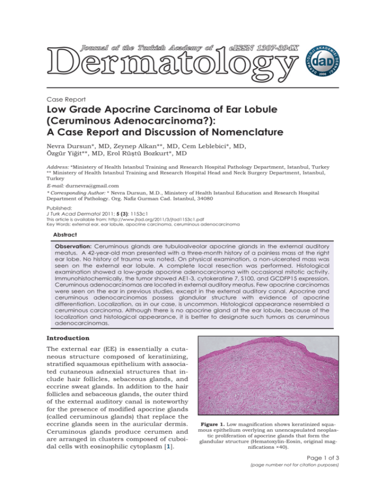
Case Report Low Grade Apocrine Carcinoma of Ear Lobule (Ceruminous Adenocarcinoma?): A Case Report and Discussion of Nomenclature Nevra Dursun*, MD, Zeynep Alkan**, MD, Cem Leblebici*, MD, Özgür Yiğit**, MD, Erol Rüştü Bozkurt*, MD Address: *Ministery of Health Istanbul Training and Research Hospital Pathology Department, Istanbul, Turkey ** Ministery of Health Istanbul Training and Research Hospital Head and Neck Surgery Department, Istanbul, Turkey E-mail: durnevra@gmail.com * Corresponding Author: * Nevra Dursun, M.D., Ministery of Health Istanbul Education and Research Hospital Department of Pathology. Org. Nafiz Gurman Cad. Istanbul, 34080 Published: J Turk Acad Dermatol 2011; 5 (3): 1153c1 This article is available from: http://www.jtad.org/2011/3/jtad1153c1.pdf Key Words: external ear, ear lobule, apocrine carcinoma, ceruminous adenocarcinoma Abstract Observation: Ceruminous glands are tubuloalveolar apocrine glands in the external auditory meatus. A 42-year-old man presented with a three-month history of a painless mass at the right ear lobe. No history of trauma was noted. On physical examination, a non-ulcerated mass was seen on the external ear lobule. A complete local resection was performed. Histological examination showed a low-grade apocrine adenocarcinoma with occasional mitotic activity. Immunohistochemically, the tumor showed AE1-3, cytokeratine 7, S100, and GCDFP15 expression. Ceruminous adenocarcinomas are located in external auditory meatus. Few apocrine carcinomas were seen on the ear in previous studies, except in the external auditory canal. Apocrine and ceruminous adenocarcinomas possess glandular structure with evidence of apocrine differentiation. Localization, as in our case, is uncommon. Histological appearance resembled a ceruminous carcinoma. Although there is no apocrine gland at the ear lobule, because of the localization and histological appearance, it is better to designate such tumors as ceruminous adenocarcinomas. Introduction The external ear (EE) is essentially a cutaneous structure composed of keratinizing, stratified squamous epithelium with associated cutaneous adnexial structures that include hair follicles, sebaceous glands, and eccrine sweat glands. In addition to the hair follicles and sebaceous glands, the outer third of the external auditory canal is noteworthy for the presence of modified apocrine glands (called ceruminous glands) that replace the eccrine glands seen in the auricular dermis. Ceruminous glands produce cerumen and are arranged in clusters composed of cuboidal cells with eosinophilic cytoplasm [1]. Figure 1. Low magnification shows keratinized squamous epithelium overlying an unencapsulated neoplastic proliferation of apocrine glands that form the glandular structure (Hematoxylin-Eosin, original magnifications ×40). Page 1 of 3 (page number not for citation purposes) J Turk Acad Dermatol 2011; 5 (3): 1153c1. http://www.jtad.org/2011/3/jtad1153c1.pdf Figure 2. Carcinoma forms glandular structures with apocrine differentiation (Hematoxylin-Eosin, original magnifications ×400). Figure 3. Tumor cells show GCDFP15 expression immunohistochemically (original magnification ×400). A malignant tumor of the EE is a rare disease, its incidence is approximately one per 1,000,000 people. The most common malignant tumor of the EE is squamous cell carcinoma (82%), and most of the remaining tumors are of glandular origin [2]. Tumors arising from ceruminous glands have been reported. Ceruminous adenocarcinomas are a malignant subtype of ceruminous glands [3, 4]. zed desmoplastic stroma was seen. There was no perineural or vascular invasion. Immunohistochemically, the tumor showed AE13, cytokeratine 7, S100, and GCDFP15 expression (Figure 3). P53 and ki-67 showed moderate positivity. There was no staining with p63. After twoyear follow-up, there was no evidence of disease. Discussion A 42-year-old man presented with a three-month history of a painless mass at the right earlobe. No history of trauma was noted. On physical examination, a non-ulcerated mass was seen on the external ear lobule. A complete resection was performed. The external ear consists of the auricula, the external auditory canal, and the outer part of the tympanic membrane. Most glandular tumors of EE originate from the external auditory canal. The normal external ear canal is an S-shaped passage measuring about 2.5 cm, lined by a very thin squamous mucosa covering scant fibrous stroma containing both sebaceous and modified apocrine-type ceruminous sweat glands. The ceruminous glands are deep within the dermis, usually close to the cartilage, which is present in the outer one-third to one-half of the canal. Ceruminous glands are absent in the auricula and the inner portion of the external auditory canal [1]. Macroscopically 2x2cm in dimension, a well-demarcated spherical mass was seen under the epidermis. Histologically the tumor consisted of stratified or single-layered tumor cells with irregular tubular architecture (Figure 1). These cells showed abundant granular eosinophilic cytoplasm and apical snouts consistent with cells of apocrine derivation (Figure 2). There were moderate atypia and occasional mitotic activity. The tumor had infiltrated to the upper dermis (Figure 1). Hyalini- Ceruminous gland tumors are uncommon lesions arising from the external auditory canal [3, 8, 9, 10]. Benign ceruminous gland tumors are less frequent than malignant tumors, but in the literature the largest number of ceruminous gland tumors reported was ceruminous gland adenomas [9]. Reports about malignant tumors are anecdotal single case reports. Only about 100 cases have been reported in the literature [3, 11, 12]. There are Primary cutaneous apocrine carcinoma (PCAC) is also a rare malignancy. Only a few cases have been reported at the ear [5, 6]. Local recurrence and locoregional lymph node metastasis are relatively frequent in PCAC [6, 7]. Case Report Page 2 of 3 (page number not for citation purposes) J Turk Acad Dermatol 2011; 5 (3): 1153c1. particular difficulties in the laboratory and in the clinical approach to ceruminous gland tumors, because the true incidence and behavior of these rare tumors are still unclear. Ceruminous gland adenomas demonstrate glands lined by tubuloglandular proliferation of the inner ceruminous cells subtended by a spindled to cuboidal myoepithelial layer [9]. In some cases, hyalinized stroma may be seen, which causes misdiagnosis as a carcinoma [9, 10]. In the literature, malignant tumors of the ceruminous glands are listed as ceruminous adenocarcinomas, adenoid cystic carcinomas, and mucoepidermoid carcinomas. Ceruminous adenocarcinomas possess a glandular structure with evidence of apocrine differentiation and infiltration not otherwise specialized. There are no myoepithelial cells at the outer part of the glandular structures. Immunohistochemically, ceruminous tumors show pan-cytokeratine, cytokeratin 7, S-100, and GCDFP15 positivity. In adenomas, the outer myoepithelial cells show p63 positivity [9]. The ki-67 and p53 expression is higher in adenocarcinomas. PCACs are rare and incompletely studied neoplasms. The largest series of PCACs consisted of 24 cases. Apocrine carcinoma of the ear is extremely rare. In the largest series, only one apocrine carcinoma was reported [5]. There are neither apocrine nor ceruminous glands at the ear lobule histologically. http://www.jtad.org/2011/3/jtad1153c1.pdf showed an apocrine nature. The histological appearance and the localization were evaluated together, and the case was diagnosed as ceruminous adenocarcinoma of the ear lobule. We speculate that the auricula and external auditory canal should all be addressed, and all of the apocrine tumors at the external ear must be diagnosed as ceruminous adenocarcinoma although, to support this thesis, it is necessary to perform studies with a wide range of series. References 1. Wenig BM, ML Urken. The ear and temporal bone. In: Histology for Pathologists. Mills SE, ed. 3rd ed. Charlottesville, VA: Lippincott Williams and Wilkins, 2007: 371-376. 2. Kuhel WI, Hume CR, Selesnick SH. Cancer of the external auditory canal and temporal bone. Otolaryngol Clin North Am 1996, 29: 827-852. PMID: 8893219 3. Wetli CV, Pardo V, Millard M, Gerston K. Tumors of ceruminous glands. Cancer 1972, 29: 1169-1178 PMID: 5021609. 4. Michel RG, Woodard BH, Shelburne JD, Bossen EH. Ceruminous gland adenocarcinoma: a light and electron microscopic study. Cancer 1978, 41: 545553. PMID: 630537 5. Robson A, Lazar AJ, Ben Nagi J et al. Primary cutaneous apocrine carcinoma: a clinico-pathologic analysis of 24 cases. Am J Surg Pathol 2008, 32: 682-690. PMID: 18347508 6. Paties C, Taccagni GL, Papotti M, Valente G, Zangrandi A, Aloi F. Apocrine carcinoma of the skin. A clinicopathologic, immunocytochemical, and ultrastructural study. Cancer 1993, 71: 375-381. PMID: 7678545 Both apocrine carcinomas and ceruminous carcinomas have a recurrence of nearly 50% [5, 6, 7, 8, 13]. Distant metastasis of apocrine carcinoma is nearly 24% [5]. Recurrence is mostly seen for low-grade ceruminous-type adenocarcinomas. Metastasis is rare. Therefore, for patient follow up and prognosis, it is very important to differentiate these two tumors. 7. Warkel RL, Helwig EB. Apocrine gland adenoma and adenocarcinoma of the axilla. Arch Dermatol 1978, 114: 198-203. PMID: 629545 The nomenclature and diagnosis of our tumor is difficult, because the tumor was low-grade and the localization was the ear lobule. In our case, histological appearance, the infiltration of the upper dermis, and a lack of myoepithelial cells at the outer edge of the glands (p63 negativity) demonstrate malignancy. The decapitation secretion into the luminal space by glandular cells with abundant granular eosinophilic cytoplasm and GCDFP15 positivity 10. Lassaletta L, Patrón M, Olóriz J, Pérez R, Gavilán J. Avoiding misdiagnosis in ceruminous gland tumours. Auris Nasus Larynx 2003, 30: 287-290. PMID:12927294 8. Hicks GW. Tumors arising from the glandular structures of the external auditory canal. Laryngoscope 1983, 93: 326-340. PMID: 6300574 9. Thompson LD, Nelson BL, Barnes EL. Ceruminous adenomas: a clinicopathologic study of 41 cases with a review of the literature. Am J Surg Pathol 2004, 28: 308-318. PMID: 20596983 11. Mills RG, Douglas-Jones T, Williams RG. Ceruminoma’--a defunct diagnosis. J Laryngol Otol 1995, 109: 180-188. PMID: 7745330 12. Iqbal A, Newman P. Ceruminous gland neoplasia. Br J Plast Surg 1998, 51: 317-320. PMID: 9771352 13. Cooper PH. Carcinomas of sweat glands. Pathol Annu 1987, 22: 83-124. PMID: 3033589 Page 3 of 3 (page number not for citation purposes)
