Science 8 Names ______ ______ Sheep's Eye Dissection Lab
advertisement
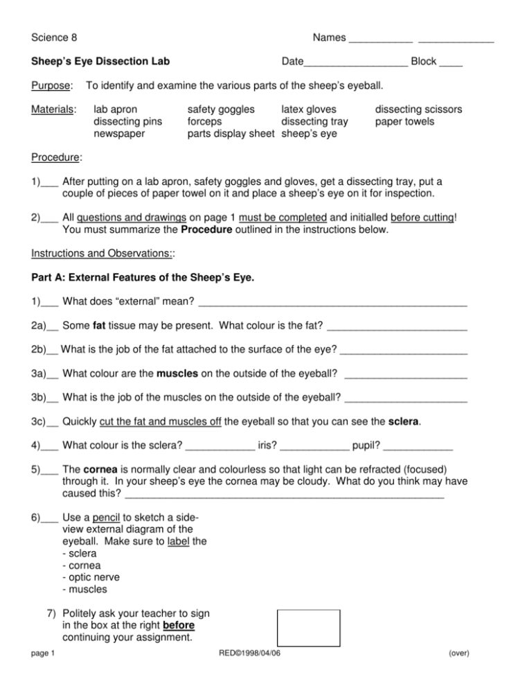
Science 8 Names ___________ _____________ Date__________________ Block ____ Sheep’s Eye Dissection Lab Purpose: Materials: To identify and examine the various parts of the sheep’s eyeball. lab apron dissecting pins newspaper safety goggles latex gloves forceps dissecting tray parts display sheet sheep’s eye dissecting scissors paper towels Procedure: 1)___ After putting on a lab apron, safety goggles and gloves, get a dissecting tray, put a couple of pieces of paper towel on it and place a sheep’s eye on it for inspection. 2)___ All questions and drawings on page 1 must be completed and initialled before cutting! You must summarize the Procedure outlined in the instructions below. Instructions and Observations:: Part A: External Features of the Sheep’s Eye. 1)___ What does “external” mean? ______________________________________________ 2a)__ Some fat tissue may be present. What colour is the fat? ________________________ 2b)__ What is the job of the fat attached to the surface of the eye? ______________________ 3a)__ What colour are the muscles on the outside of the eyeball? _____________________ 3b)__ What is the job of the muscles on the outside of the eyeball? _____________________ 3c) __ Quickly cut the fat and muscles off the eyeball so that you can see the sclera. 4)___ What colour is the sclera? ____________ iris? ____________ pupil? ____________ 5)___ The cornea is normally clear and colourless so that light can be refracted (focused) through it. In your sheep’s eye the cornea may be cloudy. What do you think may have caused this? _______________________________________________________ 6)___ Use a pencil to sketch a sideview external diagram of the eyeball. Make sure to label the - sclera - cornea - optic nerve - muscles 7) Politely ask your teacher to sign in the box at the right before continuing your assignment. page 1 RED©1998/04/06 (over) Part B: Internal Features of the Sheep’s Eye. (Place the parts on the identification sheet) 1a)__ Carefully use a razor blade or scissors to make a small incision (cut) near the centre of the eyeball. With your dissecting scissors, cut around the entire eye so that you have two equal hemispheres when you are finished. 1b)__ What did you observe about the texture of the sclera while cutting? _______________ Why is this important? ___________________________________________________ 1c) __ What colour is the sclera in your eye? _______________________________________ 1d)__ What material was the sclera made from in our model? _________________________ 2a) _ The jelly inside the eye is the vitreous humour. (It is like a jelly that gives the eyeball shape and prevents the sclera from collapsing. It does the same job as air in a beach ball.) 2b)__ Gloop your vitreous humour onto the parts identification worksheet. 2c) __ What food is like the vitreous humour? ______________________________________ 2d)__ What is the job of the vitreous humour? _____________________________________ 3a)__ Pick up the back of the sclera. It contains the optic nerve. The retina is a very thin layer containing rods and cones which detect shadows, shapes, and colour. 3b)__ Use your forceps and probe to peel the retina off the inside of the eye. Place it on the parts identification worksheet. 3c) __ What material did we make the retina from in our model? _______________________ 3d)__ Rods work well in dim light to detect _______________ ______________, while cones work especially well in bright light to detect _______________. 4a)__ You may have found it difficult to remove the retina because it was attached to the optic nerve. The attachment area is called the ______________. 4b)__ What did we make the blind spot from in our model? __________________________ 4c) __ There are no rods or cones at the blind spot due to the connection. The retina can receive no information here. What does a sheep see when an object lands exactly on the blind spot? ______________________. 5a)__ The black, shiny layer under the retina is the choroid coat. It contains blood vessels that provide nutrients for the eye. Why is its surface shiny? ____________________________________________________________________ page 2 RED©1998/04/06 (over) 5b) _ Use the forceps and probe to carefully peel the choroid coat from the sclera. It on identification worksheet. 5c) __ What was the choroid coat made from in our model? ___________________________ 5d)__ What is the job of the choroid coat? ________________________________________ 5e)__ What part of the body pumps blood to the choroid coat? ________________________ 5f) __ Now that you have removed everything from the back half of the sclera, pin it to the parts identification sheet. 6a)__ Pick up the front half of the sclera that contains the cornea, iris and lens. Remove the lens carefully with the forceps. 6b)__ Did you notice the aqueous humour between the lens and the cornea? ____________ (It is a watery fluid that carries nutrients to, and wastes away from the front of the eye. It also keeps the shape of the front of the eye and prevents the cornea from collapsing upon the lens.) 6c) __ What common food material is like the aqueous humour? _______________________ 6d)__ What is the job of the aqueous humour? _____________________________________ 7a)__ Pick up the lens and examine it carefully. Is it convex or concave? _______________ 7b)__ Why is it important that it is convex like a magnifying glass? _____________________ 7c) __ Try dropping the lens once from about shoulder height onto the lab bench. It probably bounces. Why is it important that the lens is flexible? ___________________________________ 7d)__ What material did we use in our model to represent the lens? ____________________ 7e)__ Place the lens on the parts identification worksheet. 8a)__ Use your fingers to carefully remove the iris. What is the hole in the iris called? _____ 8b)__ Make a sketch in pencil (in the space to the right) of the iris. The lines are small muscles that cause the iris to contract or dilate (get smaller or bigger). Make sure you label these muscles. 8c) __ The job of the iris is to limit the amount of light page 3 RED©1998/04/06 (over) entering the eye. Is the iris transparent, translucent or opaque? __________________ 8d)__ What part of a house window controls the amount of light entering like the iris? ______ 8e)__ What was the iris made of in our model? ____________________________________ 8f) __ Place the iris to the parts identification worksheet. 9a)__ Cut the cornea out of the front of the sclera. It acts as a lens to focus the light and also protects the eye. 9b)__ If the inside of the eye is represented by the passenger compartment of a car, what part of the car lets the light in but protects the occupants? ____________ 9c) __ What material did we use to represent the cornea in our model? __________________ 9d)__ Place the cornea on the parts identification worksheet. 10)__ Politely show your teacher your completed parts identification worksheet and ask him or her to sign this box. 11a)_ Wrap up the sheep's eye parts in the paper and throw them away. 11b)_ Carefully wash all dissecting materials and the bench top and put equipment away. Discussion: 1) page 4 Label the diagram with parts of the sheep’s eye. (Hint: See boldface words!) RED©1998/04/06 (over) 2) Write the names of the actual eye parts. (Hint: See boldface words!) a) Thick outer casing of eye _____________ b) Shiny, black with blood vessels ________ c) Thin layer registering images __________ d) Cord carrying messages to brain _______ e) Connection--no info registers __________ f) Jelly-like liquid prevents damage ________ g) Controls amount of light entering _______ h) First window protects and focuses ______ i) Opening in coloured muscle ___________ j) Final focusing transparent ball __________ k) Watery liquid feeds front of eye ________ l) Stretch and relax to move parts _________ Analysis: What are three (3) things about this lab that are similar to what doctors do? page 5 RED©1998/04/06 (over)

