Human Structure & Development 212
advertisement
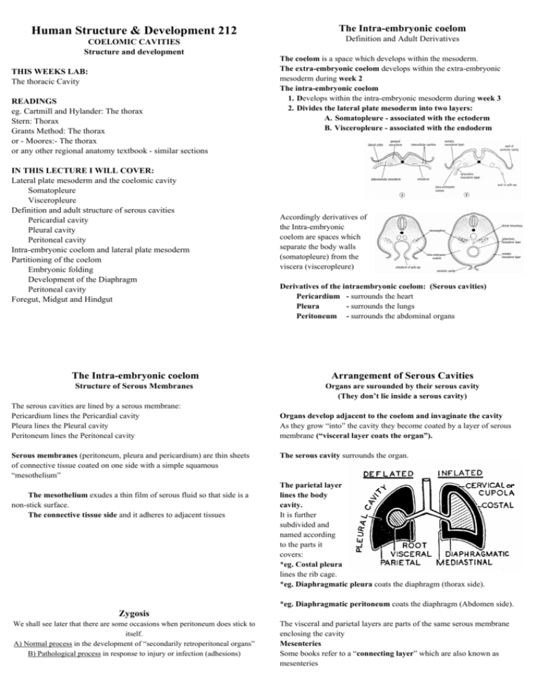
Human Structure & Development 212 COELOMIC CAVITIES Structure and development THIS WEEKS LAB: The thoracic Cavity READINGS eg. Cartmill and Hylander: The thorax Stern: Thorax Grants Method: The thorax or - Moores:- The thorax or any other regional anatomy textbook - similar sections IN THIS LECTURE I WILL COVER: Lateral plate mesoderm and the coelomic cavity Somatopleure Visceropleure Definition and adult structure of serous cavities Pericardial cavity Pleural cavity Peritoneal cavity Intra-embryonic coelom and lateral plate mesoderm Partitioning of the coelom Embryonic folding Development of the Diaphragm Peritoneal cavity Foregut, Midgut and Hindgut The Intra-embryonic coelom Definition and Adult Derivatives The coelom is a space which develops within the mesoderm. The extra-embryonic coelom develops within the extra-embryonic mesoderm during week 2 The intra-embryonic coelom 1. Develops within the intra-embryonic mesoderm during week 3 2. Divides the lateral plate mesoderm into two layers: A. Somatopleure - associated with the ectoderm B. Visceropleure - associated with the endoderm Accordingly derivatives of the Intra-embryonic coelom are spaces which separate the body walls (somatopleure) from the viscera (visceropleure) Derivatives of the intraembryonic coelom: (Serous cavities) Pericardium - surrounds the heart Pleura - surrounds the lungs Peritoneum - surrounds the abdominal organs The Intra-embryonic coelom Arrangement of Serous Cavities Structure of Serous Membranes Organs are surounded by their serous cavity (They don’t lie inside a serous cavity) The serous cavities are lined by a serous membrane: Pericardium lines the Pericardial cavity Pleura lines the Pleural cavity Peritoneum lines the Peritoneal cavity Serous membranes (peritoneum, pleura and pericardium) are thin sheets of connective tissue coated on one side with a simple squamous “mesothelium” The mesothelium exudes a thin film of serous fluid so that side is a non-stick surface. The connective tissue side and it adheres to adjacent tissues Organs develop adjacent to the coelom and invaginate the cavity As they grow “into” the cavity they become coated by a layer of serous membrane (“visceral layer coats the organ”). The serous cavity surrounds the organ. The parietal layer lines the body cavity. It is further subdivided and named according to the parts it covers: *eg. Costal pleura lines the rib cage. *eg. Diaphragmatic pleura coats the diaphragm (thorax side). *eg. Diaphragmatic peritoneum coats the diaphragm (Abdomen side). Zygosis We shall see later that there are some occasions when peritoneum does stick to itself. A) Normal process in the development of “secondarily retroperitoneal organs” B) Pathological process in response to injury or infection (adhesions) The visceral and parietal layers are parts of the same serous membrane enclosing the cavity Mesenteries Some books refer to a “connecting layer” which are also known as mesenteries Development of the Serous Cavities The coelom begins as a horseshoe shaped cavity The ends of the horseshoe are initially continuous with the extraembryonic coelom. The arch of the horseshoe passes 1. :In front of the oral membrane 2. 3. Behind the cardiogenic area (developing heart) and behind the septum transversum (developing diaphragm) Development of the pericardial cavity Heart Week 4 - Folding of the embryo (and the coelom) partitions the Coelom into Pericardium and left and right pleuro-peritoneal canals. Coelom When the head fold occurs: * The septum transversum and the developing heart and brought into their proper positions and the thorax is formed: · The coelom is folded too......... Becomes: Pericardium and two Pleuroperitoneal canals Development of the pleural cavity Lungs *The foregut (oesophagus) runs down behind the developing heart *It has the pleuroperitoneal canals on either side *The foregut gives rise to the lung buds which grow out into the left and right pleuroperitoneal canals *The Septum transversum (Developing diaphragm) receives its nerve supply (phrenic nerve) from cervical nerves (C34&5). *When folding occurs the phrenic nerve is dragged down through the thorax *The phrenic nerve comes to lie between the pericardium and the pleuro-peritoneal canals - helps to separate these parts Development of the peritoneal cavity Gut tube Initially the peritoneal cavity is in left and right parts. The gut has a dorsal and ventral mesentery The liver develops in the ventral mesentery of the foregut And the foregut retains both ventral and dorsal mesenteries Diaphragm (separates the thorax and abdomen) The pleuroperitoneal canals are partitioned (into pleural and peritoneal cavities) by the growth of the diaphragm: 1. Septum transversum (centrally) 2. Pleuroperitoneal folds (posteriorly) 3. Ingrowth of the body wall (laterally) The midgut and hindgut lose their ventral mesentery So the left and right sides become a single cavity.
