Analysis of the Humeroscapular Angular Motion Ratio using a New
advertisement

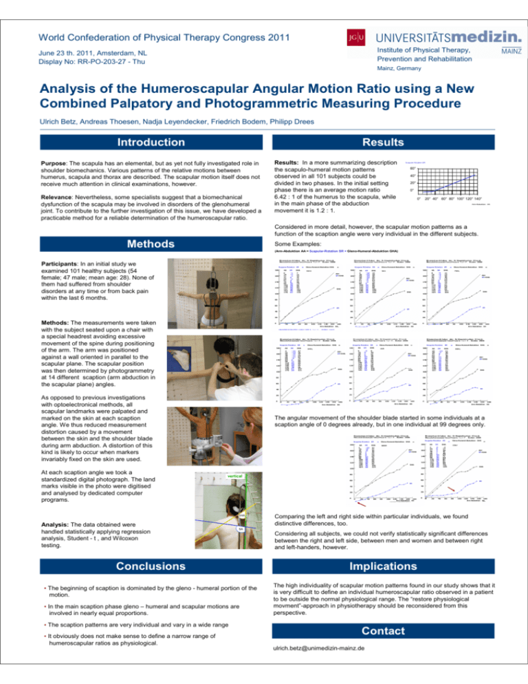
World Confederation of Physical Therapy Congress 2011 Institute of Physical Therapy, Prevention and Rehabilitation June 23 th. 2011, Amsterdam, NL Display No: RR-PO-203-27 - Thu Mainz, Germany Analysis of the Humeroscapular Angular Motion Ratio using a New Combined Palpatory and Photogrammetric Measuring Procedure Ulrich Betz, Andreas Thoesen, Nadja Leyendecker, Friedrich Bodem, Philipp Drees Introduction Results Purpose: The scapula has an elemental, but as yet not fully investigated role in shoulder biomechanics. Various patterns of the relative motions between humerus, scapula and thorax are described. The scapular motion itself does not receive much attention in clinical examinations, however. Relevance: Nevertheless, some specialists suggest that a biomechanical dysfunction of the scapula may be involved in disorders of the glenohumeral joint. To contribute to the further investigation of this issue, we have developed a practicable method for a reliable determination of the humeroscapular ratio. Results: In a more summarizing description the scapulo-humeral motion patterns observed in all 101 subjects could be divided in two phases. In the initial setting phase there is an average motion ratio 6.42 : 1 of the humerus to the scapula, while in the main phase of the abduction movement it is 1.2 : 1. Scapular-Rotation SR 60° 40° 20° 0° 0° 20° 40° 60° 80° 100° 120° 140° Arm-Abduktion AA Considered in more detail, however, the scapular motion patterns as a function of the scaption angle were very individual in the different subjects. Methods Some Examples: (Arm-Abduktion AA = Scapular-Rotation SR + Gleno-Humeral-Abduktion GHA) Participants: In an initial study we examined 101 healthy subjects (54 female; 47 male; mean age: 28). None of them had suffered from shoulder disorders at any time or from back pain within the last 6 months. Scapular-Rotation SR AA SR Gleno-Humeral-Abduktion GHA Scapular-Rotation SR GHA AA SR Gleno-Humeral-Abduktion GHA Scapular-Rotation SR AA GHA SR Gleno-Humeral-Abduktion GHA GHA AA= SR+GHA AA= SR+GHA AA= SR+GHA GHA GHA GHA SR SR Methods: The measurements were taken with the subject seated upon a chair with a special headrest avoiding excessive movement of the spine during positioning of the arm. The arm was positioned against a wall oriented in parallel to the scapular plane. The scapular position was then determined by photogrammetry at 14 different scaption (arm abduction in the scapular plane) angles. SR Arm-Abduktion AA Scapular-Rotation SR AA SR Arm-Abduktion AA Gleno-Humeral-Abduktion GHA Scapular-Rotation SR AA GHA SR Gleno-Humeral-Abduktion GHA Arm-Abduktion AA Scapular-Rotation GHA AA AA= SR+GHA SR SR Gleno-Humeral-Abduktion GHA GHA AA= SR+GHA AA= SR+GHA GHA GHA SR GHA SR SR As opposed to previous investigations with optoelectronical methods, all scapular landmarks were palpated and marked on the skin at each scaption angle. We thus reduced measurement distortion caused by a movement between the skin and the shoulder blade during arm abduction. A distortion of this kind is likely to occur when markers invariably fixed on the skin are used. Arm-Abduktion AA Arm-Abduktion AA Arm-Abduktion AA The angular movement of the shoulder blade started in some individuals at a scaption angle of 0 degrees already, but in one individual at 99 degrees only. Scapular-Rotation SR AA SR Gleno-Humeral-Abduktion GHA Scapular-Rotation SR AA GHA SR Gleno-Humeral-Abduktion GHA GHA AA= SR+GHA AA= SR+GHA GHA GHA At each scaption angle we took a standardized digital photograph. The land marks visible in the photo were digitised and analysed by dedicated computer programs. vertical SR SR Arm-Abduktion AA GHA Analysis: The data obtained were handled statistically applying regression analysis, Student - t , and Wilcoxon testing. Arm-Abduktion AA Comparing the left and right side within particular individuals, we found distinctive differences, too. SA Considering all subjects, we could not verify statistically significant differences between the right and left side, between men and women and between right and left-handers, however. Conclusions Implications • The beginning of scaption is dominated by the gleno - humeral portion of the motion. The high individuality of scapular motion patterns found in our study shows that it is very difficult to define an individual humeroscapular ratio observed in a patient to be outside the normal physiological range. The “restore physiological movment”-approach in physiotherapy should be reconsidered from this perspective. • In the main scaption phase gleno – humeral and scapular motions are involved in nearly equal proportions. • The scaption patterns are very individual and vary in a wide range • It obviously does not make sense to define a narrow range of humeroscapular ratios as physiological. Contact ulrich.betz@unimedizin-mainz.de


