Full paper in PDF - Hong Kong Medical Journal
advertisement

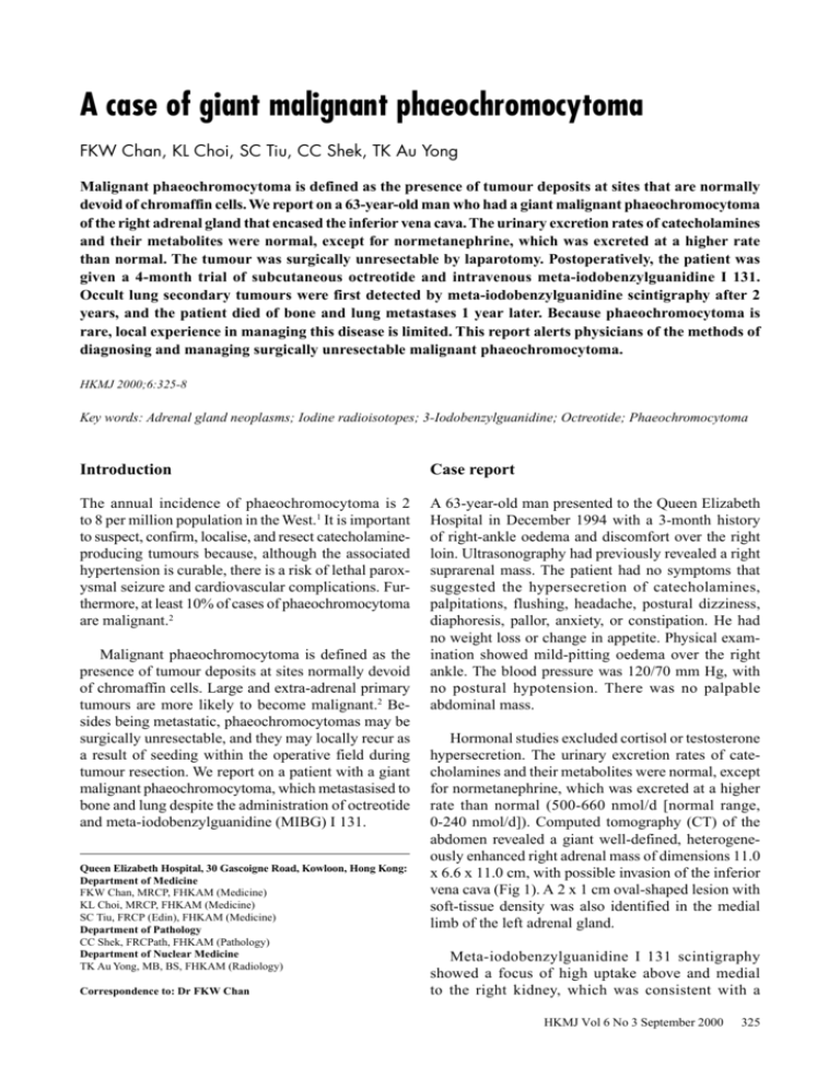
Giant malignant phaeochromocytoma A case of giant malignant phaeochromocytoma FKW Chan, KL Choi, SC Tiu, CC Shek, TK Au Yong Malignant phaeochromocytoma is defined as the presence of tumour deposits at sites that are normally devoid of chromaffin cells. We report on a 63-year-old man who had a giant malignant phaeochromocytoma of the right adrenal gland that encased the inferior vena cava. The urinary excretion rates of catecholamines and their metabolites were normal, except for normetanephrine, which was excreted at a higher rate than normal. The tumour was surgically unresectable by laparotomy. Postoperatively, the patient was given a 4-month trial of subcutaneous octreotide and intravenous meta-iodobenzylguanidine I 131. Occult lung secondary tumours were first detected by meta-iodobenzylguanidine scintigraphy after 2 years, and the patient died of bone and lung metastases 1 year later. Because phaeochromocytoma is rare, local experience in managing this disease is limited. This report alerts physicians of the methods of diagnosing and managing surgically unresectable malignant phaeochromocytoma. HKMJ 2000;6:325-8 Key words: Adrenal gland neoplasms; Iodine radioisotopes; 3-Iodobenzylguanidine; Octreotide; Phaeochromocytoma Introduction Case report The annual incidence of phaeochromocytoma is 2 to 8 per million population in the West.1 It is important to suspect, confirm, localise, and resect catecholamineproducing tumours because, although the associated hypertension is curable, there is a risk of lethal paroxysmal seizure and cardiovascular complications. Furthermore, at least 10% of cases of phaeochromocytoma are malignant.2 A 63-year-old man presented to the Queen Elizabeth Hospital in December 1994 with a 3-month history of right-ankle oedema and discomfort over the right loin. Ultrasonography had previously revealed a right suprarenal mass. The patient had no symptoms that suggested the hypersecretion of catecholamines, palpitations, flushing, headache, postural dizziness, diaphoresis, pallor, anxiety, or constipation. He had no weight loss or change in appetite. Physical examination showed mild-pitting oedema over the right ankle. The blood pressure was 120/70 mm Hg, with no postural hypotension. There was no palpable abdominal mass. Malignant phaeochromocytoma is defined as the presence of tumour deposits at sites normally devoid of chromaffin cells. Large and extra-adrenal primary tumours are more likely to become malignant.2 Besides being metastatic, phaeochromocytomas may be surgically unresectable, and they may locally recur as a result of seeding within the operative field during tumour resection. We report on a patient with a giant malignant phaeochromocytoma, which metastasised to bone and lung despite the administration of octreotide and meta-iodobenzylguanidine (MIBG) I 131. Queen Elizabeth Hospital, 30 Gascoigne Road, Kowloon, Hong Kong: Department of Medicine FKW Chan, MRCP, FHKAM (Medicine) KL Choi, MRCP, FHKAM (Medicine) SC Tiu, FRCP (Edin), FHKAM (Medicine) Department of Pathology CC Shek, FRCPath, FHKAM (Pathology) Department of Nuclear Medicine TK Au Yong, MB, BS, FHKAM (Radiology) Correspondence to: Dr FKW Chan Hormonal studies excluded cortisol or testosterone hypersecretion. The urinary excretion rates of catecholamines and their metabolites were normal, except for normetanephrine, which was excreted at a higher rate than normal (500-660 nmol/d [normal range, 0-240 nmol/d]). Computed tomography (CT) of the abdomen revealed a giant well-defined, heterogeneously enhanced right adrenal mass of dimensions 11.0 x 6.6 x 11.0 cm, with possible invasion of the inferior vena cava (Fig 1). A 2 x 1 cm oval-shaped lesion with soft-tissue density was also identified in the medial limb of the left adrenal gland. Meta-iodobenzylguanidine I 131 scintigraphy showed a focus of high uptake above and medial to the right kidney, which was consistent with a HKMJ Vol 6 No 3 September 2000 325 Chan et al R L Fig 1. Post-contrast computed tomography scan showing a giant well-defined right adrenal mass (11.0 x 6.6 x 11.0 cm), with possible invasion of the inferior vena cava phaeochromocytoma of the right adrenal gland. There was also a region of faint uptake of 131I-MIBG above the dominant mass, which suggested local metastasis. The left adrenal gland, however, did not exhibit any abnormal uptake of 131 I-MIBG. In January 1995, a giant right adrenal tumour of dimensions 12 x 10 cm was found intra-operatively. The tumour encased the inferior vena cava and infiltrated the inferoposterior aspect of the liver. It was surgically unresectable. Biopsy examination confirmed the tumour to be a phaeochromocytoma. Blood loss was approximately 500 mL during the operation and there was no hypertensive crisis during tumour manipulation. Postoperative single-photon emission CT scintigraphy using octreotide In 111 showed mild-tomoderate octreotide uptake by the tumour mass, especially at the periphery (Fig 2). Subcutaneous injection of unlabelled octreotide 100 µg every 8 hours was given for 4 months. Urinary excretion of normetanephrine became normal. Follow-up 131I-MIBG scintigraphy and CT of the adrenal glands showed no change in tumour size. The patient consented to 131 I-MIBG therapy. A trial dose of 131I-MIBG 0.5 mCi was given intravenously. Radioactive counts of the whole body, liver, and tumour were plotted against time (at 4, 24, 48, 72, and 96 hours). The estimated absorbed dose was then calculated by measuring the area under the curve. A treatment dose of 200 mCi of 131 I-MIBG was given in July 1995 and repeated in 1996. The estimated dose of radiation after each 200 mCi of 131 I-MIBG was 3544 cGy for the tumour and 173 cGy for the liver. These doses were well tolerated, and there was no clinically evident radiation-induced damage to surrounding structures. However, no change in tumour size or uptake was detectable on subsequent CT and 131 I-MIBG scintigraphy scans. Scintigraphy was repeated in December 1996 and showed faint uptake over both lung fields, despite normal chest X-rays and CT scans of the thorax. A CT scan of abdomen was performed in July 1997 and did not reveal any vertebral metastasis. Urinary excretion of catecholamines and metabolites remained normal after therapy. In November 1997, the patient started to experience lower back pain and right lower-limb weakness. A CT myelogram revealed multiple osteolytic lesions at the right ala of the upper sacrum, vertebral bodies of L3, L4, L5, T10, and T11. The right pedicle of L5 was eroded and there was compression of the cauda equina on the right side, at the level of L5. An X-ray also showed osteolytic secondary tumours over the left hip and upper shaft of the right femur. A chest X-ray revealed multiple cannon-ball–like secondary tumours over both lung fields. Bone pain was controlled with morphine and palliative external radiotherapy. The patient’s condition deteriorated rapidly during the next 2 months, and in January 1998, he died of respiratory failure as a result of the extensive lung metastases . Discussion Fig 2. Transaxial single-photon emission computed tomography scan showing mild-to-moderate uptake of octreotide by the phaeochromocytoma, especially at the periphery (arrows) 326 HKMJ Vol 6 No 3 September 2000 The natural course of malignant phaeochromocytoma is highly variable: the overall 5-year mortality rate is approximately 44%, but some patients, whose disease is indolent, can survive for 20 years or longer.3 Because of the rarity of malignant phaeochromocytoma, local experience in management of surgically unresectable or metastatic disease is limited. In addition, benign and malignant phaeochromocytoma cannot be distinguished on the basis of clinical, biochemical, Giant malignant phaeochromocytoma or histopathological characteristics. Although histological criteria for malignancy (including cellular hyperchromatism, absence of a true capsule, capsular invasion, vascular invasion, and bizarre mitotic figures) are more frequently associated with malignant tumours, they are only poorly predictive of biological behaviour.3,4 Metastasis may occur years after successful surgical excision of an apparently benign tumour. Malignant phaeochromocytoma can be diagnosed by the identification of local invasion, or metastasis to sites that do not normally contain chromaffin cells. The most common metastatic sites are the lymph nodes, bone, lung, and liver. 5 In this patient, malignant phaeochromocytoma was diagnosed on the basis of local invasion into inferoposterior aspect of the liver and inferior vena cava. Approximately 60% of patients with malignant phaeochromocytomas have been reported to have elevated levels of dopamine excretion.6 Normal levels of urinary dopamine excretion, however, do not rule out the possibility of malignant phaeochromocytoma. In this patient, urinary excretion of normetanephrine was elevated to more than twice the normal level, but urinary excretion of epinephrine, norepinephrine, and dopamine were normal. This phenomenon has been described by Crout and Sjoerdsma,7 who reported that small tumours of less than 50 g have rapid turnover rates and low catecholamine content, and mainly released unmetabolised catecholamines into the circulation. In contrast, large tumours of more than 50 g have slow turnover rates and high catecholamine content.7 These large tumours thus release mainly metabolised catecholamines into the circulation, as reflected by a high ratio of metabolites to free catecholamines in the urine. These tumours have relatively high levels of catechol-O-methyltransferase, which metabolise most of the catecholamines to metanephrines. The presence of catechol-O-methyltransferase in phaeochromocytomas has been recently confirmed by Western blot analysis, enzyme assay, and immunohistochemical staining. 8 Plasma levels of metanephrines (normetanephrine and metanephrine) in patients with phaeochromocytoma are subsequently increased because of the catecholamines produced and metabolised within the tumour.8 Furthermore, there is a significant positive association between tumour size and plasma concentrations of metanephrines, which contrasts with a complete lack of association between tumour size and plasma concentrations of catecholamines.8 The basic principles in the treatment of malignant phaeochromocytomas are to surgically resect the primary tumour and to prevent its recurrence or metastasis whenever possible, as well as to treat hypertensive symptoms by catecholamine blockade. In this patient, the presence of somatostatin receptors, as demonstrated by 111In-octreotide scintigraphy, supported a trial of octreotide treatment. Kopf et al9 reported symptomatic improvement but no mass reduction after octreotide treatment in two patients with malignant phaeochromocytoma. In this case, administration of octreotide failed to decrease tumour size, although urinary excretion of normetanephrine was normalised. The intense uptake and prolonged retention of tracer doses of 131I-MIBG by some unresectable phaeochromocytomas have provided the rationale for treating such lesions with large doses of this agent. Krempf et al10 and Shapiro et al11 have shown that local irradiation of a tumour with therapeutic doses of 131 I-MIBG produces partial and temporary responses in approximately one third of patients. In their studies, many patients in whom uptake of the agent was sufficient for obviously positive diagnostic imaging did not have an adequate uptake to deliver therapeutic doses of radiation. Increasing the dose would have increased the risk of radiation-induced enteritis and hepatitis. In this patient, there was sufficient 131I-MIBG uptake by the tumour during the tracer test, and it was estimated that a therapy with 200 mCi of 131I-MIBG would deliver a cumulative radiation dose of 3544 cGy to the tumour. The tumour size remained static for 2 years, when secondary tumours were detected in the lungs. Combination chemotherapy of cyclophosphamide, vincristine, and dacarbazine given cyclically every 21 days has been reported to be beneficial but not curative for malignant phaeochromocytoma.12 Because this patient had remained relatively asymptomatic during the course of the disease, he decided not to receive chemotherapy. This case also confirms that 131I-MIBG scintigraphy is superior to CT of the thorax and conventional chest X-ray in detecting micrometastases of phaeochromocytoma in the lung. Shapiro et al4 have shown that 131I-MIBG scintigraphy is useful in determining the extent of metastatic disease in most cases (26/30), and in some patients (16/30), scintigraphy is more sensitive than other radiological procedures. References 1. Stenstrom G, Svardsudd K. Phaeochromocytoma in Sweden, 1958-1981: an analysis of the National Cancer Registry Data. Acta Med Scand 1986;220(Suppl):225-32. 2. Young WF Jr. Phaeochromocytoma and primary aldosteronism: diagnostic approaches. Endocrinol Metab Clin North Am HKMJ Vol 6 No 3 September 2000 327 Chan et al 1997;26:801-17. 3. Scott HW Jr, Reynolds V, Green N, et al. Clinical experience with malignant phaeochromocytomas. Surg Gynecol Obstet 1982;154:801-18. 4. Shapiro B, Sisson JC, Lloyd R, et al. Malignant phaeochromocytoma: clinical, biochemical and scintigraphic characterization. Clin Endocrinol 1984;20, 189-203. 5. Bravo EL. Evolving concepts in the pathophysiology, diagnosis, and treatment of phaeochromocytoma. Endocrine Rev 1994;15:356-68. 6. Goldstein DS, Stull R, Eisenhofer G, et al. Plasma 3, 4-dihydroxyphenylalanine (Dopa) and catecholamines in neuroblastoma or phaeochromocytoma. Ann Intern Med 1986;105:887-8. 7. Crout JR, Sjoerdsma A. Turnover and metabolism of catecholamine in patients with phaeochromocytoma. J Clin Invest 1964;43:94-102. 8. Eisenhofer G, Keiser H, Friberg P, et al. Plasma metanephrines are markers of pheochromocytoma produced by catechol-Omethyltransferase within tumors. J Clin Endocrinol Metab 1998;83:2175-85. 9. Kopf D, Bockisch A, Steinert H, et al. Octreotide scintigraphy and catecholamine response to an octreotide challenge in malignant phaeochromocytoma. Clin Endocrinol 1997;46: 39-44. 10. Krempf M, Lumbroso J, Mornex R, et al. Use of m-[131I] iodobenzylguanidine in the treatment of malignant phaeochromocytoma. J Clin Endocrinol Metab 1991;72:455-61. 11. Shapiro B, Sisson JC, Wieland DM, et al. Radiopharmaceutical therapy of malignant phaeochromocytoma with [131I]metaiodobenzylguanidine: results from ten years of experience. J Nucl Biol Med 1991;35:269-76. 12. Averbuch SD, Steakley CS, Young RC, et al. Malignant phaeochromocytoma: Effective treatment with a combination of cyclophosphamide, vincristine, and dacarbazine. Ann Intern Med 1988;109:267-73. Instructions for Letters to the Editor Letters discussing a recent article in the Hong Kong Medical Journal are welcome and should be sent to the Editorial Office by e-mail to <hkmj@hkam.org.hk> within 6 weeks of the article’s publication. Original letters that do not refer to a Journal article may also be considered. All letters that relate to a Journal article will be posted on the Journal website <http://www.hkmj.org.hk> and will be eligible for publication in the paper version. Letters should not exceed 500 words, have no more than five references (in the Vancouver style), and contain only one illustration if appropriate. There should be no more than two authors; a greater number requires justification. Authors should quote their two highest qualifications and give full contact details, as well as an e-mail address for the corresponding author. Financial associations or other possible conflicts of interest should always be disclosed. Letters may be edited before publication. 328 HKMJ Vol 6 No 3 September 2000


