Leica TCS STED CW Brochure
advertisement
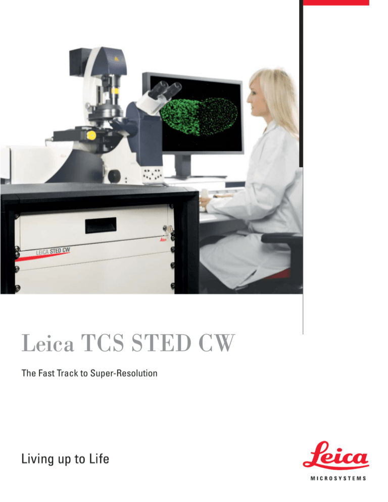
Leica TCS STED CW The Fast Track to Super-Resolution • Freedom to choose – popular fluorescent dyes and proteins • Observe what´s inside – with confocal super-resolution • Nanoscale imaging – devoid of mathematical artifacts • Upgrading, to STED – quick and affordable • Watch live cell dynamics – at the nanoscale! 2 Super-resolution light microscopy is revolutionizing life science research with increasing speed. The charm of direct visual results from an intact specimen on the nanometer scale attracts scientists from all fields of biomedical research. In its early days, super-resolution was only for biophysicists and optical specialists. Nowadays, it has become an indispensable method in many life science research institutes working with light microscopes. Structural details of synapses, ensembles of small vesicles or receptor arrangements have now become accessible for fluorescence microscopy. Leica TCS STED CW The Fast Track to Super-Resolution Subdiffraction microscopy needs to meet the requirements of daily research. But many superresolution imaging tasks require special labeling or restricted user environments. So far it was difficult to get super-resolved images with standard fluorophores, fluorescent proteins, and from living specimens. However, the access to these data means a crucial improvement of results for any researcher. Leica Microsystems, the first provider of integrated super-resolution technology, fills this gap by extending its STED portfolio with the new Leica TCS STED CW. It is a stunningly simple solution which combines the high-end confocal TCS SP5 with purely optical and patented super-resolution technology. It opens the door to the nanoworld – easy, highly affordable and as an upgrade for already installed systems! K. Willig, B. Harke, R. Medda, S.W. Hell, Nature Meth. 4, 915 (2007) 3 Dual Color STED Imaging Super-resolution in two colors is essential to put biological information into context and enables colocalization studies at the nanoscale to be performed. Combination of a large Stokes shift dye (e.g. BD Horizon V500) with a standard fluorophore (e.g. Oregon green 488) allows to obtain super-resolution in two channels applying only one STED wavelength (592 nm). Differences in excitation and emission spectra are exploited to distinguish the two dyes in sequential recordings. Applying the same STED doughnut for both super-resolution channels keeps the set up simple and also avoids problems with chromatic aberration, which other implementations of super-resolution microscopy need to compensate. Confocal STED 458 514 592 Normalized Intensity 100 80 60 40 20 0 400 450 500 Detection V500 5 μm 5 μm 550 600 nm Detection OG488 Wavelength (nm) Example of fluorophores with well separated excitation peaks and partial overlapping emission spectra (BD Horizon V500 in red and Oregon Green 488 displayed in green) suited for dual color STED imaging with the Leica TCS STED CW. In red, excitation of BD Horizon V500 at 458 nm, in green excitation of Oregon Green 488 at 514 nm, in yellow depletion at 592 nm. Please note: Suboptimal excitation can not only help to avoid crosstalk, but also to choose appropriate detection windows. 500 nm 500 nm Nuclear pore complexes and clathrin coated vesicles of adherent cell stained with BD Horizon V500 (red) and Oregon Green 488 (green), respectively. 4 Standard Dyes for STED Microscopy Confocal Alexa 488 STED 2 μm 2 μm Confocal STED 500 nm Distribution of Clathrin vesicles in HeLa cells. Fluorescent marker: Alexa 488, immunolabeling. 500 nm Oregon Green Confocal STED STED & Deconvolution 2 μm 500 nm 2 μm 500 nm 2 μm 500 nm Intermediate filamentous protein Vimentin in Vero cells. Fluorescence label: Oregon Green. 5 Super-Resolution at High Speed STED with continuous wave laser beams Continuous Wave (CW) STED is a stunningly simple way to overcome the diffraction resolution limit in light microscopy. The fundamental difference to the proven STED realization with paired pulsed lasers is the continuous excitation of fluorophores resulting in non-stop signal delivery. The benefit for the user: super-resolution without speed limits and increased fluorophore flexibility! The basic concept of pulsed STED and CW STED is the same. The spot where fluorescence is generated is scaled down to subdiffraction size by switching off the ability of the dye to fluorescence in the periphery of the excitation spot. This means genuine super-resolution pixel by pixel. Prof. Dr. Stephan Sigrist, Charité, Berlin “STED means for me: Seeing the Essential Details!” The Abbe equation decribes the achievable optical resolution. Stefan Hell extended this equation by a – super-resolution – term, breaking Abbe’s diffraction barrier. 6 The physical process behind it, stimulated emission, is well-known as being the functional principle of lasers. In addition to the excitation laser used in standard confocal microscopy, STED adds a second laser with a longer wavelength and adjustable output power. This laser keeps fluorophores at the excitation spot periphery dark by driving excited dye molecules back to the electronic ground state before they can emit fluorescence. It is necessary to restrict this fluorescence deactivation process to the periphery of the focal spot in order to make it usable for resolution improvement. The shape of the STED laser beam is modified to create a ring. This is accomplished by helical phase masks which provide an optimal laser energy distribution for STED. Pulsed lasers for maximum STED efficiency Pulsed lasers, such as Ti:Sa infrared lasers, known from two-photon microscopy, deliver high peak power intensities connected to a maximized resolution improvement. The 12 ns interpulse period exceeds the average fluorescence lifetime of a dye. This minimizes potential bleaching but limits the generation of fluorescence signal. Continuous wave lasers for persistent fluorescence emission The new, powerful CW lasers increase STED recording speeds substantially, without losing super-resolution power. The great advantage of CW STED is the abolition of dark interpulse periods. A continuous generation – and readout – of fluorescence signals become possible and result in approximately three times faster recordings! Nanoscopy with STED – the Principle Fine optics and dye photophysics break the diffraction barrier Resolution enhancement in STED microscopy requires two different lasers. One for fluorophore excitation (CW STED: any line between 458 and 514 nm) and one red shifted laser (CW: 592 nm fiber laser) to annihilate excitation by stimulated emission. This applies for pulsed STED (red and green lines in the drawing) but also for STED with continuous wave lasers (red and green faint solid areas). Both laser beams are focused through the objective onto the sample and moved, perfectly aligned, by scanning mirrors (beam scanning). The intensity distribution of the STED beam features a ring shape with zero intensity in the center. Thus, no excitation annihilation occurs in the inside of the STED doughnut. This ring shape is generated by a highly efficient helical vortex phase filter so that fluorescence spot is minimized. S1 S1 hνexc. hνem. center detected STED area hνexc. hνSTED filtered out S0 S0 Excitation and fluorescence emission Excitation and stimulated emission The involved photophysical processes are confined to different areas of the STED scanning spot. The conventional excitation of the fluorophores that is followed by spontaneous emission of photons with different energies (= wavelength) dominates inside the ring, where the STED intensity is close to zero. The STED laser depopulates the excited electronic state S1 by inducing stimulated emission in the periphery. The released photons are indistinguishable from the STED laser photons and spectrally filtered out. The process is not related to bleaching and can be repeated many thousand times. Excitation Excitationlaser laser Pulsed STED laserlaser Depletion t t t t Excitation laser Excitation laser STED laserlaser Depletion CW (Continuous Wave) Both, excitation and STED laser, are permanently active in CW STED. Thus, there is a constant competition between fluorescence emission and stimulated emission inside the doughnut. While STED with pulsed laser sources delivers pulse trains with up to 80 MHz (A), CW STED utilizes constantly emitting lasers (B). This results in a persistent delivery of fluorescence signals allowing even higher recording speeds than already possible with pulsed STED. 7 Freedom to Choose – Fluorescent Proteins & Standard Dyes for STED Fluorescent Dyes for CW STED: ✓ Alexa 488 ✓ Oregon Green 488 Standard fluorophores – for fixed and living cell investigations All researchers are attracted by the potential of super-resolution microscopy. Still, they want to rely on their well proven standard procedures and labeling strategies. ✓ FITC ✓ Chromeo 488 ✓ Chromeo 505 ✓ ATTO 488 ✓ and many more Large Stokes Shift Dyes for Dual Color CW STED: ✓ BD Horizon V500 ✓ Pacific Orange ✓ and more Usable Fluorescent Proteins for the New TCS STED CW: ✓ eYFP ✓ Citrin ✓ Venus ✓ and more 8 The expansion of the STED concept into the green range of the spectrum takes this requirement into account and opens up a wealth of new opportunities. Well known dyes such as Alexa 488, FITC and Oregon Green allow the appliance of established immunocytochemical protocols for highest resolution imaging. There is no need to get used to new, especially photo switchable markers or time-consuming statistical methods. This saves time and money and makes the Leica TCS STED CW an integrated part of the daily imaging workflow. Super-resolution experiments do not require any planning. They are simply done by activating the STED mode and turning on the depletion laser. STED and STED CW are proven, purely physical methods covering all of the sample. This is a benefit for reliability in your research. Auto-fluorescent proteins for super-resolution microscopy The development of fluorescent proteins as genetically encoded markers, established by Roger Tsien, has marked a milestone for light microscopy. Nowadays, the use of these proteins as endogenous, highly specific markers has become a standard tool in light microscopy. The expression of a fusion protein allows the selective labeling of distinct structures without the need to permeabilize the cell and to incubate it in a dye containing solution. This reduces the effort of fluorescence labeling and offers the most direct way for live cell imaging. By offering excitation lines such as 488 nm and 514 nm in combination with the depletion line at 592 nm, proteins such as eYFP and Citrin can be imaged with the Leica TCS STED CW. This enables researchers to record in order to follow structural changes on the nanoscale – live! Living Cell Imaging using Fluorescent Proteins Vesicle movement Confocal STED 3 μm Confocal t = 0 sec 3 μm STED 1 μm t = 0 sec STED 1 μm t = 18 sec STED STED STED t = 36 sec t = 54 sec t = 72 sec Time lapse experiment: movement of large dense core vesicles labeled with the fluorescent protein Venus inside of living PC12 cells. 9 Observe What’s Inside – with Confocal Super-Resolution STED CW Features • Flexible STED-excitation: any laser line between 458 and 514 nm STED: Fiber laser 592 nm; intensity modulated by AOTF • Dual Color STED imaging • XY-resolution (FWHM) < 80 nm (measured on Chromeo 488 nano-beads), depending on sample, embedding and staining • Integrated linear deconvolution • Z-resolution: confocal • Auto beam alignment of excitation and STED beam for long term stability • Vortex phase filter for maximum resolution • Available in combination with AOBS and dichroic systems • Simultaneous line sequential recording of STED and confocal possible • Recording speeds of > 20 frames per seconds with < 80 nm lateral resolution • Full range of SP5 features supported, exclusive 405/UV 10 Life is three-dimensional – and many important events which scientists are interested in happen under the surface. Thus, there is no chance to get insights with imaging technologies which are limited to the area in direct contact with the coverslip. The superior optical sectioning of the true point scanning system TCS SP5 provides super-resolution where you need it: deep in the sample. STED images of a 12 μm thick drosophila larvae featuring a thick cuticula – no problem. Complete 3D stacks can be recorded. This is the edge of confocal super-resolution! Purely Optical The well-known saying “seeing is believing” expresses it best: The success story of light microscopy is directly connected to its direct delivery of information to the researcher about the investigated specimen. Breaking the diffraction resolution barrier by STED is the logical consequence of this very fact. STED is based on a well-thought-out interplay of fine optics and well understood photophysical processes of the fluorophore. And it is currently the method to achieve super-resolution in a purely optical way. The Leica TCS STED CW microscope delivers super-resolution pixel by pixel – independent of recording speed or the dye being used. Plenty of time can be saved since time-consuming data processing steps and complex algorithms are obsolete. STED microscopy requires one single frame to generate one super-resolution image – in contrast to other localization or interference based concepts. This makes STED extremely robust against interframe-drifts and facilitates data handling due to their size. In addition, the experienced user can improve his data by applying image deconvolution on top of the STED recording. The processing steps are uncomplicated and the workflow is embedded into the Leica confocal software LAS AF. The results are immediately visible and give additional substantial improvement of image quality depending on the imaged sample. Super-Resolution Deep Inside the Sample – Without Compromises Confocal STED 3 μm 3 μm Cytoskeletal Vimentin (Chromeo 488) in HeLa cells. Top left: STED maximum projection, top right: confocal maximum projection. Below: individual optical section from a STED xyz recording (z position: 0.9 μm; 1.8 μm; 3.6 μm; 5.4 μm). Stack size (xyz): 22 x 22 x 6 μm. Sample: courtesy of Max Planck Institute for Biophysical Chemistry, Dept. Nanobiophotonics, Goettingen, Germany. 11 Green Light for Highest Resolution Software Workflow ✓ Intuitive ✓ Easy to operate ✓ Fully flexible ✓ Feedback on correct settings Pure Optics ✓ Reliable results ✓ Immediate imaging Resolution is a key issue for scientists researching biologically and medically relevant problems. The user interface of the Leica TCS STED CW takes this into account by giving access to superresolution data to all kind of users and providing maximum flexibility for image acquisition settings such as selection of laser lines and scan parameters. Leica Microsystems has created the perfect synthesis of highly developed STED technology and proven user interface. The benefit: a substantial gain in resolution that is not compromised by an increase in complexity. The established concept of “one-click usability” as known for the Leica TCS STED has been retained and adjusted. There is no fixed pairing of excitation and STED laser in the Leica TCS STED CW. The software continuously reports the suitability of the selected settings while leaving all decisions to the researcher. With the help of a comprehensive traffic light concept parameters like selection of laser lines, scan format and others are checked and reported. The balancing act between maximum flexibility and optimal user guidance has been achieved on the basis of the approved LAS AF software interface. 12 Auto-Alignment Continuous wave STED makes temporal synchronization of the lasers obsolete. As in the TCS STED with pulsed laser beams, spatial overlay accuracy of excitation laser and STED “doughnut” remains crucial to get the best results. This is granted by the patented and software controlled alignment routine which adjusts the laser beams automatically, activated by a single mouse click and completed within a minute. The entire calibration routine takes place inside the scanner chassis, not on the sample being investigated. The specimen is not illuminated during that process and the experiments can be continued immediately afterwards, since all recording parameters are restored. With the help of the newly developed Vortex phase mask it has become possible to increase the efficiency of the STED process and to reduce the time required for these alignments. The system is ready for more, exciting experiments. Deconvolution Results can be improved by applying the integrated deconvolution in simple steps: 1. Generate a point spread function based on the image to be processed with one mouse click. 2. Select the image, the according psf and define the sharpness of the deconvolution – and preview the result. 3. Apply the selected settings to generate the result image. 1 Auto-Alignment • Fully automated: calibrates by a single mouseclick • Convenient: just once every 1-2 hours during work • Time saving: duration less than 1 minute • No disturbance of ongoing experiments: settings restored after completion, no light on the sample (alignment inside the scanner) 3 2 13 Flexible Adaptation to a Broad Range of Applications Neuroscience Cell-cell interactions • Assembly of structures • Material science Nanotechnology Cell Biology Membrane Biology • • • • • Virology • • • Quantification • Vesicle transport and movement Cytoskeletal organization SR colocalization studies • Confocal Oncology • • • • • • • • • • • • • • • • • • STED YFP 4 μm 1 μm The capability to use conventional fluorescent proteins for labeling reduces preparational efforts substantially. Already well-examined structures reveal new details. YFP labeled keratin filaments in SW-13 cells. Courtesy of Reiner Windoffer. RWTH Aachen University, Institute of Molecular and Cellular Anatomy, Aachen, Germany 14 Upgrading Ready for the future: Leica Microsystems’ upgrading concept You own a Leica TCS SP5 already and you would like to enter the fluorescence nanoworld? No problem. The modular STED concept in combination with our highly educated service teams make it possible to upgrade installed Leica TC SP5 systems to STED – on site! This saves money and precious time that you can invest into your research. Contact your local Leica sales representative and discuss a tailor-made STED upgrade configuration. Upgrade to STED CW • All TCS SP5 systems can be upgraded • Dichroic and AOBS based • System less then two years old can be upgraded on site Not sure yet? The STED module is fully compatible with AOBS and with dichroic based SP5 systems. Leica grants upgradability for years, protecting your investment. You can expand the capabilities of your current system – whenever you want. The STED technology is intregrated into an ultracompact module to ensure highest long term stability. All electronics and optics for operating the 592 nm STED depletion laser and maximizing the incoupling efficiency are integrated into a stable and compact rack. 15 Confocal Super-Resolution: the Best of Both Worlds The Leica TCS SP5 is not simply the platform for excellent STED super-resolution experiments. It features plenty of elements that encourage outstanding results, not only in combination with STED, but also in conventional confocal mode. Define your requirements and configure the TCS STED CW to match your needs – starting from a dedicated super-resolution system with excellent dichroic and a minimum of two spectral detectors and ending with a fully versatile workhorse for all kinds of research, e.g. in imaging facilities. AOBS Left: conventional beam splitting by dichroic mirrors requires many optical elements with fixed properties. Right: the AOBS® is electronically adaptable to all tasks. Resonant scanner The true confocal point scanning as realized in the SP5 delivers the best optical resolution. However, to monitor dynamic events it is sometimes necessary to record with highest possible speed. Leica offers the resonant scanner that combines true confocal imaging and fast frame recordings with up to 25 frames per second for a 512 x 512 pixel image. It allows fast events to be recorded and xt scans with up to 200 lines per second. Due to the outstanding positioning accuracy of the resonant scanner it is even possible to use it for STED experiments – although not only one, but two laser beams have to be moved fast and perfectly aligned. This does not compromise the achievable spatial resolution of < 80 nm. Furthermore, the resonant scanner is a versatile tool for STED especially when performing live cell experiments. The reduced dwell time per pixel reduces potential bleaching – ideal for time lapse experiments. High flexibility in detection Leica Microsystems has developed spectral multiband detectors that offer the detection of several variable emission bands without any gaps and at the same time. The SP detector resembles a multiband spectrophotometer, based on a prism and mirror sliders. This allows optimal spectral separation of signals to do multichannel recordings and to optimize for any kind of emission-band adjustment. The dynamic range of 6 orders of magnitude in combination with fast signal recordings make them the perfect choice to record detailed images – even in combination with the resonant scanner. 16 Acousto-Optical Beam Splitter (AOBS) A critical element of incident light fluorescence microscopy is the beam splitter. Leica Microsystems has set the standard with the introduction of the AOBS. This optical device is a programmable deflection crystal, which very specifically directs narrow excitation lines onto the sample while passing the full emission onto the detection module. The efficiency is in the range of 95% transmission. As the excitation lines are computer-controlled, the system can switch the excitation regimes of various laser lines in a matter of a few microseconds. Leica TCS SP5 Features Avalanche Photodetectors Avalanche Photodetectors (APDs) are established tools for single molecule detection methods like fluorescence correlation spectroscopy (FCS) because of their substantially higher quantum efficiency compared to conventional internal photodetectors. This increase in sensitivity makes them a good choice when working with weakly fluorescent samples and the ideal complement for STED microscopy. Particularly for CW STED it is possible to achieve a significant resolution increase, depending on sample and recording settings. • 5 spectral confocal channels (max) • Precise optical sectioning with SuperZ Galvo stage • Femtosecond and picosecond IR lasers • Up to 64 Megapixels/image, field rotation 200°, also for resonant scan • Maximum transmission with prism-based Leica SP detector • Extreme sensitivity with Leica AOBS® 8 non-descanned channels (max)* • APD detection and/or up to 3 HyD SP detectors for ultimate sensitivity* • Very fast beam path configuration • Most effective channel separation *optional Tandem Scanner. By means of a motorized and computer controlled high precision device, a conventional and a resonant gavanometrically driven scan mirror are moved into the proper position for scanning, while the scan-electronics are switched simultaneously. 17 Configure Your STED System – According to Your Science! Applications/technical features Live cell imaging Various fluorophores AOBS • • Resonant scanner • • HyD • • 5 PMT Multiphoton imaging Multiple users Deep tissue imaging • • • • • • SP detectors • • Transmitted & reflected light detectors • • • Figure legends: Page 2-3 (from left to right) 1. Immunofluorescent staining of Bruchpilot in neuromuscular junctions of a drosophila larvae. Fluorescent Marker: Chromeo 488 Courtesy of Stephan Sigrist and Wernher Fouquet, Freie Universitaet Berlin. 2. ß-Tubulin in fibroblast. Immunolabeling. Marker: Chromeo 488 3. Nuclear protein in HeLa cells. Marker: Alexa 488 Page 4: Nuclear pore complexes and clathrin coated vesicles of adherent cell. Labels: BD Horizon V500 (red) and Oregon Green 488 (green). Normalized excitation and emission spectra of BD V500 (red) and Oregon Green 488 (green). Page 5: Top: Clathrin vesicles in HeLa cells. Marker: Alexa 488. Bottom: Vimentin in Vero cells. Marker: Oregon Green. Page 9: Large density core vesicles inside living PC12 cells. Marker: fluorescent protein Venus. Page 11: Vimentin in HeLa cells. Marker: Chromeo 488. Courtesy of Max Planck Institute for Biophysical Chemistry, Dept. Nanobiophotonics, Goettingen, Germany. Page 14: YFP labeled keratin filaments in SW-13 cells. Sample: courtesy of Reiner Windoffer, RWTH Aachen University, Institute of Molecular and Cellular Anatomy, Aachen, Germany. 18 System Components 18 20 APD 2 APD 1 19 17 16 15 13 14 10 10 IR- La ser 4 10 er 12 11 10 10 VIS- La s 6 3 5 7 9 21 2 8 1 2 3 4 5 6 7 8 9 10 11 12 Housing for 592 nm STED laser, AOTF, electronics Fiber Helical vortex phase filter Incoupling STED dichroic Tandem Scanner Field rotation optics Quarter wave plate Transmitted light detector Reflected light detectors Photomultipliers Multi-function port IR EOM LEICA ST ED CW 13 14 15 16 17 18 19 20 21 1 Visible range AOTF AOBS Confocal detection pinhole Filter- and polarizer wheel incl. notch filters X1 emission port APD filter cubes Avalanche photodetectors Spectrophotometer prism STED objective lens visible and ultraviolet radiation: infrared radiation: 19 “With the user, for the user” Leica Microsystems • Life Science Division The Leica Microsystems Life Science Division supports the imaging needs of the scientific community with advanced innovation and technical expertise for the visualization, measurement, and analysis of microstructures. Our strong focus on understanding scientific applications puts Leica Microsystems’ customers at the leading edge of science. The statement by Ernst Leitz in 1907, “with the user, for the user,” describes the fruitful collaboration with end users and driving force of innovation at Leica Microsystems. We have developed five brand values to live up to this tradition: Pioneering, High-end Quality, Team Spirit, Dedication to Science, and Continuous Improvement. For us, living up to these values means: Living up to Life. Active worldwide Australia: North Ryde Tel. +61 2 8870 3500 Fax +61 2 9878 1055 Austria: Vienna Tel. +43 1 486 80 50 0 Fax +43 1 486 80 50 30 Belgium: Groot Bijgaarden Tel. +32 2 790 98 50 Fax +32 2 790 98 68 Canada: Concord/Ontario Tel. +1 800 248 0123 Fax +1 847 236 3009 Denmark: Ballerup Tel. +45 4454 0101 Fax +45 4454 0111 France: Nanterre Cedex Tel. +33 811 000 664 Fax +33 1 56 05 23 23 Germany: Wetzlar Tel. +49 64 41 29 40 00 Fax +49 64 41 29 41 55 Italy: Milan Tel. +39 02 574 861 Fax +39 02 574 03392 Japan: Tokyo Tel. +81 3 5421 2800 Fax +81 3 5421 2896 Korea: Seoul Tel. +82 2 514 65 43 Fax +82 2 514 65 48 Netherlands: Rijswijk Tel. +31 70 4132 100 Fax +31 70 4132 109 The Leica Microsystems Biosystems Division brings histopathology labs and researchers the highest-quality, most comprehensive product range. From patient to pathologist, the range includes the ideal product for each histology step and high-productivity workflow solutions for the entire lab. With complete histology systems featuring innovative automation and Novocastra™ reagents, Leica Microsystems creates better patient care through rapid turnaround, diagnostic confidence, and close customer collaboration. People’s Rep. of China: Hong Kong Tel. +852 2564 6699 Fax +852 2564 4163 Shanghai Tel. +86 21 6387 6606 Fax +86 21 6387 6698 Lisbon Tel. +351 21 388 9112 Fax +351 21 385 4668 Tel. +65 6779 7823 Fax +65 6773 0628 • Medical Division • Industry Division The Leica Microsystems Industry Division’s focus is to support customers’ pursuit of the highest quality end result. Leica Microsystems provide the best and most innovative imaging systems to see, measure, and analyze the microstructures in routine and research industrial applications, materials science, quality control, forensic science investigation, and educational applications. • Biosystems Division The Leica Microsystems Medical Division’s focus is to partner with and support surgeons and their care of patients with the highest-quality, most innovative surgical microscope technology today and into the future. www.leica-microsystems.com Portugal: Singapore Spain: Barcelona Tel. +34 93 494 95 30 Fax +34 93 494 95 32 Sweden: Kista Tel. +46 8 625 45 45 Fax +46 8 625 45 10 Switzerland: Heerbrugg Tel. +41 71 726 34 34 Fax +41 71 726 34 44 United Kingdom: Milton Keynes Tel. +44 800 298 2344 Fax +44 1908 246312 USA: Buffalo Grove/lllinois Tel. +1 800 248 0123 Fax +1 847 236 3009 and representatives in more than 100 countries Order no.: English 1593002008 • X/11/AX/Br.H. • Copyright © by Leica Microsystems CMS GmbH, Mannheim, Germany, 2011. Subject to modifications. LEICA and the Leica Logo are registered trademarks of Leica Microsystems IR GmbH. Leica Microsystems operates globally in four divisions, where we rank with the market leaders.
