N otfor - Osteocom
advertisement
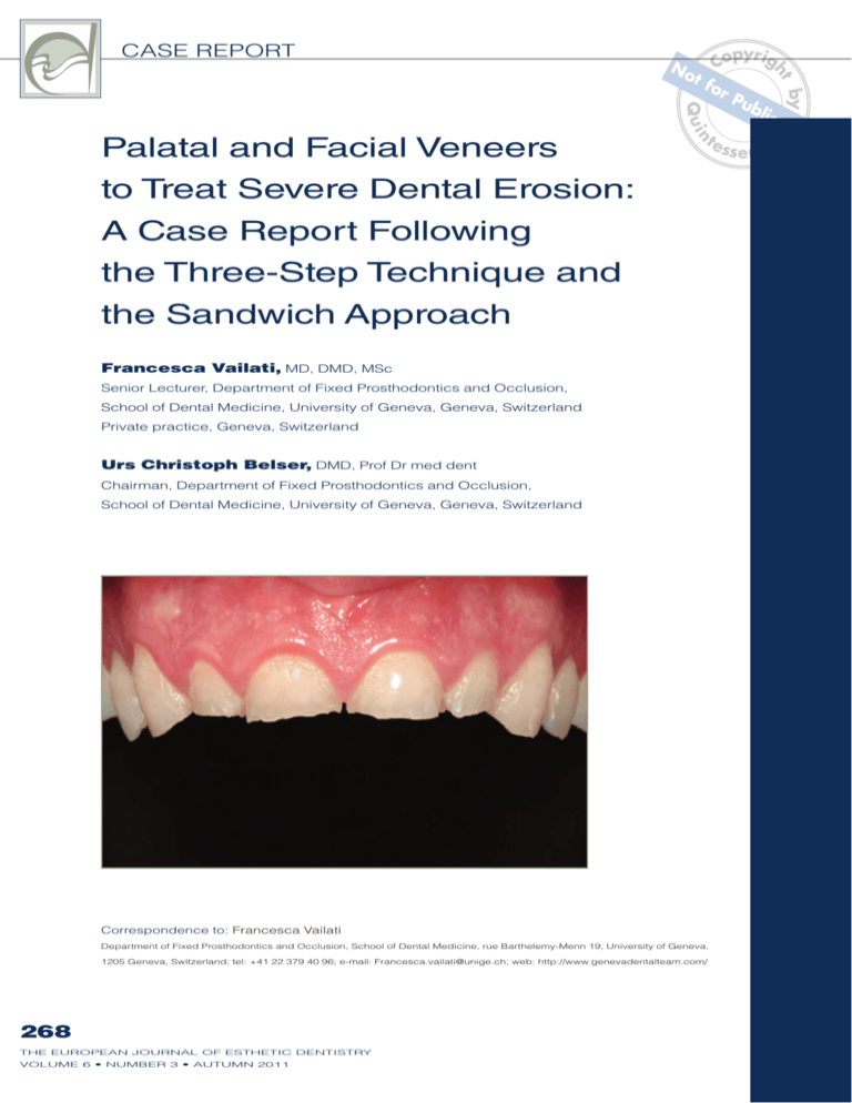
Q ui by N ht pyrig No Co t fo rP ub lica tio n te ss e n c e n to Treat Severe Dental Erosion: A Case Report Following the Three-Step Technique and the Sandwich Approach Francesca Vailati, MD, DMD, MSc Senior Lecturer, Department of Fixed Prosthodontics and Occlusion, School of Dental Medicine, University of Geneva, Geneva, Switzerland Private practice, Geneva, Switzerland Urs Christoph Belser, DMD, Prof Dr med dent Chairman, Department of Fixed Prosthodontics and Occlusion, School of Dental Medicine, University of Geneva, Geneva, Switzerland Correspondence to: Francesca Vailati Department of Fixed Prosthodontics and Occlusion, School of Dental Medicine, rue Barthelemy-Menn 19, University of Geneva, 1205 Geneva, Switzerland; tel: +41 22 379 40 96; e-mail: Francesca.vailati@unige.ch; web: http://www.genevadentalteam.com/ 268 THE EUROPEAN JOURNAL OF ESTHETIC DENTISTRY VOLUME 6 • NUMBER 3 • AUTUMN 2011 fo r Palatal and Facial Veneers ot CASE REPORT n Abstract fo r Minimally invasive principles should be crown lengthening. To preserve the pulp the driving force behind rehabilitating vitality, six palatal resin composite ven- young individuals affected by severe eers and four facial ceramic veneers dental erosion. The maxillary anterior were delivered instead with minimal, if teeth of a patient, class ACE IV, has been any, removal of tooth structure. In this treated following the most conservatory article, the details about the treatment approach, are described. the Sandwich Approach. These teeth, if restored by conventional ot Q ui by N ht VAILATI/BELSERopyrig No C t fo rP ub lica tio n te redentistry (eg, crowns) would have ss e n c e quired elective endodontic therapy and (Eur J Esthet Dent 2011;6:268–278) 269 THE EUROPEAN JOURNAL OF ESTHETIC DENTISTRY VOLUME 6 • NUMBER 3 • AUTUMN 2011 CASE REPORT Q ui by N ht pyrig No Co t fo rP ub lica tio n te (composite palatal veneers), followed by ss e n c e ot n Introduction fo r restoration of the facial aspect (ceramic Due to the work of several authors, such facial veneers). The treatment objective as Lussi and Jaeggi,1 Milosevic and was attained using the most conserva- O’Sullivan,2 Bartlett,3 and Schmidlin et tive approach possible, as the remain- al,4 more awareness about dental ero- ing tooth structure was preserved and sion is finally being raised. Many clin- located in the center between the two icians are evaluating their patients with different restorations.6-8 a fresh outlook, discovering cases in which treatment has been postponed too long, and cases where it was started Case presentation but in a too aggressive manner (conventional dentistry). A 30-year-old Caucasian male present- Since 2006 at the University of Geneva, ed at the School of Dental Medicine at patients affected by dental erosion are the University of Geneva. His chief com- treated as soon as possible after iden- plaint was the deterioration of his anter- tification of dentin exposure through the ior teeth. Since he could not afford to Geneva Erosion Study. Only adhesive receive crowns, as proposed by his clin- techniques are implemented, with mini- ician, he had fractured his incisal edges mal (if any) tooth preparation (principle significantly over the past seven years. of minimal invasiveness). Despite the The clinical examination revealed that tendency for adhesive modalities to sim- the patient had severe and generalized plify the involved clinical and laboratory dental erosion involving both the anterior procedures, the therapy of such patients and posterior teeth. All the teeth were still remains a challenge because of the vital and not at all sensitive to tempera- number of teeth affected in the same ture. He was not wearing an occlusal dentition. guard, and he did not relate his dental To simplify the dental treatment and reduce financial costs, an innovative problem to dental erosion. The gastroenterological evaluation approach termed the “three-step tech- used to establish the etiology of the nique” was developed in connection with dental erosion confirmed the presence the Geneva Erosion Study. This article of gastric reflux, and the patient started describes the full-mouth adhesive reha- a medical therapy based on histamine bilitation of one of the study patients, who H2-receptor antagonists. was affected by severe dental erosion According to the ACE classification, (ACE class IV).5 Since emphasis should the patient was considered ACE class always be placed on removing only the IV,5 since the palatal dentin was largely minimal amount of tooth structure when exposed and the loss of length of the restoring the teeth, the patient’s maxil- clinical crowns was more than two mil- lary anterior teeth were treated follow- limeters, while the facial enamel and the ing the “Sandwich Approach,” which pulp vitality were still preserved. consists of reconstruction of the lingual During the first visit (Fig 1), photos, aspect with resin composite restorations radiographs, and full-arch impressions 270 THE EUROPEAN JOURNAL OF ESTHETIC DENTISTRY VOLUME 6 • NUMBER 3 • AUTUMN 2011 n fo r a ot Q ui by N ht VAILATI/BELSERopyrig No C t fo rP ub lica tio n te ss e n c e b Fig 1 Initial status. (a) The four maxillary incisors’ incisal edges were compromised. The severe dental erosion also affected the posterior teeth, especially the maxillary premolars. (b) All of the teeth, however, kept their vitality. a b Fig 2 First clinical step: maxillary vestibular mock-up. (a) To achieve the harmony between the incisal edge plane and the occlusal plane (correction of the reverse smile), the incisors were lengthened. (b) Note how the patient’s ability to smile improves when the shape of the teeth is corrected by the mock-up. were taken. The initial visit was conclud- aspect of the maxillary teeth (from #15 ed with a face bow record. to #25) and the information obtained The maxillary and mandibular casts from the maxillary waxup was regis- were mounted in maximum intercuspal tered by means of a precise silicone position (MIP) using a semi-adjustable key. articulator. Since the patient had a very During a second clinical appointment, prominent reverse smile, to determine a maxillary mock-up was fabricated di- the lengthening of the anterior maxil- rectly in the mouth. The clinician loaded lary teeth and the related esthetic po- the silicone key with a tooth-colored sition of the occlusal plane, a maxillary auto-polymerizing resin composite ma- labial and buccal mock-up visit was terial (Telio, Ivoclar/Vivadent, Schaan, planned (first step). The technician Liechtenstein) and positioned it in the waxed up only the labial and buccal patient’s mouth. 271 THE EUROPEAN JOURNAL OF ESTHETIC DENTISTRY VOLUME 6 • NUMBER 3 • AUTUMN 2011 CASE REPORT Q ui by N ht pyrig No Co t fo rP ub lica ti testage, on teeth were not yet restored at this ss e n c e fo r an anterior open bite was created. ot n After the removal of the key, all labial and buccal surfaces of the involved maxillary teeth were covered by a thin Since the second step of the three- layer of resin composite, reproducing step technique was performed without the shape defined for the future restor- anesthesia, the patient could fully co- ations by the laboratory technician. The operate in checking and adjusting the reverse smile was corrected by length- occlusion (Fig 3). ening the anterior teeth. After the clinical validation of the posi- The protocol of the Geneva Erosion Study recommends an observation tion of the future plane of occlusion (first period of approximately 1 month to as- step), the increase of the vertical dimen- sess the patient’s adaptation to the newly sion of occlusion (VDO), mandatory for established VDO. After 1 month the pa- the restoration in this patient, was de- tient felt comfortable with the new occlu- termined subsequently on the articulator sion, and two alginate impressions and a (Fig 2). new facebow record were taken. In order The technician was asked to produce to mount the casts in MIP, an anterior oc- the waxup of the occlusal surfaces of clusal bite registration was also required. the posterior teeth, the two premolars, Since the interocclusal distance be- and the first molar in each sextant. Four tween the anterior teeth was significant, translucent silicone keys were then fab- it was decided to restore the palatal as- ricated, each duplicating the waxup of pect of the maxillary anterior teeth with one posterior quadrant (Elite Transparent, indirect restorations (resin composite Zhermack, Badia Polesine (RO), Italy). palatal veneers). The patient was then scheduled for a The interproximal contacts between third appointment. Without any anesthe- the maxillary anterior teeth were slight- sia, the exposed dentin in the four poster- ly opened by means of stripping us- ior quadrants was roughened and after ing thin diamond strips, and the incisal etching for 30 seconds the enamel, and edges were smoothened by removing for 15 seconds the dentin, the primer and the unsupported enamel prisms. The bond were applied (Optibond FL, Kerr, palatal dentin was also cleaned with Orange, CA, USA). Then the clinician non-fluoride-containing loaded each translucent key with nano- the most superficial layer was removed hybrid resin composite (Miris, Coltène with diamond burs. The exposed scle- Whaledent, Altstätten, Switzerland), po- rotic dentin was immediately sealed with sitioned the key in the patient’s mouth, Optibond FL and flowable resin com- and light-cured the resin composite. As posite (Tetric flow T, Ivoclar Vivadent) a consequence, in the single visit, with- before the final impression.9-13 For this out any tooth preparation, the occlusal patient, the preparation of the teeth for surfaces of all the premolars and the first the palatal veneers did not require local molars were restored at an increased anesthesia, and the removal of the most VDO with a layer of resin composite, superficial layer of sclerotic dentin did reproducing the respective diagnostic not involve any sensitivity. No provisional waxup (second step). Since the anterior restorations were delivered. 272 THE EUROPEAN JOURNAL OF ESTHETIC DENTISTRY VOLUME 6 • NUMBER 3 • AUTUMN 2011 pumice, and n fo r Fig 3 ot Q ui by N ht VAILATI/BELSERopyrig No C t fo rP ub lica tio n te ss e n c e Second clinical step: the provisional posterior resin composite restorations. The VDO was in- creased and an open bite was created to allow restoring the palatal aspect of the maxillary anterior teeth. a b Fig 4 Third step: resin composite palatal veneers. (a) Note the fracture of the palatal cusp of the provi- sional posterior resin composite on tooth 24. (b) Since the contact point was not missing and a final restoration was previewed anyway, the tooth was not repaired. After 1 week, the palatal veneers silane were applied (Silicup, Heraeus were bonded, one at a time, using rub- Kulzer, Hanau, Germany). A final layer ber dam. The palatal sealed dentin was of bond (Optibond FL) was used with- air abraded (Cojet, 3M, Espe, Seefeld, out curing. A warmed-up resin compos- Germany), the surrounding enamel was ite was then applied to the restorations etched (37% phosphoric acid) for 30 (Miris) before they were placed on the seconds, and the bond (Optibond FL) teeth and light cured. was applied but not cured. The resin The open contact points facilitated composite veneers were also sand- the bonding procedures, from position- blasted (Cojet) and cleaned in alcohol ing of the veneers to excess removal. and with ultrasound, and three coats of Thanks to the presence of a resin com- 273 THE EUROPEAN JOURNAL OF ESTHETIC DENTISTRY VOLUME 6 • NUMBER 3 • AUTUMN 2011 CASE REPORT Q ui by N ht pyrig No Co t fo rP ub lica ti te this on distance ss e n c e ot n posite “hook” at the level of the incisal a close view, at a social edges of the veneers, it was easier to was largely acceptable, so the patient achieve correct positioning, even on the decided to have only the four maxillary “slippery” palatal surfaces. The hooks incisors restored. ishing and polishing (Fig 4). fo r were subsequently removed during fin- The veneer preparation was carried out without local anesthesia, due to the The restoration of the palatal aspect minimal removal of tooth structure and of the maxillary anterior teeth concluded the lack of dentin exposure. The inter- the three-step technique. At this stage, proximal contact areas, already open, the patient reached a stable occlusion were further adjusted with a metallic in the anterior and posterior sextants. strip. A light chamfer was prepared at The VDO was clinically tested, and the the cervical level, following the curve of anterior guidance was re-established the marginal gingiva, with no need to (Fig 5). extend the preparation to the gingival The patient was satisfied with the sulcus (in contrast to the crown prep- esthetic of the palatal veneers even aration of devitalized teeth), since the though the incisal edges were lighter color of the underlying tooth structure compared to the remainder of the teeth, was ideal. Since the palatal aspects, and a translucent band was present at restored with resin composite veneers, the level of the junction with the veneers, were considered an integral part of the due to the intentional lack of preparation respective teeth, no particular effort was of the facial surface (eg, no facial bevel). made to place the preparation margins It was decided not to rush into the com- on tooth structure. At the incisal level, all pletion of the Sandwich Approach and the length created by the palatal veneer to bleach the teeth. was removed, and a flat preparation was However, the patient had a nail-biting habit and fractured the incisal edge of performed, paying attention to smoothing all the line angles. tooth 11 several times. The decision was After the impression, a provisional made to use the ceramic facial veneers, veneer was fabricated with the same sili- and to push the patient to stop the nail con key used for the mock-up. The key biting habit (Fig 6). was loaded with provisional resin com- Following the principle of minimal in- posite material (Telios, Ivoclar Vivadent, vasiveness, the option of leaving the fa- Schaan, Liechtenstein), and retention cial surface of the canines unrestored was achieved by both the contraction of was discussed with the patient. Since the product and the presence of minimal the facial aspect of the canines was interproximal excess. mostly intact, including these teeth in The bonding of the veneers was car- the veneer preparation would have led ried out after 2 weeks without anesthe- either to veneer preparation that was too sia, and followed the protocol developed aggressive or to final canines that were and published by Pascal Magne (Figs 7 too bulky. Although the margins be- and 8).14-18 tween the palatal veneers and the tooth The patient was clearly satisfied with structure of the canines were visible at the overall treatment. The restorations 274 THE EUROPEAN JOURNAL OF ESTHETIC DENTISTRY VOLUME 6 • NUMBER 3 • AUTUMN 2011 n fo r a Fig 5 ot Q ui by N ht VAILATI/BELSERopyrig No C t fo rP ub lica tio n te ss e n c e b (a) At the completion of the three-step technique the patient had stable occlusion, comprising posterior support at a new clinically tested VDO and anterior guidance. (b) The incisal edges added with the palatal veneers presented a lighter shade, since it was planned to bleach the patient’s teeth after protecting the exposed dentin. a Fig 6 b (a) Due to the patient’s nail biting habit, the incisal edge of one the resin composite palatal veneers was deteriorating at a faster rate. The decision was made to proceed to the fabrication of four maxillary incisor ceramic veneers. (b) Patient stated later that he had stopped using the incisal edges during his parafunctional habit after the ceramic veneers were bonded. integrated nicely with the surrounding After the completion of the Sandwich dentition (color and shape), and the soft Approach (palatal resin composite ven- tissues were healthy (esthetic success). eers and facial ceramic veneers), the Finally, the amount of tooth structure re- treatment continued with the replace- moved was minimal, and all the teeth re- ment of the posterior provisional resin tained their vitality (biological success) composite (Fig 9). the maxillary premolars and first molars restorations. Whereas all 275 THE EUROPEAN JOURNAL OF ESTHETIC DENTISTRY VOLUME 6 • NUMBER 3 • AUTUMN 2011 CASE REPORT Q ui by N ht pyrig No Co t fo rP ub lica tio n te ss e n c e fo r Fig 7 ot n a b Initial status and after veneer preparation. (a) The original tooth length was maintained, since the space necessary for the fabrication of the veneers (1.5 mm) was obtained by removing the length added by the palatal veneers. (b) Note that the rubber dam is not yet in place, since the veneer try-in with glycerin should be done as quickly as possible to verify the color match before the teeth may become dehydrated. Fig 8 Intraoral view of the final restorations at 1-year follow-up. All of the teeth retained their vitality. 276 THE EUROPEAN JOURNAL OF ESTHETIC DENTISTRY VOLUME 6 • NUMBER 3 • AUTUMN 2011 n fo r a Fig 9 ot Q ui by N ht VAILATI/BELSERopyrig No C t fo rP ub lica tio n te ss e n c e b Final result of the patient restored with the “Sandwich approach.” (a) The esthetic and biological success (all teeth vital) could not have been achieved with any other type of restoration (eg, conventional crowns). (b) Note the correction of the reverse smile, which is one of the predictable results of restoring patients following the three-step technique. a Fig 10 b (a) Occlusal view of the maxillary incisors restored with two veneers, and the canines with only one palatal veneer 1 week after facial veneer bonding. (b) Follow-up at 1 year, note that the posterior provisional restorations have been replaced by indirect resin composite restorations (full-mouth adhesive rehabilitation). were restored with indirect restorations Conclusion (resin composite onlays), the maxillary second molars and all the mandibular Dental erosion is increasing, but the den- posterior teeth were restored with direct tal community often appears to under- restorations, due to a lack of interoc- estimate the extent of the problem. The clusal space. Finally, an occlusal guard frequent lack of timely intervention is re- was given to the patient, who was en- lated not only to the slow progression of tered in the Geneva Erosion Study and the disease, which can take years before re-examined every year as part of the becoming evident to patients, but also to protocol (Fig 10). clinicians’ hesitation to propose restor- 277 THE EUROPEAN JOURNAL OF ESTHETIC DENTISTRY VOLUME 6 • NUMBER 3 • AUTUMN 2011 CASE REPORT Q ui by N ht pyrig No Co t fo rP ub lica tio n could bet ea reass e n c e ot n ative treatments based on non-invasive that early intervention adhesive procedures in asymptomatic sonable solution even for very young pa- patients. tients affected by dental erosion. fo r In this article the treatment of a 30-yearold ACE class IV patient was successfully completed. The two main goals – mini- Acknowledgements mal tooth preparation and tooth vitality The authors would like to thank Mr Alwin Schonen- preservation – were achieved, showing berger CCT, for his excellent laboratory work. References 1. 2. 3. 4. 5. 6. Lussi A, Jaeggi T. Erosion – diagnosis and risk factors. Clin Oral Investig 2008;12 (Suppl 1):S5–S13. Milosevic A, O’Sullivan E. Diagnosis, prevention and management of dental erosion: summary of an updated national guideline. Prim Dent Care 2008;15:11–12. Bartlett D. Intrinsic causes of erosion. Monogr Oral Sci 2006;20:119–139. Schmidlin PR, Filli T, Imfeld C, Tepper S, Attin T. Threeyear evaluation of posterior vertical bite reconstruction using direct resin composite – a case series. Oper Dent 2009;34:102–108. Vailati F, Belser UC. Classification and treatment of the anterior maxillary dentition affected by dental erosion: the ACE classification. Int J Periodontics Restorative Dent 2010;30:559–571. Vailati F, Belser UC. Fullmouth adhesive rehabilitation of a severely eroded dentition: the three-step technique. Part 3. Eur J Esthet Dent 2008; 3:236–257. 7. Vailati F, Belser UC. Fullmouth adhesive rehabilitation of a severely eroded dentition: the three-step technique. Part 2. Eur J Esthet Dent 2008; 3:128–146. 8. Vailati F, Belser UC. Fullmouth adhesive rehabilitation of a severely eroded dentition: the three-step technique. Part 1. Eur J Esthet Dent 2008;3:30–44. 9. Magne P, So WS, Cascione D. Immediate dentin sealing supports delayed restoration placement. J Prosthet Dent 2007;98:166–174. 10. Magne P, Kim TH, Cascione D, Donovan TE. Immediate dentin sealing improves bond strength of indirect restorations. J Prosthet Dent 2005;94:511–519. 11. Magne P. Immediate dentin sealing: a fundamental procedure for indirect bonded restorations. J Esthet Restor Dent 2005;17:144–154. 12. Paul SJ, Schärer P. The dual bonding technique: a modified method to improve adhesive luting procedures. Int J Periodontics Restorative Dent 1997;17:537–545. 278 THE EUROPEAN JOURNAL OF ESTHETIC DENTISTRY VOLUME 6 • NUMBER 3 • AUTUMN 2011 13. Bertschinger C, Paul SJ, Lüthy H, Schärer P. Dual application of dentin bonding agents: Effect on bond strength. Am J Dent 1996;9:115–119. 14. Magne P, Douglas WH. Porcelain veneers: dentin bonding optimization and biomimetic recovery of the crown. Int J Prosthodont 1999;12:111–121. 15. Belser UC, Magne P, Magne M. Ceramic laminate veneers: continuous evolution of indications. J Esthet Dent 1997;9:197–207. 16. Magne P, Belser UC. Novel porcelain laminate preparation approach driven by a diagnostic mock-up. J Esthet Restor Dent 2004;6:7–16. 17. Magne P, Perroud R, Hodges JS, Belser UC. Clinical performance of novel-design porcelain veneers for the recovery of coronal volume and length. Int J Periodontics Restorative Dent 2000; 20:440–457. 18. Magne P, Douglas WH. Additive contour of porcelain veneers: a key element in enamel preservation, adhesion, and esthetics for aging dentition. J Adhes Dent 1999;1:81–92.


