Outline Major Brain Subdivisions Optic Chiasm
advertisement
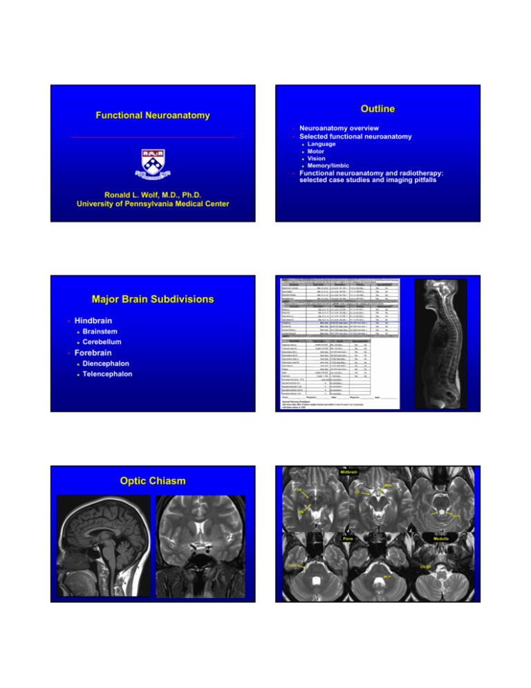
Outline Functional Neuroanatomy Neuroanatomy overview Selected functional neuroanatomy • • z z z z Language Motor Vision Memory/limbic Functional neuroanatomy and radiotherapy: selected case studies and imaging pitfalls • Ronald L. Wolf, M.D., Ph.D. University of Pennsylvania Medical Center Major Brain Subdivisions • Hindbrain z z • Brainstem Cerebellum Forebrain z z Diencephalon Telencephalon Midbrain Optic Chiasm MBs SN OT RN SCPs Pons Medulla CN V CN XII MCP V Cord, Optic Chiasm, Brainstem Hypertrophic Olivary Degeneration Level 1 Injury in triangle of Guillain and Mollaret Cord: weakness/paresis, sensory disruption Optic chiasm: blindness, field cut Pons, medulla, midbrain • • • z z z z Ipsilateral cranial nerve(s), contralateral body Disruption afferents, efferents, circuits Ataxia, tremor, swallowing, breathing, etc. Small lesions can cause big problems z z Wallenberg – N/V, vertigo, swallowing d/o Paramedian midbrain/thalamic infarcts - Akinetic mutism Goyal et al, AJNR 2000; 21: 1073 Paramedian Midbrain/Thalamic Infarcts • • • • CN III palsy Lethargic, minimally arousable R > L side weakness Memory impairment Cerebellum Primary Fissure Flocculus AL 4V PL T Nodulus Tonsil 4V Uvula (vermis ), Nodulus (vermis), Cerebellum • • • Ataxia, abnormalities in tone, equilibrium and speech Cerebellar hemispheres - ipsilateral ataxia, intention tremor, dysmetria, hypotonia, dysdiadochokinesia, nystagmas Vermis, flocculus – truncal ataxia Optic Nerves, Retina, Lacrimal Glands Level 2 Cochlea Major Brain Subdivisions Level 2 • Hindbrain z z • Forebrain z z • Neurosarcoid: Neurosarcoid: Hypothalamic Involvement HypothalamicHypothalamic-Pituitary Dysfunction Other Diencephalic Structures Hypothalamic-neurohypophysial damage • Diabetes insipidus Hypopituitarism Nonhypophysial structure damage z Disruption of autonomic functions like temperature regulation, eating, sleeping, emotion, and memory Thalamus z Hypothalamic-infundibular-portal damage z • Diencephalon Telencephalon Hypothalamus and Pituitary z • Brainstem Cerebellum Many possible disorders, eg, thermal or proprioceptive, hemiataxia, involuntary movements, pain syndromes • Subthalamic nucleus and globus pallidus • Metathalamus (LGN, MGN) • Epithalamus z z z Hemiballismus Unilateral LGN lesion – field cut Pineal, chroid plexus, stria medullaris, habenular nuclei Telencephalon Thalamus • Cortical z z z • Subcortical nuclei z T T STN T • z RN Hippocampus Amygdala, striatum (caudate nucleus, putamen, n. accumbens), septal nuclei, claustrum Important telencephalic/diencephalic interactions z SN Paleocortex – olfactory structures Archicortex – hippocampus, dentate gyrus, etc. Neocortex – most of the cortex in major lobes Basal ganglia: striatum + globus pallidus (dienceph.) Limbic system: hippocampus, cingulate gyrus, septal nuclei, etc. + hypothalamus, ant. thalamic nucleus, etc. (dienceph.) Hippocampus (Coronal, Right) a: amygdala b: hipp. body h: hipp. head s: subiculum u: uncal part arrowhead: hipp. sulcus Hippocampus: Selective Vulnerability CO Poisoning FLAIR DWI Telencephalon: Telencephalon: Major Cerebral Lobes e-Anatomy, Micheau A, Hoa D, www.imaios.com Telencephalon: Telencephalon: Nuclei Telencephalon: Telencephalon: Nuclei NA C GP P claustrum C C P P GP GP C GP T1WT1W-IR P claustrum NA PDW Brain MRI for Neoplasm Outline • • Overview basic neuroanatomy Focus on selected functional neuroanatomy z z z z • Motor Language Vision Memory/limbic • • • • • Functional neuroanatomy and radiotherapy: selected case studies and imaging pitfalls • • • Detection, location, F/U Navigation Functional anatomy, eloquent tissue Hemorrhage Cellularity Angiogenesis z Vascularity z Vascular integrity Metabolism Biochemical pathways • • • • • • • • T2, DWI, FLAIR, pre/post T1 3D T1, hires FLAIR or T2 fMRI, diffusion tensor imaging (DTI), MEG GRE, SWI Diffusion imaging (DWI, DTI) Perfusion imaging z Blood flow, volume (DSC) z Permeability (DCE) Spectroscopy Molecular imaging Sensorimotor Cortex Voluntary movements • • Primary motor – precentral gyrus (area 4) Participating z z z Supplemental motor area (SMA, area 6) Premotor – precentral gyrus (area 6) Somatosensory - parietal cortex (area 5) SMA PaCL Glioma in SMA Sup. frontal s. Precentral s. Central s. Corticospinal Tract (CST) Corticospinal Tract: DTI Tractography Courtesy Paolo Nucifora, MD, PhD, HUP CST: Wallerian Degeneration DTI: Displaced CST Oligoastrocytoma, Oligoastrocytoma, WHO II/IV Control Circuits Primary Sensory Dorsal column – medial lemniscus (vibration, proprioception) proprioception) Spinothalamic (pain, temperature) • Components z z z z • • Main circuit: neocortex > striatum > VAN, VLN thalamus > premotor and motor cortex Accessory z z z Control Circuits Striatum Globus pallidus (GP) Subthalamic nucleus (STN) and substantial nigra (SN) Red nucleus, lateral vestibular nucleus, thalamic nuclei (1) striatum > GP > thalamus > striatum (2) GP > STN > GP (3) striatum > SN > striatum DBS Placement in Subthalamic Nucleus Patterned Finger Tapping Sensorimotor cortex and control circuits Language Repeating written (or spoken) word, WernickeWernicke-Geschwind model • Right handed • Non-Right handed z z z z • Primary sensorimotor cortex SMA Putamen Thalamus Bilateral cerebellum >95% L dominant 60-65% L dominant 15-20% R dominant Rest bilateral Non-dominant hemisphere has language functions Language – Practical • z z z Broca’s area Wernicke’s area Inferior parietal lobule z z z • • Neuroanatomy and Eloquence Perisylvian areas • Supramarginal and angular gyri Associative or processing area Auditory, visual, somatosensory input z z SLF/AF connecting Additional areas z z Interindividual anatomic variations, mass effect, infiltration and reorganization Functional eloquence unpredictable based on anatomical features alone Individualized functional maps and treatment plans using techniques like fMRI, DTI, and/or MEG in addition to structural imaging SMA Head of caudate nucleus Pouratian and Bookheimer, Neurosurg Focus 2010 Ojemann et al, J Neurosurg 1989; 71: 316-326 Broca’ Broca’s Area fMRI: fMRI: Perisylvian Neoplasm Infiltrating recurrent glioma Rhyming, word generation, and sentence completion tasks FLAIR T1 T1 Combined fMRI and DTI Superior Longitudinal/Arcuate Longitudinal/Arcuate Fasciculus fMRI: fMRI: Bilateral finger tapping Tractography: Tractography: 1. Cortical spinal tracts 2. SLF/arcuate SLF/arcuate fasciculus T2 Vision fMRI: fMRI: Visual (Checkerboard) Parietal occipital sulcus (POS) POS (crosshairs) CS (arrowheads) Calcarine sulcus (CS) Optic Radiations Optic Radiations LGN LGN Optic Raditions LGN Optic Raditions Optic Raditions Optic Raditions Optic Raditions LGN Optic Tracts Optic Raditions Memory and Limbic System • Cortical z z z z • Subcortical z z z z z Yoneoka AJNR 2004; 25: 964 • Septal nuclei Preoptic area Hypothalamic nuclei and mammillary bodies Anterior thalamic nucleus Midbrain nuclei Tracts/connections z z z Hofer et al, al, FNANA 2010;4:1 Amygdala, olfactory connections Hippocampus, dentate gyrus, subiculum Parahippocampal gyrus Cingulate gyrus Fornix Mammillothalamic tract Stria medullaris, terminalis Limbic System Limbic System Amygdala, Amygdala, hippocampus: left cortical dysplasia, dysplasia, MB atrophy Mammillary bodies (MB), mammillothalamic tract (MT), fornix (F) MB F F MT tract AC F F Amygdala Pes hippocampus Mid hippocampus Limbic System Function and Injuries Septal nuclei (arrowhead), nucleus accumbens (NA), cingulate gyrus • Cingulate gyrus. gyrus. • • Emotional reactions – anger, fear, desire Homeostasis, self preservation Memory and learning AC NA MB z z Limbic Encephalitis: HSV Wernicke-Korsakoff syndrome TLE Wernicke Encephalopathy fMRI Prediction of Memory Outcome after Surgery for TLE Injury to which may impair memory? A. L TLE GOOD OUTCOME B. BAD OUTCOME C. D. R TLE E. Hippocampus Fornices, mammillary bodies, mamillothalamic tracts Septal nuclei Caudate nucleus All of the above Mesial temporal memory activation detected by fMRI during complex visual scene encoding correlates with postsurgical memory outcome Motor Mapping: Finger Tapping Outline • • Overview basic neuroanatomy Focus on selected functional neuroanatomy z z z z • LUE deficit after resection, fMRI and intraop mapping Motor Language Vision Memory/limbic Functional neuroanatomy and radiotherapy: selected case studies and imaging pitfalls z Motor and Language Central sulcus Hand and Face Motor Mapping fMRI: fMRI: GB Distorting Anatomy Atypical ganglioglioma progressing after incomplete resection T1W FLAIR fMRI: Face (G), Hand (R), Foot (B) motor mapping DTI: CST (G), AF (Y) RTOG Grade 4 Toxicity S/P GK Sept. Oct. Infiltrating Glioma Dec. fMRI: Foot (Red), Hand (Blue) motor mapping • • GK in Oct. (before middle scan) Status epilepticus soon after For the targeted volume, which is true? Surg Path: Astrocytoma WHO II 0 mo. 2 mo. (SIM) 3 mo. • A. Targeted volume B. C. D. E. Resection will not be a problem, no motor activation close enough fMRI is irrelevant, the anatomy is clear on the structural images Patient will be weak but will likely recover Patient will be permanently weak Language deficits are unlikely z z After SIM: • Decision z z Focal therapy? 59 Gy Chemo Considerations for Clinical fMRI STG • Significance of activation (or lack of activation) z Bx site z • Sentence completion 54 Gy No chemo • z “Inoperable” Inoperable” GBM: Language Mapping Passive listening Initial Can fMRI determine if an activated lesion is critical (or if a non-activating region is not critical)? Relationship of statistical test “significance” to neuronal function? Patients and pathology z z z Task performance altered by pathologic function like aphasia, or simply vision or hearing impairment Pathophysiology alters activation-flow coupling – AVM alters flow, hemosiderin destroys signal Task relevance to function in location of lesion Diffuse Infiltration Premotor LGG Surgical vs. radiation plan Speech arrest adjacent to glioma Right handed: Broca’ Broca’s Area Uncertain Significance of Poor or No Activation? No speech arrest adjacent to oligoastrocytoma (WHO II) Early case: Aphasic after resection LGG, rightright-handed patient Verbal Fluency Bilateral Motor Right handed: Progressive Astrocytoma (WHO III) February 2008 – initial preop December 2010 - progression In this right handed patient, which of the following is true? A. B. C. D. E. fMRI indicates reorganization of language to right hemisphere Pathology may create false negative left frontal activation The right hemisphere in general has no role in language If left handed, I wouldn’t be worried No problem articulating preference for Beatles over Stones False Negative Potential Cavernoma • • Preop • Caudate has role in memory and language Appropriate task? Lack of activation near cavernoma not useful Postop infarct - aphasic No Activation ≠ No Function 7y, Male AVM AVM, 7 YM Cavernoma SWI mIP Courtesy Erin Simon Schwartz, MD, CHOP Motor fMRI, fMRI, RD2: No Activation MEG: Motor Beta Band LD2 Courtesy Erin Simon Schwartz, MD, CHOP RD2 Courtesy Erin Simon Schwartz, MD, CHOP Summary • • • • Basic neuroanatomy already included in planning More detailed neuroanatomic knowledge increasingly important with radiosurgery and proton therapy Conventional anatomic imaging not sufficient due to individual variation, pathology, distortion, etc. Mapping techniques like fMRI, DTI, MEG needed T1 Atlas Stacks Acknowledgements • • • • • Gul Moonis, Beth Israel Deaconess Joe Maldjian, Wake Forest Erin Simon Schwartz, CHOP Selected images created using Cerefy Neuroradiology Atlas (www.cerefy.com) Selected images from references cited in talk Atlas Overlays
