LPS detection protocol
advertisement
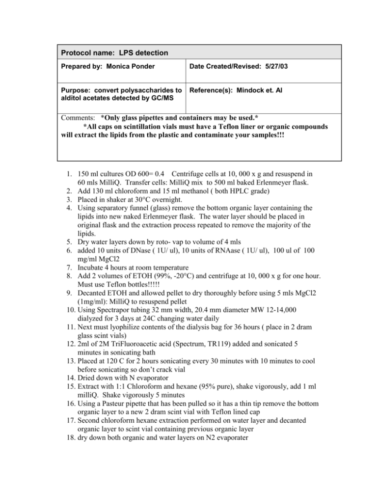
Protocol name: LPS detection Prepared by: Monica Ponder Date Created/Revised: 5/27/03 Purpose: convert polysaccharides to alditol acetates detected by GC/MS Reference(s): Mindock et. Al Comments: *Only glass pipettes and containers may be used.* *All caps on scintillation vials must have a Teflon liner or organic compounds will extract the lipids from the plastic and contaminate your samples!!! 1. 150 ml cultures OD 600= 0.4 Centrifuge cells at 10, 000 x g and resuspend in 60 mls MilliQ. Transfer cells: MilliQ mix to 500 ml baked Erlenmeyer flask. 2. Add 130 ml chloroform and 15 ml methanol ( both HPLC grade) 3. Placed in shaker at 30°C overnight. 4. Using separatory funnel (glass) remove the bottom organic layer containing the lipids into new naked Erlenmeyer flask. The water layer should be placed in original flask and the extraction process repeated to remove the majority of the lipids. 5. Dry water layers down by roto- vap to volume of 4 mls 6. added 10 units of DNase ( 1U/ ul), 10 units of RNAase ( 1U/ ul), 100 ul of 100 mg/ml MgCl2 7. Incubate 4 hours at room temperature 8. Add 2 volumes of ETOH (99%, -20°C) and centrifuge at 10, 000 x g for one hour. Must use Teflon bottles!!!!! 9. Decanted ETOH and allowed pellet to dry thoroughly before using 5 mls MgCl2 (1mg/ml): MilliQ to resuspend pellet 10. Using Spectrapor tubing 32 mm width, 20.4 mm diameter MW 12-14,000 dialyzed for 3 days at 24C changing water daily 11. Next must lyophilize contents of the dialysis bag for 36 hours ( place in 2 dram glass scint vials) 12. 2ml of 2M TriFluoroacetic acid (Spectrum, TR119) added and sonicated 5 minutes in sonicating bath 13. Placed at 120 C for 2 hours sonicating every 30 minutes with 10 minutes to cool before sonicating so don’t crack vial 14. Dried down with N evaporator 15. Extract with 1:1 Chloroform and hexane (95% pure), shake vigorously, add 1 ml milliQ. Shake vigorously 5 minutes 16. Using a Pasteur pipette that has been pulled so it has a thin tip remove the bottom organic layer to a new 2 dram scint vial with Teflon lined cap 17. Second chloroform hexane extraction performed on water layer and decanted organic layer to scint vial containing previous organic layer 18. dry down both organic and water layers on N2 evaporater 19. Add 0.5 ml MilliQ to the water layer ( top layers) to remove last traces of TFA 20. Blow to dryness 21. Made NaBorohydride solution: 0.2mg to 100 ul MilliQ. Add 2 drops from Pasteur pipette into water layers. Should see a visible fizzing. If no fizz add more drops. Next add 300 ul to remaining NaBorohydride solution in test tube mix and add 6 drops of this to water layers. Incubate overnight at room temperature. 22. Add 6 drops HCl to water layer and incubate 2 hours room temp. dry down by evaporation overnight or use N evaporator 23. Resuspend in 0.3 ml milliQ making sure all carbohydrates are resuspended by flicking sides gently. Evaporate again 24. Resuspend in 4 drops glacial acetic acid followed by 4 drops of methanol mixing thoroughly by flicking. Dried down. 25. Resuspend in 1 ml methanol, dry down, repeat 3 times to completely remove proteins and contaminants of carbohydrates. 26. Sugars are now cleaved and washed so must convert to alditiol acetates. Also create a control using 3 drops of pure glycerol that is treated same as all samples 27. Add 0.3 mls of pyridine, sonicate 10 minutes in sonicating bath 28. Add 0.1 ml of acetic anhydride, sonicated 5 minutes in bath 29. Reaction to go 24 hours at room temperature then dried down 30. Chloform: water extraction (3:1). Added 1.5 mls CHCl3 and 0.5 mls MilliQ to glass scint vials then shaken vigorously for 5 minutes. 31. Decanted bottom organic layer to new 2 dram glass scint vial with Teflon cap using a long thin Pasteur pipette. These contain the alditol acetates that are analyzed by GC:MS
