HEAD AND NECK CONTOURING TEMPLATE:
advertisement
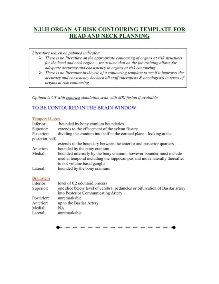
N.U.H ORGAN AT RISK CONTOURING TEMPLATE FOR HEAD AND NECK PLANNING Literature search on pubmed indicates: There is no literature on the appropriate contouring of organs at risk structures for the head and neck region – we assume that on the job training allows for adequate accuracy and consistency in organs at risk contouring There is no literature in the use of a contouring template to see if it improves the accuracy and consistency between all staff (therapists & oncologists) in terms of organs at risk contouring Optimal is CT with contrast simulation scan with MRI fusion if available TO BE CONTOURED IN THE BRAIN WINDOW Temporal Lobes Inferior: bounded by bony cranium boundaries. Superior: extends to the effacement of the sylvan fissure Posterior: dividing the cranium into half in the coronal plane - looking at the posterior half, extends to the boundary between the anterior and posterior quarters Anterior: bounded by the bony cranium Medial: bounded inferiorly by the bony cranium, however bounder must include medial temporal including the hippocampus and move laterally thereafter to not volume basal ganglia Lateral: bounded by the bony cranium. Brainstem Inferior: Superior: Posterior: Anterior: Medial: Lateral: level of C2 odontoid process one slice below level of cerebral peduncles or bifurcation of Basilar artery into Posterior Communicating Artery unremarkable up to the Basilar Artery NA unremarkable TO BE CONTOURED IN THE SOFT TISSUE WINDOW Optic Chiasm Mid-line middle cranial fossa structure located anteriorly and superiorly (by one slice) to the pituitary gland and stalk Optic Nerves Clearly identifiable – ensure it is contoured all the way to the posterior eye and optic foramen where visible. Do not visualize a nerve on a slice where it might be Lens & Eyes Unremarkable Parotid Gland This wedge shaped organ has contains the external carotid artery and some of its contents identifiable on a contrast simulation scan. These vessels should not be considered the medial border of the parotid gland but should be include in the contour. MRI fusion is of significant help. Even in the context of poor matching, it allows identification of medial and superior extent and organ shape, which can be visually match to the simulation scan. Superior border is often obscured by artifact from the reference markers placed at the mastoid process. Inferior: variable but does not extend beyond the angle of the mandible Superior: zeugmatic arch Posterior: skin and superiorly by the external auditory canal and mastoid process Anterior: rami of the mandible and variable over the masseter muscle Medial: extends deeply into the neck medial to the glenoid fossa (temporal bone) of the TMJ and medial to the styloid process beneath the mastoid process Lateral: to subcutaneous tissue Thyroid Easy to identify and contour, especially with a contrast scan, from a radiology point of view. Pituitary Attempt to identify this soft-tissue density ensuring all countours do not extend beyond the bony pituitary fossa. TO BE CONTOURED IN THE BONE WINDOW Inner Ear This includes: 1. Vestibule 2. Cochlea and 3. Semi-circular canals 4. Bony part of Cranial Nerve 8 The vestibule is the common junction of the cochlea and semi-circular canals and receives fibers from cranial nerve 8. Due to the close proximity of cranial nerve 8 and its clinical importance, it is not practical to attempt to exclude it from being contoured. Its soft-tissue - bony interface provides a consistent contouring landmark. All contours must remain within the Piteous part of the Temporal Bone. TMJ The whole joint will not be visualized on any one slice due to angulation of the neck at simulation +/- jaw opening. The joint space is convex from anterior to posterior and right to left with the joint space extending more posteriorly than anteriorly. It is for this reason that the joint space appears more prominent posteriorly rather than anteriorly (Grey’s anatomy) Inferior: one slice superior to the appearance of the sigmoid notch connecting to the coronoid process Superior: appearance of joint cavity Posterior: include glenoid cavity of temporal bone Anterior: include condyloid process of mandible Medial: joint cavity and inferiorly condyloid process Lateral: condyloid process of mandible ORGAN CHALLENGES TEMPORAL LOBES Posterior/superior extent 1) Sylvan fissure 2) Posterior/middle third boundary BRAINSTEM Superior extent 1) Bifurcation of Basilar Artery 2) 1 Slice below bifurcation of cerebral peduncles 1) Contour posterior eye to orbital foramen 1) CT-MRI fusion 2) 2 slices in width 3) sup/ant to pituitary 1) Attempt to contour pituitary, not fossa 1) Contour in CT with bone windows OPTIC NERVES OPTIC CHIASM Location PITUITARY Exact pituitary INNER EAR Bony demarcation TMJ Contour of glenoid cavity & inferior extent PAROTID Medial/superior border METHODS TO OVERCOME 1) 2) 1) 2) 3) Contour in CT with bone windows Only until sigmoid notch CT-MRI fusion Parotid can have deep medial margin Contour gland beneath external auditory meatus N.U.H ORGAN AT RISK CONTOURING TEMPLATE FOR HEAD AND NECK PLANNING Optimal is CT with contrast simulation scan with MRI fusion if available TO BE CONTOURED IN THE BRAIN WINDOW Temporal Lobes Inferior: bounded by bony cranium boundaries. Superior: extends to the effacement of the sylvan fissure Posterior: dividing the cranium into half in the coronal plane - looking at the posterior half, extends to the boundary between the anterior and posterior quarters Anterior: bounded by the bony cranium Medial: bounded inferiorly by the bony cranium, however bounder must include medial temporal including the hippocampus and move laterally thereafter to not volume basal ganglia Lateral: bounded by the bony cranium.

