Amygdala Subregion Reactivity to Social Signals of Threat in
advertisement
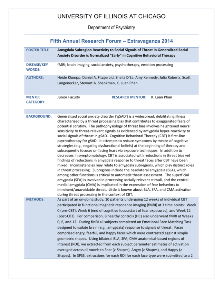
UNIVERSITY OF ILLINOIS AT CHICAGO Department of Psychiatry Fifth Annual Research Forum – Extravaganza 2014 POSTER TITLE Amygdala Subregion Reactivity to Social Signals of Threat in Generalized Social Anxiety Disorder is Normalized “Early” in Cognitive Behavioral Therapy DISEASE/KEY WORDS: fMRI; brain imaging, social anxiety, psychotherapy, emotion processing AUTHORS: Heide Klumpp, Daniel A. Fitzgerald, Sheila D’Sa, Amy Kennedy, Julia Roberts, Scott Langenecker, Stewart A. Shankman, K. Luan Phan MENTEE CATEGORY: Junior Faculty BACKGROUND: Generalized social anxiety disorder (‘gSAD’) is a widespread, debilitating illness characterized by a threat processing bias that contributes to exaggerated fears of potential scrutiny. The pathophysiology of threat bias involves heightened neural sensitivity to threat-relevant signals as evidenced by amygdala hyper-reactivity to social signals of threat in gSAD. Cognitive Behavioral Therapy (CBT) is first-line psychotherapy for gSAD. It attempts to reduce symptoms by means of cognitive strategies (e.g., negating dysfunctional beliefs) at the beginning of therapy and subsequently focuses on facing fears via exposure techniques. In addition to decreases in symptomatology, CBT is associated with reductions in threat bias yet findings of reductions in amygdala response to threat faces after CBT have been mixed. Inconsistencies may relate to amygdala subregions, which play distinct roles in threat processing. Subregions include the basolateral amygdala (BLA), which among other functions is critical to automatic threat assessment. The superficial amygdala (SFA) is involved in processing socially relevant stimuli, and the central medial amygdala (CMA) is implicated in the expression of fear behaviors to imminent/unavoidable threat. Little is known about BLA, SFA, and CMA activation during threat processing in the context of CBT. As part of an on-going study, 10 patients undergoing 12 weeks of individual CBT participated in functional magnetic resonance imaging (fMRI) at 3 time points: Week 0 (pre-CBT), Week 6 (end of cognitive focus/start of fear exposures), and Week 12 (post-CBT). For comparison, 8 healthy controls (HC) also underwent fMRI at Weeks 0, 6, and 12. During fMRI all subjects completed an Emotional Face Matching Task designed to isolate brain (e.g., amygdala) response to signals of threat. Faces comprised angry, fearful, and happy faces which were contrasted against simple geometric shapes. Using bilateral BLA, SFA, CMA anatomical-based regions of interest (ROI), we extracted from each subject parameter estimates of activation averaged across all voxels to Fear (> Shapes), Angry (> Shapes), and Happy (> Shapes). In SPSS, extractions for each ROI for each face type were submitted to a 2 METHODS: RESEARCH MENTOR: K. Luan Phan UNIVERSITY OF ILLINOIS AT CHICAGO Department of Psychiatry (Group) x 2 (Laterality) x 3 (Time) Analysis of Variance with time as a repeated measure. Significant main effects for group or group-related interactions were followed-up by two-tailed t-tests (independent, paired). The clinician-administered Liebowitz Social Anxiety Scale (“LSAS”) was used to examine symptom severity. RESULTS: CONCLUSIONS: Significant Group effects emerged for fearful faces but not for angry or happy faces. In BLA, there was a significant main effect for Time that was moderated by a Group x Time interaction. The gSAD group exhibited exaggerated bilateral amygdala reactivity to fearful faces compared to HC at Week 0 but not at Weeks 6 or 12. Within the gSAD group, BLA reactivity to fearful faces was significantly decreased by Week 6 with no further change noted at Week 12. In HC, no significant changes in BLA response over the course of time were observed. In SFA, a significant main effect for Time was moderated by a Group x Time interaction. There was also a main effect for Laterality with activation greater for right than left SFA across subjects. No Group x Time x Laterality interaction was observed. Follow-up analysis revealed a non-significant trend towards greater SFA reactivity in gSAD relative to HC at Week 0. No group differences emerged at Week 6, however, at Week 12 the gSAD group showed a significant reduction in SFA response relative to HC. Within the gSAD group, there was a significant decrease in SFA reactivity to fearful faces at Week 6 with no further decrease at Week 12. Again, in HC there were no significant changes in SFA response over time. For CMA, no group-related findings were revealed. Regarding symptom severity, LSAS was significantly reduced at Weeks 6 and 12 in gSAD. No correlations between LSAS and significant findings were observed. Preliminary findings indicate pre-CBT exaggerated bilateral BLA and SFA reactivity to fearful faces in gSAD relative to HC. By the mid-point of 12 weeks of treatment, activation in these amygdala subregions in gSAD was analogous to BLA and SFA response in HC. Notably, the HC group did not show changes in response in these regions over time indicating reliable activation to fearful faces in controls. In CBT for gSAD Week 6 largely marks the end of the cognitive phase of CBT and start of systematic exposures to fears. The BLA and SFA are in general input regions for sensory information and results suggest cognitive therapy may reduce neural sensitivity to threat-relevant signals potentially by engaging prefrontal regions. Thus, future analyses will include psychophysiological interactions analyses to examine amygdala-prefrontal interactions. Also, further study is needed to rule out nonspecific effects such as time spent in psychotherapy. Lastly, a lack of power may have reduced our ability to detect group effects for angry faces or correlations between significant results and symptom severity. Nevertheless, data suggests BLA and SFA reactivity is modulated by CBT relatively early in the course of treatment.
