1. ACLS algorithms. Cardiac arrest
advertisement
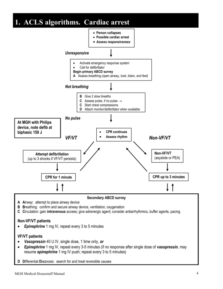
1. ACLS algorithms. Cardiac arrest Person collapses Possible cardiac arrest Assess responsiveness Unresponsive Activate emergency response system Call for defibrillator Begin primary ABCD survey A Assess breathing (open airway, look, listen, and feel) Not breathing B C C D At MGH with Philips device, note defib at biphasic 150 J Give 2 slow breaths Assess pulse, if no pulse Start chest compressions Attach monitor/defibrillator when available No pulse VF/VT CPR continues Assess rhythm Attempt defibrillation (up to 3 shocks if VF/VT persists) CPR for 1 minute Non-VF/VT 1 x 360 J (or Non-VF/VT equivalent (asystole or PEA) biphasic) within 30 to 60 seconds CPR up to 3 minutes Secondary ABCD survey A Airway: attempt to place airway device B Breathing: confirm and secure airway device, ventilation, oxygenation C Circulation: gain intravenous access; give adrenergic agent; consider antiarrhythmics, buffer agents, pacing Non-VF/VT patients Epinephrine 1 mg IV, repeat every 3 to 5 minutes VF/VT patients Vasopressin 40 U IV, single dose, 1 time only, or Epinephrine 1 mg IV, repeat every 3-5 minutes (if no response after single dose of vasopressin, may resume epinephrine 1 mg IV push; repeat every 3 to 5 minutes) D Differential Diagnosis: search for and treat reversible causes MGH Medical Housestaff Manual 4 1. ACLS algorithms. Ventricular fibrillation/Pulseless VT Primary ABCD Survey Focus: basic CPR and defibrillation Check responsiveness Activate emergency response system Call for defibrillator A Airway: open the airway B Breathing: provide positive-pressure ventilations C Circulation: give chest compressions At MGH with Philips device, note defib at biphasic 150 J D Defibrillation: assess for and shock VF/pulseless VT, up to 3 times (200 J, 200-300 J, 360 J, or equivalent biphasic) if necessary Rhythm after first 3 shocks? Persistent or recurrent VF/VT A B B B C C C D Secondary ABCD Survey Focus: more advanced assessments and treatments Airway: place airway device as soon as possible Breathing: confirm airway device placement by exam plus confirmation device Breathing: secure airway device; purpose-made tube holders preferred Breathing: confirm effective oxygenation and ventilation Circulation: establish IV access Circulation: identify rhythm monitor Circulation: administer drugs appropriate for rhythm and condition Differential diagnosis: search for and treat defined reversible causes Epinephrine 1 mg IV push, repeat every 3-5 minutes or Vasopressin 40 U IV, single dose, 1 time only Resume attempts to defibrillate 1 x 360 J (or equivalent biphasic) within 30 to 60 seconds Consider antiarrhythmics Amiodarone (IIb for persistent or recurrent VF/pulseless VT Lidocaine (Indeterminate for persistent or recurrent VF/pulseless VT) Magnesium (IIb if known hypomagnesemic state) Procainamide (Indeterminate for persistent VF/pulseless VT; IIb for recurrent VF/pulseless VT) Resume attempts to defibrillate MGH Medical Housestaff Manual 5 1. ACLS algorithms. Pulseless electrical activity Pulseless Electrical Activity (PEA = rhythm on monitor, without detectable pulse) A B C D A B B B C C C C D Primary ABCD Survey Focus: basic CPR and defibrillation ● Check responsiveness ● Activate emergency response system ● Call for defibrillator Airway: open the airway Breathing: provide positive-pressure ventilations Circulation: give chest compressions Defibrillation: assess for and shock VF/pulseless VT Secondary ABCD Survey Focus: more advanced assessments and treatments Airway: place airway device as soon as possible Breathing: confirm airway device placement by exam plus confirmation device Breathing: secure airway device; purpose-made tube holders preferred Breathing: confirm effective oxygenation and ventilation Circulation: establish IV access Circulation: identify rhythm monitor Circulation: administer drugs appropriate for rhythm and condition Circulation: assess for occult blood flow (“pseudo-EMD”) Differential diagnosis: search for and treat defined reversible causes ● ● ● ● ● ● ● ● ● ● Review for most frequent causes Hypovolemia Hypoxia Hydrogen ion—acidosis Hyper-/hypokalemia Hypothermia “Tablets” (drug OD, accidents) Tamponade, cardiac Tension, pneumothorax Thrombosis, coronary (ACS) Thrombosis, pulmonary (embolism) Epinephrine 1 mg IV push, repeat every 3 to 5 minutes Atropine 1 mg IV (if PEA rate is slow), repeat every 3 to 5 minutes as needed, to a total dose of 0.04 mg/kg MGH Medical Housestaff Manual 6 1. ACLS algorithms. Asystole Asystole Primary ABCD Survey Focus: basic CPR and defibrillation A B C C D Check responsiveness Activate emergency response system Call for defibrillator Airway: open the airway Breathing: provide positive-pressure ventilations Circulation: give chest compressions Confirm true asystole Defibrillation: assess for VF/pulseless VT; shock if indicated Rapid scene survey: any evidence personnel should not attempt resuscitation? A B B B C C C C D Secondary ABCD Survey Focus: more advanced assessments and treatments Airway: place airway device as soon as possible Breathing: confirm airway device placement by exam plus confirmation device Breathing: secure airway device; purpose-made tube holders preferred Breathing: confirm effective oxygenation and ventilation Circulation: confirm true asystole Circulation: establish IV access Circulation: identify rhythm monitor Circulation: give medications appropriate for rhythm and condition Differential diagnosis: search for and treat identified reversible causes Transcutaneous pacing If considered, perform immediately Epinephrine 1 mg IV push, Repeat every 3 to 5 min Atropine 1 mg IV, Repeat every 3 to 5 minutes up to a total of 0.04 mg/kg Asystole persists Withhold or cease resuscitation efforts? Consider quality of resuscitation? Atypical clinical features present? Support for cease-effort protocols in place? MGH Medical Housestaff Manual 7 1. ACLS algorithms. Bradycardia Bradycardias Slow (absolute bradycardia = rate <60 bpm) or ● Relatively slow (rate less than expected relative to underlying condition or cause) Primary ABCD Survey Assess ABCs Secure airway noninvasively Ensure monitor/defibrillator is available Secondary ABCD survey Assess secondary ABCs (invasive airway management needed?) Oxygen—IV access—monitor—fluids Vital signs, pulse oximeter, monitor BP Obtain and review 12-lead ECG Obtain and review portable chest x-ray Problem-focused history Problem-focused physical examination Consider causes (differential diagnoses) Serious signs or symptoms? Due to the bradycardia? No Yes Intervention sequence Atropine 0.5 to 1.0 mg Transcutaneous pacing if available Dopamine 5-20 mcg/kg/min Epinephrine 2-10 mcg/min Isoproterenol 2-10 mcg/min Type II second-degree AV block or Third-degree AV block? No Yes Observe MGH Medical Housestaff Manual Prepare for transvenous pacer If symptoms develop, use transcutaneous pacemaker until transvenous pacemaker placed 8 1. ACLS algorithms. Tachycardias overview Evaluate patient Is patient stable or unstable? Are there serious signs or symptoms? Are signs and symptoms due to tachycardia ● ● ● Stable Unstable patient: serious signs or symptoms Stable patient: no serious signs or symptoms ● ● ● Initial assessment identifies 1 of 4 types of tachycardias Establish rapid heart rate as cause of signs and symptoms Rate-related signs and symptoms occur at many rates, seldom <150 bpm ● 1. Atrial fibrillation, atrial flutter 2. Narrow-complex tachycardias Evaluation focus: 4 clinical features Treatment focus: clinical evaluation 1. Treat unstable patients urgently 2. Control the heart rate 3. Convert the rhythm 4. Provide anticoagulation Prepare for immediate cardioversion 3. Stable wide-complex tachycardia: unknown type Attempt to establish a specific diagnosis ● ● ● ● ● ● Ectopic atrial tachycardia Multifocal atrial tachycardia Paroxysmal SVT Treatment of SVT (see narrow complex tachycardia algorithm) MGH Medical Housestaff Manual 12-lead ECG Esophageal lead Clinical information Diagnostic efforts yield Confirmed SVT Treatment of atrial fibrillation/atrial flutter 4. Stable monomorphic VT and/or polymorphic VT Attempt to establish a specific diagnosis 12-lead ECG Clinical information Vagal maneuvers Adenosine ● ● ● ● 1. Patient clinically unstable? 2. Cardiac function impaired? 3. WPW present? 4. Duration <48 or >48 hours Unstable Preserved cardiac function DC cardioversion or Procainamide or Amiodarone Wide-complex tachycardia of unknown type Confirmed stable VT EF <40% Clinical CHF DC cardioversion or Amiodarone Treatment of stable monomorphic and polymorphic VT (see stable VT algorithm) 9 1. ACLS algorithms. Stable ventricular tachycardia Stable ventricular tachycardia Monomorphic or polymorphic? Monomorphic VT ● Is cardiac function impaired? Preserved heart function Note! May go directly to cardioversion Poor ejection fraction Normal baseline QT interval Medications any one ● Procainamide ● Sotalol Others acceptable ● Amiodarone ● Lidocaine Normal baseline QT interval ● Treat ischemia ● Correct electrolytes Medications: any one ● Beta-blockers or ● Lidocaine or ● Amiodarone or ● Procainamide or ● Sotalol Polymorphic VT ● Is QT baseline prolonged? Prolonged baseline QT interval (suggests torsades) Long baseline QT interval ● Correct abnormal electrolytes Therapies: any one ● Magnesium ● Overdrive pacing ● Isoproterenol ● Phenytoin ● Lidocaine Cardiac function impaired ● ● MGH Medical Housestaff Manual Amiodarone 150 mg IV over 10 minutes or Lidocaine ● 0.5-0.75 mg/kg IV push Then use Synchronized cardioversion 10 1. ACLS algorithms. Stable supraventricular tachycardia Attempt therapeutic diagnostic maneuver ● Vagal stimulation ● Adenosine Beta-blocker Ca+ channel blocker ● Amiodarone NO DC cardioversion! ● ● Preserved heart function ● Amiodarone NO DC cardioversion! Junctional tachycardia EF <40%, CHF Preserved heart function Paroxysmal supraventricular tachycardia EF <40%, CHF Preserved heart function Ectopic or multifocal atrial tachycardia Priority order: ● AV nodal blockade Beta-blocker Ca+ channel blocker Digoxin ● DC cardioversion ● Antiarrhythmics: consider procainamide, amiodarone, sotalol Priority order: ● DC cardioversion ● Digoxin ● Amiodarone ● Diltiazem Beta-blocker Ca+ channel blocker ● Amiodarone NO DC cardioversion! ● ● Amiodarone ● Diltiazem NO DC cardioversion! ● EF <40%, CHF MGH Medical Housestaff Manual 11 1. ACLS algorithms. Cardioversion Tachycardia With serious signs and symptoms related to the tachycardia If ventricular rate is >150 bpm, prepare for immediate cardioversion. May give brief trial of medications based on specific arrhythmia. Immediate cardioversion is generally not needed if heart rate if <150 bpm. Have available at bedside ● Oxygen saturation monitor ● Suction device ● IV line ● Intubation equipment Premedicate whenever possible Synchronized cardioversion ● Ventricular tachycardia ● Paroxysmal SVT ● Atrial fibrillation ● Atrial flutter 100 J, 200 J, 300 J, 360 J monophasic energy dose (or clinically equivalent biphasic energy dose Notes Effective regimens have included a sedative (e.g. diazepam, midazolam, barbiturate, etomidate, ketamine, methohexital) with or without an analgesic agent (e.g. fentanyl, morphine, meperidine). Many experts recommend anesthesia if service is readily available Both monophasic and biphasic waveforms are acceptable if documented as clinically equivalent to reports of monophasic shock success. Note possible need to resynchronize after each cardioversion If delays in synchronization occur and clinical condition is critical, go immediately to unsynchronized shocks. Treat polymorphic ventricular tachycardia (irregular form and rate) like ventricular fibrillation; see ventricular fibrillation/pulseless ventricular tachycardia algorithm. Paroxysmal supraventricular tachycardia and atrial flutter often respond to lower energy levels (start with 50 J). MGH Medical Housestaff Manual Steps for synchronized cardioversion 1. Consider sedation. 2. Turn on defibrillator (monophasic or biphasic). 3. Attach monitor leads to the patient (“white to right, red to ribs, what’s left over to the left shoulder”) and ensure proper display of the patient’s rhythm. 4. Engage the synchronization mode by pressing the “sync” control button. 5. Look for markers on R waves indicating sync mode 6. If necessary, adjust monitor gain until sync markers occur with each R wave. 7. Select appropriate energy level. 8. Position conductor pads on patient (or apply gel to paddles). 9. Position pads on patient (sternum-apex). 10. Announce to team members: “Charging defibrillator—stand clear!” 11. Press “charge” button on apex paddle (right hand). 12. When the defibrillator is charged, begin the final clearing chant. State firmly in a forceful voice the following chant before each shock: ● “I am going to shock on three. One, I’m clear.” (Check to make sure you are clear of contact with the patient, the stretcher, and the equipment.) ● “Two, you are clear.” (Make a visual check to ensure that no one continues to touch the patient or stretcher. In particular, do not forget about the person providing ventilations. That person’s hands should not be touching the ventilatory adjuncts, including the tracheal tube!) ● “Three, everybody’s clear.” (Check yourself one more time before pressing the “shock” buttons.) 13. Apply 25 lb pressure to both paddles. 14. Press the “discharge” buttons simultaneously. 15. Check the monitor. If tachycardia persists, increase the joules according to the electrical cardioversion algorithm. 16. Reset the sync mode after each synchronized cardioversion because most defibrillators default back to unsynchronized mode. This default allow an immediate defibrillation if the cardioversion produces VF. 12 1. ACLS algorithms. Antiarrhythmic drugs Adenosine Amiodarone Atenolol Atropine Diltiazem Labetalol Lidocaine Magnesium sulfate Metoprolol Procainamide Verapamil IV rapid push: Initial bolus: 6 mg over 1-3 sec, followed by normal saline bolus 20 mL; then elevate the extremity. Repeat 12 mg in 1-2 min if needed. Third dose of 12 mg may be given in 1-2 min. Cardiac arrest: 300 mg IV push. Consider repeating 150 mg IV push in 3-5 min. Max cumulative dose 2.2 g IV/24 h. Wide complex tachycardia (stable): Rapid infusion: 150 mg IV over 1st 10 min (15 mg/min). May repeat rapid infusion (150 mg IV) q10 min as needed. Slow infusion: 360 mg IV over 6 h (1 mg/min). Maintenance infusion: 540 mg IV over 18 h (0.5 mg/min) 5 mg slow IV (over 5 min). Wait 10 min, then 2nd dose 5mg slow IV (over 5 min). Asystole or PEA: 1 mg IV push. Repeat q3-5 min if asystole persists to max 0.03-0.04 mg/kg. Bradycardia: 0.5-1 mg q3-5 min as needed, max 0.04 mg/kg. Tracheal: 2-3 mg in 10 mL NS. Acute rate control: 15-20 mg (0.25 mg/kg) IV over 2 min. May repeat in 15 min at 20-25 mg (0.35 mg/kg) over 2 min. Maintenance infusion: 5-15 mg/h, titrated to heart rate. 10 mg labetalol IV push over 1-2 min. May repeat or double labetalol q10 min to max dose of 150 mg. Cardiac arrest from VF/VT: Initial 1.0-1.5 mg/kg IV. For refractory VF may give additional 0.5-0.75 mg/kg IV push, repeat in 5-10 min; max total: 3 mg/kg Stable VT, wide-complex tachycardia: 1-1.5 mg/kg IV push. Repeat 0.5-0.75 mg/kg q5-10 min. Max total 3 mg/kg. Maintenance infusion: 1-4 mg/min (30-50 mcg/kg/min). Cardiac arrest (for hypomagnesemia or torsades de pointes): 1-2 g (2-4 mL of 50% solution) diluted in 10 mL D5W IV push. Torsades de pointes (not in cardiac arrest): Load dose of 1-2 g in 50-100 mL D5W, over 560 min IV. Follow with 0.5-1 g/h IV (titrate to control torsades). Initial: 5 mg slow IV at 5 min intervals to total 15 mg. Recurrent VF/VT: 20 mg/min IV infusion (max total: 17 mg/kg). In urgent situations, up to 50 mg/min may be given in a total dose 17 mg/kg. IV infusion: 2.5-5 mg IV bolus over 2 min. 2nd dose: 5-10 mg, if needed, in 15-30 min. Max dose: 20 mg. MGH Medical Housestaff Manual 13
