Developing a Laboratory Workflow
advertisement
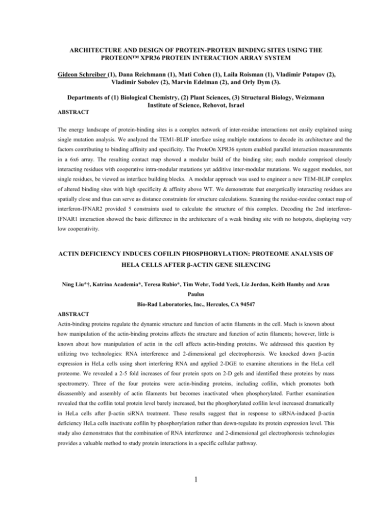
ARCHITECTURE AND DESIGN OF PROTEIN-PROTEIN BINDING SITES USING THE PROTEON™ XPR36 PROTEIN INTERACTION ARRAY SYSTEM Gideon Schreiber (1), Dana Reichmann (1), Mati Cohen (1), Laila Roisman (1), Vladimir Potapov (2), Vladimir Sobolev (2), Marvin Edelman (2), and Orly Dym (3). Departments of (1) Biological Chemistry, (2) Plant Sciences, (3) Structural Biology, Weizmann Institute of Science, Rehovot, Israel ABSTRACT The energy landscape of protein-binding sites is a complex network of inter-residue interactions not easily explained using single mutation analysis. We analyzed the TEM1-BLIP interface using multiple mutations to decode its architecture and the factors contributing to binding affinity and specificity. The ProteOn XPR36 system enabled parallel interaction measurements in a 6x6 array. The resulting contact map showed a modular build of the binding site; each module comprised closely interacting residues with cooperative intra-modular mutations yet additive inter-modular mutations. We suggest modules, not single residues, be viewed as interface building blocks. A modular approach was used to engineer a new TEM-BLIP complex of altered binding sites with high specificity & affinity above WT. We demonstrate that energetically interacting residues are spatially close and thus can serve as distance constraints for structure calculations. Scanning the residue-residue contact map of interferon-IFNAR2 provided 5 constraints used to calculate the structure of this complex. Decoding the 2nd interferonIFNAR1 interaction showed the basic difference in the architecture of a weak binding site with no hotspots, displaying very low cooperativity. ACTIN DEFICIENCY INDUCES COFILIN PHOSPHORYLATION: PROTEOME ANALYSIS OF HELA CELLS AFTER β-ACTIN GENE SILENCING Ning Liu*†, Katrina Academia*, Teresa Rubio*, Tim Wehr, Todd Yeck, Liz Jordan, Keith Hamby and Aran Paulus Bio-Rad Laboratories, Inc., Hercules, CA 94547 ABSTRACT Actin-binding proteins regulate the dynamic structure and function of actin filaments in the cell. Much is known about how manipulation of the actin-binding proteins affects the structure and function of actin filaments; however, little is known about how manipulation of actin in the cell affects actin-binding proteins. We addressed this question by utilizing two technologies: RNA interference and 2-dimensional gel electrophoresis. We knocked down β-actin expression in HeLa cells using short interfering RNA and applied 2-DGE to examine alterations in the HeLa cell proteome. We revealed a 2-5 fold increases of four protein spots on 2-D gels and identified these proteins by mass spectrometry. Three of the four proteins were actin-binding proteins, including cofilin, which promotes both disassembly and assembly of actin filaments but becomes inactivated when phosphorylated. Further examination revealed that the cofilin total protein level barely increased, but the phosphorylated cofilin level increased dramatically in HeLa cells after β-actin siRNA treatment. These results suggest that in response to siRNA-induced β-actin deficiency HeLa cells inactivate cofilin by phosphorylation rather than down-regulate its protein expression level. This study also demonstrates that the combination of RNA interference and 2-dimensional gel electrophoresis technologies provides a valuable method to study protein interactions in a specific cellular pathway. 1

